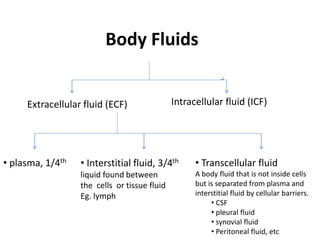
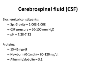
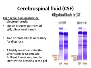
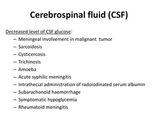
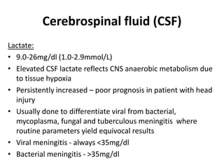
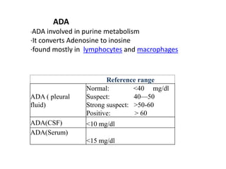
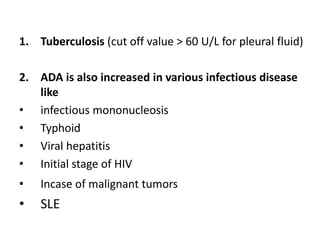
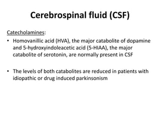
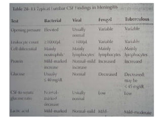
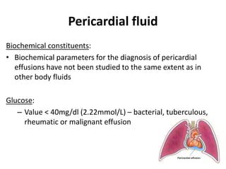
SlideShare a Scribd company logo
BODY FLUIDS
Mr. Rajesh Kumar Gupta
PG Cl. Biochemistry
class ppt
Body Fluids
Extracellular fluid (ECF) Intracellular fluid (ICF)
• plasma, 1/4th • Interstitial fluid, 3/4th
liquid found between
the cells or tissue fluid
Eg. lymph
• Transcellular fluid
A body fluid that is not inside cells
but is separated from plasma and
interstitial fluid by cellular barriers.
• CSF
• pleural fluid
• synovial fluid
• Peritoneal fluid, etc
Cerebrospinal fluid (CSF)
Cerebrospinal Fluid (CSF)
• Produced at the Choroid plexus of the 4 ventricles by
modified Ependymal cells
• At rate @20 ml / hr (adults)
• CSF flows through the Subarachnoid
space
• Where a volume of 90 – 150 ml is
maintained (adults)
• Neonate volume 10-60 mL
• Reabsorbed at the Arachnoid villus /
granulation
• to be eventually reabsorbed into the
blood
FUNCTIONS OF CSF
▪As shock absorber
▪ As mechanical buffer
▪ Act as cushion between the brain and cranium
▪ Act as a reservoir and regulates the contents of the cranium
▪ Serves as a medium for nutritional exchange in CNS
▪ Transport hormones and hormone releasing factors
▪ Removes the metabolic waste products through absorption
CSF Evaluation
• Tube 1- for cell count and differential
• Tube 2- for glucose, protein & enzymes
• Tube 3- for culture, Gram stain, AFB stain,
India ink etc
• Tube 4- for cytology
Composition of CSF
Appearance Cells DLC Protein glucose
Clear/colorless
0-5/ul
(lymphocytes
predominant)
∙Adults: 70% lymps, 30%
monos.
∙Children / newborns:
monocyte
15-45 mg/dl 45-75 mg/dl
Typical Viral Meningitis
• CSF WBC elevated, but < 250 (PMNs in early
disease, then lymphocytes)
• CSF protein elevated, but < 150 mg/dl
• Glucose > 25 mg/dlof serum concentration
Typical Bacterial Meningitis
• CSF WBC > 1000, PMN predominance
• CSF protein > 500mg/dl
• CSF glucose < 25 mg/dl
Cerebrospinal fluid (CSF)
Biochemical constituents:
– Sp. Gravity – 1.003-1.008
– CSF pressure – 60-100 mm H2O
– pH – 7.28-7.32
Proteins:
– 15-45mg/dl
– Newborn (0-1mth) – 60-120mg/dl
– Albumin/globulin – 3.1
Cerebrospinal fluid (CSF)
Mechanism of increased CSF protein:
– increased permeability of the blood brain barrier d/t damage
– Decreased reabsorption at the arachnoid villi
– Mechanical obstruction of CSF flow due to spinal block above
the puncture site
– Increased in inthrathecal immunoglobulin synthesis
• inflammatory meningitis - ↑ to about 125mg-1gm/dl
• Neurosyphilis, encephalitis, abscess, tumor - ↑ to 20-300mg/dl
• Spinal cord tumor – 100 – 2000mg/dl
Cerebrospinal fluid (CSF)
Low CSF protein:
• May normally occur in young children between 6 months to
12 years
• Patient with increased CSF turnover
– removal of large volume of CSF
– CSF leak induced by trauma or lumbar puncture
– increased intracranial pressure, probably due to an
increased rate of protein reabsorption by the arachnoid
vili
Cerebrospinal fluid (CSF)
CSF protein estimation:
1) Turbidimetric method:
– Uses trichloroacetic acid (TCA) or sulfosalicylic acid (SSA) and
sodium sulphate for protein precipitation
– Benzethonium chloride or benzalkonium chloride
2) Colorimetric method:
– Uses Lowry method (Folin phenol reagent) or
– Dye binding method using Coomassie brilliant blue (CBB) or
Ponceau S and
– Biuret method
Cerebrospinal fluid (CSF)
Pyrogallol red technique:
• Protein present in CSF will quantitatively bind with pyrogallol
red molybdate reagent dye at pH 2.5  violet colored
complex
• intensity of this colored complex is measured at 600 nm in a
spectrophotometer
• higher the concentration of protein, more intense or darker
will be the color of the CSF solution
Cerebrospinal fluid (CSF)
Serum and CSF Albumin &IgG ratio:
• assess permeability of blood brain barrier
CSF/Se. albumin index = CSF albumin (mg/dl)
Se. albumin (g/dl)
• Normal ratio: 1: 230
Cerebrospinal fluid (CSF)
CSF IgG:
• 3-5%
• MS - ↑ to about 15-18%
• Increased intrathecal IgG assessed by
CSF/Se. IgG ratio = CSF IgG (mg/dl)
Se. IgG (g/dl)
• Normal ratio: 1:390
Cerebrospinal fluid (CSF)
Electrophoresis of CSF:
• Using cellulose acetate or agarose
• Pre-albumin, albumin, alpha1, alpha2, beta1, beta2 and
gammaglobulins
• CSF always contain pre-albumin and plasma does not
• Pre-albumin or Transthyretin is thyroxine (T4) and retinol-
binding protein
Cerebrospinal fluid (CSF)
• Pattern is abnormal when IgG synthesis increases
• MS – gammaglobulin fraction↑(ologoclonal bands seen)
• Multiple sclerosis(MS): is an inflammatory disease in
which the insulating covers of nerve cells in
the brain and spinal cord are damaged
• CSF total protein/Gammaglobulin fraction exceeds 0.12 in
about 65% of cases of MS
Cerebrospinal fluid (CSF)
High resolution agarose gel
electrophoresis:
• Shows discrete patterns of
IgG, oligoclonal bands
• Two or more bands necessary
for diagnosis
• A highly sensitive stain like
silver stain or Coomassie
Brilliant Blue is required to
identify the proteins in the gel
Cerebrospinal fluid (CSF)
• detection of oligoclonal bands is performed if there is suspicion
of an inflammatory or demyelinating condition
• Concomitant serum sample for elecrophoresis and protein
estimation is mandatory
• presence of oligoclonal bands in CSF combined with their
absence in blood serum often indicates that immunoglobulins
are produced in CNS
• Oligoclonal bands are detected in upto 90% of MS
Cerebrospinal fluid (CSF)
• oligoclonal bands are also seen in:
– Panencephilitis - Various viral CNS infection
– Neurosyphilis - Neurobrucellosis
– Cryptococcal meningitis - Guillian-Barre syndrome
– Transverse myelitis - Meningial carcinomatosis
– Burkitt’s lymphoma - Chronic relapsing
polyneuropathy
– Cysticercosis - Trypanosomasis
Cerebrospinal fluid (CSF)
Glucose:
– 50-85mg/dl (2.8-4.4mmol/L), about 2/3rd of plasma value
– normal CSF/plasma glucose ratio = 0.3-0.9
– Hypoglycorrhachia = < 35mg/dl (characteristic of
bacterial, tuberculous and fungal meningitis)
– Some viral meningoencephalitis have low CSF glucose but
not to that extent as in bacterial meningoencephalitis
Cerebrospinal fluid (CSF)
Decreased level of CSF glucose:
– Meningeal involvement in malignant tumor
– Sarcoidosis
– Cysticercosis
– Trichinosis
– Amoeba
– Acute syphilic meningitis
– Intrathecial administration of radoiodinated serum albumin
– Subarachonoid haemorrhage
– Symptomatic hypoglycemia
– Rheumatoid meningitis
Cerebrospinal fluid (CSF)
• Decreased CSF glucose results from increased anaerobic
glycolysis in brain tissue and leucocytes and impaired
transport to CSF
• CSF glucose normalize before protein levels and cells count
during recovery of meningitis, making it a useful parameter
in assessing response to treatment
Cerebrospinal fluid (CSF)
Lactate:
• 9.0-26mg/dl (1.0-2.9mmol/L)
• Elevated CSF lactate reflects CNS anaerobic metabolism due
to tissue hypoxia
• Persistently increased – poor prognosis in patient with head
injury
• Usually done to differentiate viral from bacterial,
mycoplasma, fungal and tuberculous meningitis where
routine parameters yield equivocal results
• Viral meningitis - always <35mg/dl
• Bacterial meningitis - >35mg/dl
Cerebrospinal fluid (CSF)
F2 isoprostanes:
• Increased in Alzheimer’s disease
Urea:
• Level is slightly lower than in blood
• In uremia, urea conc. in CSF rises in parallel with that in
blood
Cerebrospinal fluid (CSF)
Enzymes:
Reference range
ADA ( pleural
fluid)
Normal: <40 mg/dl
Suspect: 40—50
Strong suspect: >50-60
Positive: > 60
ADA(CSF) <10 mg/dl
ADA(Serum)
<15 mg/dl
ADA
∙ADA involved in purine metabolism
∙It converts Adenosine to inosine
∙found mostly in lymphocytes and macrophages
1. Tuberculosis (cut off value > 60 U/L for pleural fluid)
2. ADA is also increased in various infectious disease
like
• infectious mononucleosis
• Typhoid
• Viral hepatitis
• Initial stage of HIV
• Incase of malignant tumors
• SLE
Cerebrospinal fluid (CSF)
Creatine kinase (CK):
• Increased CSF CK activity are seen in numerous CSF disorders:
– Hydrocephalus, cerebral infraction, primary brain tumors and
subarachnoid hemorrhage
• In patient with head trauma, CSF CK levels correlate directly with
the severity of the Concussion
• CK-BB isoenzyme – better than CK-total
Cerebrospinal fluid (CSF)
• CK-BB isoenzyme increases about 6 hour following an
ischemic or anoxic insult
CK-BB:
< 5U/L - minimum neurological damage
5-20U/L - mild to moderate neurological
damage
21-50U/L - commonly correlated with death
Cerebrospinal fluid (CSF)
LDH:
• < 40 U/L
• used as the marker in estimating the potential outcome during the
early stages of ischemic brain injury
• Also elevated in bacterial meningitis but not in aseptic or viral
meningitis
Ammonia:
• Increased levels are generally proportional to the degree of
existing hepatic encephalopathy
• Generally correlates with blood value
• Also increases in Reye’s syndrome, inherited hyperammonemias
Cerebrospinal fluid (CSF)
Catecholamines:
• Homovanillic acid (HVA), the major catabolite of dopamine
and 5-hydroxyindoleacetic acid (5-HIAA), the major
catabolite of serotonin, are normally present in CSF
• The levels of both catabolites are reduced in patients with
idiopathic or drug induced parkinsonism
Cerebrospinal fluid (CSF)
Tumor marker
• Various tumor markers have been seen increased in CSF
of patients with both primary and metastatic tumors
• eg. CEA, HCG, ALP
PLEURAL FLUID
Pleural Fluid
• pleural cavity normally contains small amount of fluid that
facilitates movement of two membranes against each other
• Plasma filtrate derived from capillaries of the parietal pleura
• Produced continuously at the rate dependent on capillary
hydrostatic pressure, plasma oncotic pressure and capillary
permeability
• Reabsorbed – lymphatics and venules of visceral pleura
• Volume – about 10ml each
Pleural Fluid
• Accumulation of fluid – an effusion, results from imbalance
between the fluid production and reabsorption
• Fluid accumulation in pleural, pericardial and peritoneal
cavities  serous effusion
Transudate
• Clear, pale yellow, watery substance
• Influenced by systemic factors that alter the
formation or absorption of fluid
• Increase in hydrostatic pressure
• Decrease in plasma oncotic pressure
• Contains few protein cells
• Common causes: CHF and liver or kidney disease
Exudate
• Pale yellow and cloudy substance
• Influenced by local factors where fluid absorption is altered
(inflammation, infection, cancer)
• Rich in protein (serum protein greater than 0.5)
• Ratio of pleural fluid LDH and serum LDH is >0.6
• Pleural fluid LDH is more the two-thirds normal upper limit
for serum
• Rich in white blood cells and immune cells
• Always has a low pH
• Common causes: pneumonia, cancer, and trauma
Pleural Fluid
According to Light’s criteria exudate meets one or more of
following criteria:
1. Pleural fluid protein/serum protein > 0.5
2. Pleural fluid LDH/serum LDH > 0.6
3. Pleural fluid LDH more than two-thirds normal upper
limit for serum
Pleural Fluid
Biochemical constituents:
Protein:
– contain < 50% of serum protein level
– Estimation helps in differentiating transudate or exudate
– Protein electrophoresis shows pattern similar to serum except
for higher proportion of albumin
Glucose:
– Similar to serum glucose level
– Low pleural fluid glucose – malignancy, TB, rheumatoid
pleuritis, non purulent bacterial infections, lupus pleuritis, etc
Pleural Fluid
Lactate:
– Useful adjunct - rapid diagnosis of infectious pleuritis
(>90mg/dl)
– Levels are significantly high in bacterial and tuberculous
pleural infections
Enzymes:
Adenosine Deaminase:
– Normal – about 36U/L
– Significantly increased in tuberculous pleuritis
Pleural Fluid
Amylase:
– Elevations above the serum level (usually 1.5-2 or more
times greater) – pancreatitis, esophageal rupture or
malignant effusion
LDH:
– Levels rise in proportion to degree of inflammation
– Declining LDH level in course of an effusion – resolving of
inflammatory process
Pleural Fluid
Interferon – gamma (INF-gamma):
– Useful diagnostic modality for TB pleural effusion
– Levels increases significantly in pleural fluid of patient
with tuberculous pleuritis (>136pg/ml)
Lipids:
– Pleural fluid Tg level > 110mg/dl – a chylous effusion
– Pleural fluid/Se. cholesterol ≥ 0.32 – exudate
Pleural Fluid
Tuberculostearic acid (TSA):
– Structural component of Mycobacteriun tuberculosis, not
present normally in human tissue
– Using gas chromatography or mass spectroscopy TSA is
measured in sputum, bronchial aspirates, washings or pleural
fluid
Tumor markers:
– Not recommended routinely
– May be useful in diagnosing cases with negative cytology or
unexplained effusions
PERITONEAL FLUID
Peritoneal fluid
• Ascites – pathologic accumulation of excess fluid in the
peritoneal cavity
• Normal volume – 50ml
• Produced as an ultrafiltrate of plasma dependent on vascular
permeability, hydrostatic and oncotic pressure
Peritoneal fluid
Biochemical constituents:
Protein:
• serum-ascites albumin gradient or gap (SAAG)
SAAG = albumin conc. of serum - albumin conc. of ascitic fluid
• high gradient:
– > 1.1 g/dL – due to portal hypertension
• Important causes of high SAAG (> 1.1 g/dL) include:
– high protein : heart failure, Budd Chiari syndrome
– low protein : cirrhosis of the liver
Peritoneal fluid
• Low gradient:
– < 1.1 g/dL - causes of ascites not associated with
increased portal pressure such
as tuberculosis, pancreatitis, nephrotic syndrome and
various types of peritoneal cancer
Glucose:
– Decreased level in tuberculous ascites (< 50mg/dl)
– Ascites glucose estimation are of little value
Peritoneal fluid
Enzymes:
Amylase:
– Amylase activity in normal peritoneal fluid is similar to
plasma level
– Level greater than three times the plasma value is
good evidence of pancreas related ascites
– Also increases in gastroduodenal perforation, acute
mesenteric vein thrombosis, intestinal strangulation
or necrosis
Peritoneal fluid
ALP:
– levels >10U/L – predicts hollow visceral injury
LDH:
– increased in malignant effusions
Telomerase:
– Increased in malignant ascites
Peritoneal fluid
ADA:
– Increases in tuberculous peritonitis
Lactate:
– Increased in malignant and tuberculous ascites
Creatinine and urea:
– Essential in differentiating peritoneal fluid from urine
– Increased peritoneal fluid urea and creatinine along with
increased serum urea but normal serum creatinine 
urinary bladder rupture
Peritoneal fluid
Bilirubin:
– Ascitic fluid bilirubin > 6mg/dl and ascitic fluid/serum
bilirubin > 1.0  choleperitoneum
Tuberculostearic acid: helpful
Tumor markers:
– little value, however, CEA, PSA, α – fetoprotein found to
be very specific for serous fluid malignancies
PERICARDIAL FLUID
Pericardial fluid
• Normal volume: 10-50ml
• Pericardial effusion – excess accumulation
• Often caused by viral infection, most common by enterovirus
Pericardial fluid
• It may be:
– transudative (congestive heart failure, myxoedema, nephrotic
syndrome),
– exudative (tuberculosis, spread from empyema)
– haemorrhagic (trauma, rupture of aneurysms, malignant
effusion).
– malignant (due to fluid accumulation caused by metastasis)
• Light’s criteria - reliable diagnostic tool for identifying pericardial
exudates and transudates
• Other indicators suggestive of exudate -
Specific gravity >1.015, total protein >3.0
mg/dL, LDH >300 U/dL, glucose fluid-to-serum
ratio < 1
Pericardial fluid
Biochemical constituents:
• Biochemical parameters for the diagnosis of pericardial
effusions have not been studied to the same extent as in
other body fluids
Glucose:
– Value < 40mg/dl (2.22mmol/L) – bacterial, tuberculous,
rheumatic or malignant effusion
Pericardial fluid
Enzymes:
LDH:
– Level > 200U/L suggests pericardial exudate
• Significantly increased pericardial fluid levels of CK-MB,
myoglobin and Troponin I in postmortem pericardial fluid –
myocardial injury
ADA :
– Useful adjunctive test for tuberculous pericardits
Pericardial fluid
Interferon-gamma:
– Increased in tuberculous pericarditis
– Cutoff value – 200pg/L
PCR:
– More specific than ADA in diagnosing tuberculous
pericarditis but
– Negative test does not rule out tuberculous pericarditis
since some pericardial fluids from patients with large
tuberculous effusions may not contain M. tuberculosis
SYNOVIAL FLUID
SYNOVIAL FLUID
• Ultrafiltrate of blood plasma combined with hyaluronic acid
produced in the joints space by the synovial cells lining
synovial tendon sheaths, joints, etc
• Composition similar to plasma as small ions and molecules
readily pass into the joint space
• Reabsorption – lymphatics
• Acts as a lubricant and adhesive, and provides nutrients for
the avascular articular cartilage
SYNOVIAL FLUID
• Examination of synovial fluid is essential to differentiate infectious
from non-infectious arthritis
Biochemical parameters:
• Adds only supportive information
to the routine test
Mucin clot test:
• Add acetic acid to SF  precipitates hyaluronate into a mucin clot
which may be graded as good, fair or poor
• Fair to poor mucin clot  reflects dilution and depolymerization
of hyaluronic acid, a non-specific finding of several inflammatory
arthrites
SYNOVIAL FLUID
Glucose:
• Proper interpretation of SF glucose values requires comparison
with serum levels, ideally preceded by eight hours fast to allow
glucose to equilibrate across the synovial membrane
• Normally, Serum – synovial = < 10mg/dl, also in many non-
inflammatory conditions
• In septic arthritis, this difference increases from 20-60mg/dl
SYNOVIAL FLUID
Protein:
• Mean normal – 1.0-3.0 g/dl
• Total protein estimation is not generally useful
• With increasing inflammation, larger proteins enter the synovial
space
Enzymes:
LDH:
• Increased in RA, gout, failed arthroplasties and infectious arthritis
reflecting neutrophilic infiltration
SYNOVIAL FLUID
Acid phosphatase:
• Elevated acid phosphatase may have negative prognostic
value in RA but is non-specific
Organic acids:
Lactic acid:
• increased in septic arthritis
• Increased > 30mg/dl  septic arthritis due to gram +ve
cocci and gram –ve bacilli
SYNOVIAL FLUID
• Using gas-liquid chromatography, presence of other organic acids
not normally present in SF (eg. n-valeric, n-hexanoic and succinic
acids) may be helpful in differentiating septic from non-septic
arthritis
Uric acid:
• Increased SF uric acid level supports a diagnosis of gout
Lipids:
• Contains extremely low conc. of lipids than in plasma
• Helps when cholesterol crystals of SF resemble MSU or CPPD
Reference intervals for synovial fluid constituents
Constituents Synovial fluid Plasma
Total protein 1 - 3 g/dl 6 – 8 g/dl
• albumin 55 – 70 % 50 – 65 %
• α1- globulin 6 - 8 % 3 – 5 %
• α2- globulin 5 – 7 % 7 – 13 %
• β – globulin 8 - 10 % 8 – 14 %
• γ – globulin 10 - 14 % 12 – 22 %
Hyaluronic acid 0.3 - 0.4 g/dl -
Glucose 70 - 110 mg/dl 70 – 110 mg/dl
Uric acid 2 – 8 mg/dl 2 – 8 mg/dl
Lactate 9 - 29 mg/dl 9 – 29 mg/dl
AMNIOTIC FLUID
AMNIOTIC FLUID
• fluid surrounds, protects, and nourishes a growing fetus
during pregnancy
• allows the baby to move relatively freely and helps maintain a
stable temperature
• increases in volume as fetus grows
• Highest – 34wks
• At first, it is mainly water with electrolytes, by about 12-14th week
- proteins, carbohydrates, lipids and phospholipids and urea(all of
which aid in the growth of the fetus)
AMNIOTIC FLUID
• detect and diagnose some birth defects, genetic diseases, and
chromosome abnormalities in a fetus, especially if pregnancy
screening tests are abnormal
• to evaluate fetal lung maturity
• obtained through a procedure
- amniocentesis
AMNIOTIC FLUID
• 15 and 20 weeks - for genetic diseases, chromosome
abnormalities and open neural tube defects
• after 32 weeks - to evaluate fetal lung maturity, when
there is an increased risk of premature delivery
AMNIOTIC FLUID
• For genetic testing and chromosome analysis, fetal cells in the
amniotic fluid are cultured and grown for several days in the
laboratory, then are analyzed
• Biochemical tests, such as bilirubin and alpha-fetoprotein, and
sometimes genetic tests can be performed directly on the
amniotic fluid
AMNIOTIC FLUID
• Chromosome analysis, a cytogenetics test that may also be called
karyotyping - detect chromosome abnormalities associated with a
variety of disorders. (It evaluates the 22 paired chromosomes and
the sex chromosomes (XY) in the nucleus of cells cultured from
those collected in the sample of amniotic fluid and can be used to
diagnose a variety of chromosomal disorders)(down’s, klinefelter,
edward, patau’s, turner)
• Genetic testing, also called molecular testing.( It looks at
fetal DNA to identify specific gene mutations and diagnose a
variety of inherited diseases)(cystic fibrosis, tay sach’s disease,
sickle cell anemia, thalessemia)
AMNIOTIC FLUID
• AFP (alpha-fetoprotein)—increased with neural tube defects
• Acetylcholinesterase—increased with neural tube defects and
also other anatomic abnormalities
• Testing to evaluate fetal lung maturity- (tests are based upon
the presence of adequate protective liquid substances called
surfactants in the lungs, which are necessary for proper lung
function)
• Tests for bilirubin may be performed on a regular basis, starting
at about 25 weeks of pregnancy, to detect, evaluate and
monitor the severity of the hemolytic anemia in the fetus
SEMINAL FLUID
SEMINAL FLUID
• Semi-gelatinous or liquid suspension containing spermatozoa and
secretions from male accessory organ
• Net fluid formed by mixing of the testicular fluid, prostatic fluid
and secretion from the seminal vesicles
• Composition is most suitable for the maintenance and survival of
spermatozoa
SEMINAL FLUID
• Has same pH as blood plasma
• Conc. of lactate, phosphate and citrate is higher than in
blood
• Chloride and cholesterol are lower
• Sugar content is high –fructose
• If fructose is low – infertility (spermatozoa can’t survive)
SEMINAL FLUID
• semen analysis is used to determine whether a man might
be infertile
Components Reference range
Volume (ml) 2.3 – 2.99
pH 7.19 – 8.47
Osmolality (mosm) 254 – 423
Fructose (mg/dl) 136 – 628
Glucose (mg/dl) 5 – 295
Total protein (mg/dl) 3700 – 7460
Albumin (mg/dl) 1100 – 2000
Urea (mg/dl) 13 – 98
Lactic acid (mg/dl) 22 – 136
Citrate (mg/dl) 304 – 751
Ca (mg/dl) 16 – 53
Cl (mg/dl) 130 – 158
K (mg/dl) 50 – 248
Mg (mg/dl) 7.89 – 31.8
Na (mg/dl) 236 – 512
Zn (mg/dl) 6.78 – 69.29
THANK-YOU

More Related Content

What's hot

Hematocrit determination
Hematocrit determinationHematocrit determination
Hematocrit determination
Hussein Al-tameemi
 
Hematology auto analyzer
Hematology auto analyzerHematology auto analyzer
Hematology auto analyzer
benazeer fathima
 
Quality control in clinical biochemistry
Quality control in clinical biochemistryQuality control in clinical biochemistry
Quality control in clinical biochemistry
Ashok Katta
 
Esr, pcv, blood indices copy
Esr, pcv, blood indices   copyEsr, pcv, blood indices   copy
Esr, pcv, blood indices copy
Janani Mathialagan
 
Haematocrit
HaematocritHaematocrit
Haematocrit
Saiduzzaman Sayid
 
PLATELET COUNT by Dr. Pandian M .pptx
PLATELET COUNT by Dr. Pandian M .pptxPLATELET COUNT by Dr. Pandian M .pptx
PLATELET COUNT by Dr. Pandian M .pptx
Pandian M
 
Automated cell counters: principle and types
Automated cell counters: principle and typesAutomated cell counters: principle and types
Automated cell counters: principle and types
Sivaranjini N
 
Blood component preparation blood banking
Blood component preparation blood bankingBlood component preparation blood banking
Blood component preparation blood banking
Appy Akshay Agarwal
 
Demonstration of le cells
Demonstration of le cellsDemonstration of le cells
Demonstration of le cells
SHRUTHI VASAN
 
Blood component, sample collection, storage, preservation
Blood component, sample collection, storage, preservationBlood component, sample collection, storage, preservation
Blood component, sample collection, storage, preservation
Nityanand Upadhyay
 
Routine examination of stool
Routine examination of stoolRoutine examination of stool
Routine examination of stool
Priyanka Buragohain
 
Osmotic fragility test
Osmotic fragility testOsmotic fragility test
Osmotic fragility test
fateh11
 
Reticulocyte count
Reticulocyte countReticulocyte count
Reticulocyte countPrbn Shah
 
Blood collection and anticoagulants
Blood collection and anticoagulantsBlood collection and anticoagulants
Blood collection and anticoagulants
Janani Mathialagan
 
Rbc count
Rbc countRbc count
Rbc count
ruchivss
 
CSF BIOCHEMICAL EXAMINATION
CSF BIOCHEMICAL EXAMINATIONCSF BIOCHEMICAL EXAMINATION
CSF BIOCHEMICAL EXAMINATION
Hussein Al-tameemi
 
Cytochemical staining checked
Cytochemical staining checkedCytochemical staining checked
Cytochemical staining checked
BALRAM KRISHAN
 
Automated cell counters
Automated cell countersAutomated cell counters
Automated cell counters
Kasturba Medical College
 

What's hot (20)

Hematocrit determination
Hematocrit determinationHematocrit determination
Hematocrit determination
 
Hematology auto analyzer
Hematology auto analyzerHematology auto analyzer
Hematology auto analyzer
 
Quality control in clinical biochemistry
Quality control in clinical biochemistryQuality control in clinical biochemistry
Quality control in clinical biochemistry
 
Esr, pcv, blood indices copy
Esr, pcv, blood indices   copyEsr, pcv, blood indices   copy
Esr, pcv, blood indices copy
 
stains
stainsstains
stains
 
Haematocrit
HaematocritHaematocrit
Haematocrit
 
PLATELET COUNT by Dr. Pandian M .pptx
PLATELET COUNT by Dr. Pandian M .pptxPLATELET COUNT by Dr. Pandian M .pptx
PLATELET COUNT by Dr. Pandian M .pptx
 
Automated cell counters: principle and types
Automated cell counters: principle and typesAutomated cell counters: principle and types
Automated cell counters: principle and types
 
Blood component preparation blood banking
Blood component preparation blood bankingBlood component preparation blood banking
Blood component preparation blood banking
 
Demonstration of le cells
Demonstration of le cellsDemonstration of le cells
Demonstration of le cells
 
Blood component, sample collection, storage, preservation
Blood component, sample collection, storage, preservationBlood component, sample collection, storage, preservation
Blood component, sample collection, storage, preservation
 
Routine examination of stool
Routine examination of stoolRoutine examination of stool
Routine examination of stool
 
Introduction to Body fluids Analysis
Introduction to Body fluids AnalysisIntroduction to Body fluids Analysis
Introduction to Body fluids Analysis
 
Osmotic fragility test
Osmotic fragility testOsmotic fragility test
Osmotic fragility test
 
Reticulocyte count
Reticulocyte countReticulocyte count
Reticulocyte count
 
Blood collection and anticoagulants
Blood collection and anticoagulantsBlood collection and anticoagulants
Blood collection and anticoagulants
 
Rbc count
Rbc countRbc count
Rbc count
 
CSF BIOCHEMICAL EXAMINATION
CSF BIOCHEMICAL EXAMINATIONCSF BIOCHEMICAL EXAMINATION
CSF BIOCHEMICAL EXAMINATION
 
Cytochemical staining checked
Cytochemical staining checkedCytochemical staining checked
Cytochemical staining checked
 
Automated cell counters
Automated cell countersAutomated cell counters
Automated cell counters
 

Viewers also liked

CSF Examination
CSF ExaminationCSF Examination
CSF Examination
Gaurav S
 
Clinical applications of Synovial (joint ) fluid analysis Dr.Sandeep C Agraw...
Clinical applications of Synovial (joint ) fluid analysis  Dr.Sandeep C Agraw...Clinical applications of Synovial (joint ) fluid analysis  Dr.Sandeep C Agraw...
Clinical applications of Synovial (joint ) fluid analysis Dr.Sandeep C Agraw...
AGRASEN Fracture Arthritis Hospital, Ganesh Nagar,Gondia,Maharashtra,INDIA
 
cytology of body fluid
 cytology of body fluid cytology of body fluid
cytology of body fluidMusa Khan
 
ppt on Joint
ppt on Jointppt on Joint
Body fluid & electrolytes........Dr.Muhammad Anwarul Kabir,FCPS(Medicine)
Body fluid & electrolytes........Dr.Muhammad Anwarul Kabir,FCPS(Medicine)Body fluid & electrolytes........Dr.Muhammad Anwarul Kabir,FCPS(Medicine)
Body fluid & electrolytes........Dr.Muhammad Anwarul Kabir,FCPS(Medicine)
kabirshiplu
 
Bones,joints ppt
Bones,joints pptBones,joints ppt
Bones,joints ppt
Sachin Goyal
 
Steps of Glycolysis in a brief
Steps of Glycolysis in a brief Steps of Glycolysis in a brief
Steps of Glycolysis in a brief
Rahman Hillol
 
Glycolysis
GlycolysisGlycolysis
Glycolysis
Tamil Silambarasan
 
Serology - Prac. Microbiology
Serology - Prac. MicrobiologySerology - Prac. Microbiology
Serology - Prac. MicrobiologyCU Dentistry 2019
 
Hb elect
Hb electHb elect
Osteoarthritis Diagnosis and Treatment
Osteoarthritis Diagnosis and TreatmentOsteoarthritis Diagnosis and Treatment
Osteoarthritis Diagnosis and Treatment
Rachmat Gunadi Wachjudi
 
Agglutination reactions
Agglutination reactionsAgglutination reactions
Agglutination reactions
Yeyeh Santos
 
parametric test of difference z test f test one-way_two-way_anova
parametric test of difference z test f test one-way_two-way_anova parametric test of difference z test f test one-way_two-way_anova
parametric test of difference z test f test one-way_two-way_anova
Tess Anoza
 
Synovial fluid
Synovial fluidSynovial fluid
Synovial fluid
Asif Zeb
 
Examination of cerebrospinal fluid presentation mode
Examination of cerebrospinal fluid presentation modeExamination of cerebrospinal fluid presentation mode
Examination of cerebrospinal fluid presentation modePavulraj Selvaraj
 
Synovial fluid analysis
Synovial fluid analysisSynovial fluid analysis
Synovial fluid analysis
Patnaik Gourishankar
 
Plasma Cell Dyscrasias & The Kidney (How they affect the kidney? When to susp...
Plasma Cell Dyscrasias & The Kidney (How they affect the kidney? When to susp...Plasma Cell Dyscrasias & The Kidney (How they affect the kidney? When to susp...
Plasma Cell Dyscrasias & The Kidney (How they affect the kidney? When to susp...
NephroTube - Dr.Gawad
 

Viewers also liked (20)

CSF Examination
CSF ExaminationCSF Examination
CSF Examination
 
Clinical applications of Synovial (joint ) fluid analysis Dr.Sandeep C Agraw...
Clinical applications of Synovial (joint ) fluid analysis  Dr.Sandeep C Agraw...Clinical applications of Synovial (joint ) fluid analysis  Dr.Sandeep C Agraw...
Clinical applications of Synovial (joint ) fluid analysis Dr.Sandeep C Agraw...
 
cytology of body fluid
 cytology of body fluid cytology of body fluid
cytology of body fluid
 
ppt on Joint
ppt on Jointppt on Joint
ppt on Joint
 
Body fluid & electrolytes........Dr.Muhammad Anwarul Kabir,FCPS(Medicine)
Body fluid & electrolytes........Dr.Muhammad Anwarul Kabir,FCPS(Medicine)Body fluid & electrolytes........Dr.Muhammad Anwarul Kabir,FCPS(Medicine)
Body fluid & electrolytes........Dr.Muhammad Anwarul Kabir,FCPS(Medicine)
 
Bones,joints ppt
Bones,joints pptBones,joints ppt
Bones,joints ppt
 
Steps of Glycolysis in a brief
Steps of Glycolysis in a brief Steps of Glycolysis in a brief
Steps of Glycolysis in a brief
 
Glycolysis
GlycolysisGlycolysis
Glycolysis
 
Synovial fluid
Synovial fluidSynovial fluid
Synovial fluid
 
Serology - Prac. Microbiology
Serology - Prac. MicrobiologySerology - Prac. Microbiology
Serology - Prac. Microbiology
 
Hb elect
Hb electHb elect
Hb elect
 
Osteoarthritis Diagnosis and Treatment
Osteoarthritis Diagnosis and TreatmentOsteoarthritis Diagnosis and Treatment
Osteoarthritis Diagnosis and Treatment
 
Agglutination reactions
Agglutination reactionsAgglutination reactions
Agglutination reactions
 
parametric test of difference z test f test one-way_two-way_anova
parametric test of difference z test f test one-way_two-way_anova parametric test of difference z test f test one-way_two-way_anova
parametric test of difference z test f test one-way_two-way_anova
 
Synovial fluid
Synovial fluidSynovial fluid
Synovial fluid
 
Examination of cerebrospinal fluid presentation mode
Examination of cerebrospinal fluid presentation modeExamination of cerebrospinal fluid presentation mode
Examination of cerebrospinal fluid presentation mode
 
Approach knee pain
Approach knee painApproach knee pain
Approach knee pain
 
Pericardial Disease
Pericardial DiseasePericardial Disease
Pericardial Disease
 
Synovial fluid analysis
Synovial fluid analysisSynovial fluid analysis
Synovial fluid analysis
 
Plasma Cell Dyscrasias & The Kidney (How they affect the kidney? When to susp...
Plasma Cell Dyscrasias & The Kidney (How they affect the kidney? When to susp...Plasma Cell Dyscrasias & The Kidney (How they affect the kidney? When to susp...
Plasma Cell Dyscrasias & The Kidney (How they affect the kidney? When to susp...
 

Similar to Body fluids

Csf in clinico laboratory diagnoses
Csf in clinico laboratory diagnosesCsf in clinico laboratory diagnoses
Csf in clinico laboratory diagnoses
Basil "Lexi" Bruno
 
Cerebral Spinal Fluid.pptx
Cerebral Spinal Fluid.pptxCerebral Spinal Fluid.pptx
Cerebral Spinal Fluid.pptx
NamanMishra87
 
Csf analysis
Csf analysis Csf analysis
Csf analysis
excitingpoint
 
CEREBROSPINAL FLUID IN HEALTH AND DISEASE.pptx
CEREBROSPINAL FLUID IN HEALTH AND  DISEASE.pptxCEREBROSPINAL FLUID IN HEALTH AND  DISEASE.pptx
CEREBROSPINAL FLUID IN HEALTH AND DISEASE.pptx
Emmanuellaodia
 
Chemical components of CSF analysis
Chemical components of CSF analysisChemical components of CSF analysis
Cerebrospinal fluid
Cerebrospinal fluidCerebrospinal fluid
Cerebrospinal fluid
Farhad Jahanfar
 
Csf composition and significance by Dr. Ashok KUmar J
Csf composition and significance by Dr. Ashok KUmar JCsf composition and significance by Dr. Ashok KUmar J
Csf composition and significance by Dr. Ashok KUmar J
International Medical School Malaysia
 
Cerebrospinal fluid (CSF) and interpreting lumbar puncture
Cerebrospinal fluid (CSF) and interpreting lumbar punctureCerebrospinal fluid (CSF) and interpreting lumbar puncture
Cerebrospinal fluid (CSF) and interpreting lumbar puncturemeducationdotnet
 
Csf seminar
Csf seminarCsf seminar
Csf seminar
Nilesh Chandra
 
CSF ANALYSIS presentation OTT 5th Semester
CSF ANALYSIS presentation OTT 5th SemesterCSF ANALYSIS presentation OTT 5th Semester
CSF ANALYSIS presentation OTT 5th Semester
Usama156008
 
body fluid analysis ppt.pptx
body fluid analysis ppt.pptxbody fluid analysis ppt.pptx
body fluid analysis ppt.pptx
FenembarMekonnen
 
Cerebral edema
Cerebral edemaCerebral edema
Cerebral edema
ShreyaYadav35
 
Central nervous system physiology and cerebral blood flow2012
Central nervous system physiology and cerebral blood flow2012Central nervous system physiology and cerebral blood flow2012
Central nervous system physiology and cerebral blood flow2012Siti Azila
 
Cerebrospinal fluid
Cerebrospinal fluidCerebrospinal fluid
Cerebrospinal fluid
SKYFALL
 
ICP_Smith.ppt
ICP_Smith.pptICP_Smith.ppt
ICP_Smith.ppt
Jigar Mehta
 
Anatomy and physiology of cns anupama
Anatomy and physiology of cns anupamaAnatomy and physiology of cns anupama
Anatomy and physiology of cns anupama
Anupama Nagar
 
Cerebrospinal fluid (CSF).pptx
Cerebrospinal fluid (CSF).pptxCerebrospinal fluid (CSF).pptx
Cerebrospinal fluid (CSF).pptx
SanjayYadav20565
 
cerebro spinal fluid analysis
 cerebro spinal fluid analysis cerebro spinal fluid analysis
cerebro spinal fluid analysis
Saint Vincent Hospital
 

Similar to Body fluids (20)

Csf in clinico laboratory diagnoses
Csf in clinico laboratory diagnosesCsf in clinico laboratory diagnoses
Csf in clinico laboratory diagnoses
 
Cerebral Spinal Fluid.pptx
Cerebral Spinal Fluid.pptxCerebral Spinal Fluid.pptx
Cerebral Spinal Fluid.pptx
 
Csf analysis
Csf analysis Csf analysis
Csf analysis
 
CEREBROSPINAL FLUID (CSF)
CEREBROSPINAL FLUID (CSF)CEREBROSPINAL FLUID (CSF)
CEREBROSPINAL FLUID (CSF)
 
CEREBROSPINAL FLUID IN HEALTH AND DISEASE.pptx
CEREBROSPINAL FLUID IN HEALTH AND  DISEASE.pptxCEREBROSPINAL FLUID IN HEALTH AND  DISEASE.pptx
CEREBROSPINAL FLUID IN HEALTH AND DISEASE.pptx
 
Chemical components of CSF analysis
Chemical components of CSF analysisChemical components of CSF analysis
Chemical components of CSF analysis
 
Cerebrospinal fluid
Cerebrospinal fluidCerebrospinal fluid
Cerebrospinal fluid
 
Csf composition and significance by Dr. Ashok KUmar J
Csf composition and significance by Dr. Ashok KUmar JCsf composition and significance by Dr. Ashok KUmar J
Csf composition and significance by Dr. Ashok KUmar J
 
Cerebrospinal Fluid
Cerebrospinal FluidCerebrospinal Fluid
Cerebrospinal Fluid
 
Cerebrospinal fluid (CSF) and interpreting lumbar puncture
Cerebrospinal fluid (CSF) and interpreting lumbar punctureCerebrospinal fluid (CSF) and interpreting lumbar puncture
Cerebrospinal fluid (CSF) and interpreting lumbar puncture
 
Csf seminar
Csf seminarCsf seminar
Csf seminar
 
CSF ANALYSIS presentation OTT 5th Semester
CSF ANALYSIS presentation OTT 5th SemesterCSF ANALYSIS presentation OTT 5th Semester
CSF ANALYSIS presentation OTT 5th Semester
 
body fluid analysis ppt.pptx
body fluid analysis ppt.pptxbody fluid analysis ppt.pptx
body fluid analysis ppt.pptx
 
Cerebral edema
Cerebral edemaCerebral edema
Cerebral edema
 
Central nervous system physiology and cerebral blood flow2012
Central nervous system physiology and cerebral blood flow2012Central nervous system physiology and cerebral blood flow2012
Central nervous system physiology and cerebral blood flow2012
 
Cerebrospinal fluid
Cerebrospinal fluidCerebrospinal fluid
Cerebrospinal fluid
 
ICP_Smith.ppt
ICP_Smith.pptICP_Smith.ppt
ICP_Smith.ppt
 
Anatomy and physiology of cns anupama
Anatomy and physiology of cns anupamaAnatomy and physiology of cns anupama
Anatomy and physiology of cns anupama
 
Cerebrospinal fluid (CSF).pptx
Cerebrospinal fluid (CSF).pptxCerebrospinal fluid (CSF).pptx
Cerebrospinal fluid (CSF).pptx
 
cerebro spinal fluid analysis
 cerebro spinal fluid analysis cerebro spinal fluid analysis
cerebro spinal fluid analysis
 

Recently uploaded

THOA 2.ppt Human Organ Transplantation Act
THOA 2.ppt Human Organ Transplantation ActTHOA 2.ppt Human Organ Transplantation Act
THOA 2.ppt Human Organ Transplantation Act
DrSathishMS1
 
Physiology of Special Chemical Sensation of Taste
Physiology of Special Chemical Sensation of TastePhysiology of Special Chemical Sensation of Taste
Physiology of Special Chemical Sensation of Taste
MedicoseAcademics
 
Maxilla, Mandible & Hyoid Bone & Clinical Correlations by Dr. RIG.pptx
Maxilla, Mandible & Hyoid Bone & Clinical Correlations by Dr. RIG.pptxMaxilla, Mandible & Hyoid Bone & Clinical Correlations by Dr. RIG.pptx
Maxilla, Mandible & Hyoid Bone & Clinical Correlations by Dr. RIG.pptx
Dr. Rabia Inam Gandapore
 
Pulmonary Thromboembolism - etilogy, types, medical- Surgical and nursing man...
Pulmonary Thromboembolism - etilogy, types, medical- Surgical and nursing man...Pulmonary Thromboembolism - etilogy, types, medical- Surgical and nursing man...
Pulmonary Thromboembolism - etilogy, types, medical- Surgical and nursing man...
VarunMahajani
 
Superficial & Deep Fascia of the NECK.pptx
Superficial & Deep Fascia of the NECK.pptxSuperficial & Deep Fascia of the NECK.pptx
Superficial & Deep Fascia of the NECK.pptx
Dr. Rabia Inam Gandapore
 
ANATOMY AND PHYSIOLOGY OF URINARY SYSTEM.pptx
ANATOMY AND PHYSIOLOGY OF URINARY SYSTEM.pptxANATOMY AND PHYSIOLOGY OF URINARY SYSTEM.pptx
ANATOMY AND PHYSIOLOGY OF URINARY SYSTEM.pptx
Swetaba Besh
 
heat stroke and heat exhaustion in children
heat stroke and heat exhaustion in childrenheat stroke and heat exhaustion in children
heat stroke and heat exhaustion in children
SumeraAhmad5
 
New Directions in Targeted Therapeutic Approaches for Older Adults With Mantl...
New Directions in Targeted Therapeutic Approaches for Older Adults With Mantl...New Directions in Targeted Therapeutic Approaches for Older Adults With Mantl...
New Directions in Targeted Therapeutic Approaches for Older Adults With Mantl...
i3 Health
 
POST OPERATIVE OLIGURIA and its management
POST OPERATIVE OLIGURIA and its managementPOST OPERATIVE OLIGURIA and its management
POST OPERATIVE OLIGURIA and its management
touseefaziz1
 
The Normal Electrocardiogram - Part I of II
The Normal Electrocardiogram - Part I of IIThe Normal Electrocardiogram - Part I of II
The Normal Electrocardiogram - Part I of II
MedicoseAcademics
 
ARTIFICIAL INTELLIGENCE IN HEALTHCARE.pdf
ARTIFICIAL INTELLIGENCE IN  HEALTHCARE.pdfARTIFICIAL INTELLIGENCE IN  HEALTHCARE.pdf
ARTIFICIAL INTELLIGENCE IN HEALTHCARE.pdf
Anujkumaranit
 
Antiulcer drugs Advance Pharmacology .pptx
Antiulcer drugs Advance Pharmacology .pptxAntiulcer drugs Advance Pharmacology .pptx
Antiulcer drugs Advance Pharmacology .pptx
Rohit chaurpagar
 
Hemodialysis: Chapter 3, Dialysis Water Unit - Dr.Gawad
Hemodialysis: Chapter 3, Dialysis Water Unit - Dr.GawadHemodialysis: Chapter 3, Dialysis Water Unit - Dr.Gawad
Hemodialysis: Chapter 3, Dialysis Water Unit - Dr.Gawad
NephroTube - Dr.Gawad
 
micro teaching on communication m.sc nursing.pdf
micro teaching on communication m.sc nursing.pdfmicro teaching on communication m.sc nursing.pdf
micro teaching on communication m.sc nursing.pdf
Anurag Sharma
 
KDIGO 2024 guidelines for diabetologists
KDIGO 2024 guidelines for diabetologistsKDIGO 2024 guidelines for diabetologists
KDIGO 2024 guidelines for diabetologists
د.محمود نجيب
 
HOT NEW PRODUCT! BIG SALES FAST SHIPPING NOW FROM CHINA!! EU KU DB BK substit...
HOT NEW PRODUCT! BIG SALES FAST SHIPPING NOW FROM CHINA!! EU KU DB BK substit...HOT NEW PRODUCT! BIG SALES FAST SHIPPING NOW FROM CHINA!! EU KU DB BK substit...
HOT NEW PRODUCT! BIG SALES FAST SHIPPING NOW FROM CHINA!! EU KU DB BK substit...
GL Anaacs
 
Report Back from SGO 2024: What’s the Latest in Cervical Cancer?
Report Back from SGO 2024: What’s the Latest in Cervical Cancer?Report Back from SGO 2024: What’s the Latest in Cervical Cancer?
Report Back from SGO 2024: What’s the Latest in Cervical Cancer?
bkling
 
Prix Galien International 2024 Forum Program
Prix Galien International 2024 Forum ProgramPrix Galien International 2024 Forum Program
Prix Galien International 2024 Forum Program
Levi Shapiro
 
Alcohol_Dr. Jeenal Mistry MD Pharmacology.pdf
Alcohol_Dr. Jeenal Mistry MD Pharmacology.pdfAlcohol_Dr. Jeenal Mistry MD Pharmacology.pdf
Alcohol_Dr. Jeenal Mistry MD Pharmacology.pdf
Dr Jeenal Mistry
 
Tom Selleck Health: A Comprehensive Look at the Iconic Actor’s Wellness Journey
Tom Selleck Health: A Comprehensive Look at the Iconic Actor’s Wellness JourneyTom Selleck Health: A Comprehensive Look at the Iconic Actor’s Wellness Journey
Tom Selleck Health: A Comprehensive Look at the Iconic Actor’s Wellness Journey
greendigital
 

Recently uploaded (20)

THOA 2.ppt Human Organ Transplantation Act
THOA 2.ppt Human Organ Transplantation ActTHOA 2.ppt Human Organ Transplantation Act
THOA 2.ppt Human Organ Transplantation Act
 
Physiology of Special Chemical Sensation of Taste
Physiology of Special Chemical Sensation of TastePhysiology of Special Chemical Sensation of Taste
Physiology of Special Chemical Sensation of Taste
 
Maxilla, Mandible & Hyoid Bone & Clinical Correlations by Dr. RIG.pptx
Maxilla, Mandible & Hyoid Bone & Clinical Correlations by Dr. RIG.pptxMaxilla, Mandible & Hyoid Bone & Clinical Correlations by Dr. RIG.pptx
Maxilla, Mandible & Hyoid Bone & Clinical Correlations by Dr. RIG.pptx
 
Pulmonary Thromboembolism - etilogy, types, medical- Surgical and nursing man...
Pulmonary Thromboembolism - etilogy, types, medical- Surgical and nursing man...Pulmonary Thromboembolism - etilogy, types, medical- Surgical and nursing man...
Pulmonary Thromboembolism - etilogy, types, medical- Surgical and nursing man...
 
Superficial & Deep Fascia of the NECK.pptx
Superficial & Deep Fascia of the NECK.pptxSuperficial & Deep Fascia of the NECK.pptx
Superficial & Deep Fascia of the NECK.pptx
 
ANATOMY AND PHYSIOLOGY OF URINARY SYSTEM.pptx
ANATOMY AND PHYSIOLOGY OF URINARY SYSTEM.pptxANATOMY AND PHYSIOLOGY OF URINARY SYSTEM.pptx
ANATOMY AND PHYSIOLOGY OF URINARY SYSTEM.pptx
 
heat stroke and heat exhaustion in children
heat stroke and heat exhaustion in childrenheat stroke and heat exhaustion in children
heat stroke and heat exhaustion in children
 
New Directions in Targeted Therapeutic Approaches for Older Adults With Mantl...
New Directions in Targeted Therapeutic Approaches for Older Adults With Mantl...New Directions in Targeted Therapeutic Approaches for Older Adults With Mantl...
New Directions in Targeted Therapeutic Approaches for Older Adults With Mantl...
 
POST OPERATIVE OLIGURIA and its management
POST OPERATIVE OLIGURIA and its managementPOST OPERATIVE OLIGURIA and its management
POST OPERATIVE OLIGURIA and its management
 
The Normal Electrocardiogram - Part I of II
The Normal Electrocardiogram - Part I of IIThe Normal Electrocardiogram - Part I of II
The Normal Electrocardiogram - Part I of II
 
ARTIFICIAL INTELLIGENCE IN HEALTHCARE.pdf
ARTIFICIAL INTELLIGENCE IN  HEALTHCARE.pdfARTIFICIAL INTELLIGENCE IN  HEALTHCARE.pdf
ARTIFICIAL INTELLIGENCE IN HEALTHCARE.pdf
 
Antiulcer drugs Advance Pharmacology .pptx
Antiulcer drugs Advance Pharmacology .pptxAntiulcer drugs Advance Pharmacology .pptx
Antiulcer drugs Advance Pharmacology .pptx
 
Hemodialysis: Chapter 3, Dialysis Water Unit - Dr.Gawad
Hemodialysis: Chapter 3, Dialysis Water Unit - Dr.GawadHemodialysis: Chapter 3, Dialysis Water Unit - Dr.Gawad
Hemodialysis: Chapter 3, Dialysis Water Unit - Dr.Gawad
 
micro teaching on communication m.sc nursing.pdf
micro teaching on communication m.sc nursing.pdfmicro teaching on communication m.sc nursing.pdf
micro teaching on communication m.sc nursing.pdf
 
KDIGO 2024 guidelines for diabetologists
KDIGO 2024 guidelines for diabetologistsKDIGO 2024 guidelines for diabetologists
KDIGO 2024 guidelines for diabetologists
 
HOT NEW PRODUCT! BIG SALES FAST SHIPPING NOW FROM CHINA!! EU KU DB BK substit...
HOT NEW PRODUCT! BIG SALES FAST SHIPPING NOW FROM CHINA!! EU KU DB BK substit...HOT NEW PRODUCT! BIG SALES FAST SHIPPING NOW FROM CHINA!! EU KU DB BK substit...
HOT NEW PRODUCT! BIG SALES FAST SHIPPING NOW FROM CHINA!! EU KU DB BK substit...
 
Report Back from SGO 2024: What’s the Latest in Cervical Cancer?
Report Back from SGO 2024: What’s the Latest in Cervical Cancer?Report Back from SGO 2024: What’s the Latest in Cervical Cancer?
Report Back from SGO 2024: What’s the Latest in Cervical Cancer?
 
Prix Galien International 2024 Forum Program
Prix Galien International 2024 Forum ProgramPrix Galien International 2024 Forum Program
Prix Galien International 2024 Forum Program
 
Alcohol_Dr. Jeenal Mistry MD Pharmacology.pdf
Alcohol_Dr. Jeenal Mistry MD Pharmacology.pdfAlcohol_Dr. Jeenal Mistry MD Pharmacology.pdf
Alcohol_Dr. Jeenal Mistry MD Pharmacology.pdf
 
Tom Selleck Health: A Comprehensive Look at the Iconic Actor’s Wellness Journey
Tom Selleck Health: A Comprehensive Look at the Iconic Actor’s Wellness JourneyTom Selleck Health: A Comprehensive Look at the Iconic Actor’s Wellness Journey
Tom Selleck Health: A Comprehensive Look at the Iconic Actor’s Wellness Journey
 

Body fluids

  • 1. BODY FLUIDS Mr. Rajesh Kumar Gupta PG Cl. Biochemistry class ppt
  • 2. Body Fluids Extracellular fluid (ECF) Intracellular fluid (ICF) • plasma, 1/4th • Interstitial fluid, 3/4th liquid found between the cells or tissue fluid Eg. lymph • Transcellular fluid A body fluid that is not inside cells but is separated from plasma and interstitial fluid by cellular barriers. • CSF • pleural fluid • synovial fluid • Peritoneal fluid, etc
  • 4. Cerebrospinal Fluid (CSF) • Produced at the Choroid plexus of the 4 ventricles by modified Ependymal cells • At rate @20 ml / hr (adults) • CSF flows through the Subarachnoid space • Where a volume of 90 – 150 ml is maintained (adults) • Neonate volume 10-60 mL • Reabsorbed at the Arachnoid villus / granulation • to be eventually reabsorbed into the blood
  • 5. FUNCTIONS OF CSF ▪As shock absorber ▪ As mechanical buffer ▪ Act as cushion between the brain and cranium ▪ Act as a reservoir and regulates the contents of the cranium ▪ Serves as a medium for nutritional exchange in CNS ▪ Transport hormones and hormone releasing factors ▪ Removes the metabolic waste products through absorption
  • 6.
  • 7. CSF Evaluation • Tube 1- for cell count and differential • Tube 2- for glucose, protein & enzymes • Tube 3- for culture, Gram stain, AFB stain, India ink etc • Tube 4- for cytology
  • 8. Composition of CSF Appearance Cells DLC Protein glucose Clear/colorless 0-5/ul (lymphocytes predominant) ∙Adults: 70% lymps, 30% monos. ∙Children / newborns: monocyte 15-45 mg/dl 45-75 mg/dl
  • 9. Typical Viral Meningitis • CSF WBC elevated, but < 250 (PMNs in early disease, then lymphocytes) • CSF protein elevated, but < 150 mg/dl • Glucose > 25 mg/dlof serum concentration
  • 10. Typical Bacterial Meningitis • CSF WBC > 1000, PMN predominance • CSF protein > 500mg/dl • CSF glucose < 25 mg/dl
  • 11. Cerebrospinal fluid (CSF) Biochemical constituents: – Sp. Gravity – 1.003-1.008 – CSF pressure – 60-100 mm H2O – pH – 7.28-7.32 Proteins: – 15-45mg/dl – Newborn (0-1mth) – 60-120mg/dl – Albumin/globulin – 3.1
  • 12. Cerebrospinal fluid (CSF) Mechanism of increased CSF protein: – increased permeability of the blood brain barrier d/t damage – Decreased reabsorption at the arachnoid villi – Mechanical obstruction of CSF flow due to spinal block above the puncture site – Increased in inthrathecal immunoglobulin synthesis • inflammatory meningitis - ↑ to about 125mg-1gm/dl • Neurosyphilis, encephalitis, abscess, tumor - ↑ to 20-300mg/dl • Spinal cord tumor – 100 – 2000mg/dl
  • 13. Cerebrospinal fluid (CSF) Low CSF protein: • May normally occur in young children between 6 months to 12 years • Patient with increased CSF turnover – removal of large volume of CSF – CSF leak induced by trauma or lumbar puncture – increased intracranial pressure, probably due to an increased rate of protein reabsorption by the arachnoid vili
  • 14. Cerebrospinal fluid (CSF) CSF protein estimation: 1) Turbidimetric method: – Uses trichloroacetic acid (TCA) or sulfosalicylic acid (SSA) and sodium sulphate for protein precipitation – Benzethonium chloride or benzalkonium chloride 2) Colorimetric method: – Uses Lowry method (Folin phenol reagent) or – Dye binding method using Coomassie brilliant blue (CBB) or Ponceau S and – Biuret method
  • 15. Cerebrospinal fluid (CSF) Pyrogallol red technique: • Protein present in CSF will quantitatively bind with pyrogallol red molybdate reagent dye at pH 2.5  violet colored complex • intensity of this colored complex is measured at 600 nm in a spectrophotometer • higher the concentration of protein, more intense or darker will be the color of the CSF solution
  • 16. Cerebrospinal fluid (CSF) Serum and CSF Albumin &IgG ratio: • assess permeability of blood brain barrier CSF/Se. albumin index = CSF albumin (mg/dl) Se. albumin (g/dl) • Normal ratio: 1: 230
  • 17. Cerebrospinal fluid (CSF) CSF IgG: • 3-5% • MS - ↑ to about 15-18% • Increased intrathecal IgG assessed by CSF/Se. IgG ratio = CSF IgG (mg/dl) Se. IgG (g/dl) • Normal ratio: 1:390
  • 19. Cerebrospinal fluid (CSF) Electrophoresis of CSF: • Using cellulose acetate or agarose • Pre-albumin, albumin, alpha1, alpha2, beta1, beta2 and gammaglobulins • CSF always contain pre-albumin and plasma does not • Pre-albumin or Transthyretin is thyroxine (T4) and retinol- binding protein
  • 20. Cerebrospinal fluid (CSF) • Pattern is abnormal when IgG synthesis increases • MS – gammaglobulin fraction↑(ologoclonal bands seen) • Multiple sclerosis(MS): is an inflammatory disease in which the insulating covers of nerve cells in the brain and spinal cord are damaged • CSF total protein/Gammaglobulin fraction exceeds 0.12 in about 65% of cases of MS
  • 21. Cerebrospinal fluid (CSF) High resolution agarose gel electrophoresis: • Shows discrete patterns of IgG, oligoclonal bands • Two or more bands necessary for diagnosis • A highly sensitive stain like silver stain or Coomassie Brilliant Blue is required to identify the proteins in the gel
  • 22. Cerebrospinal fluid (CSF) • detection of oligoclonal bands is performed if there is suspicion of an inflammatory or demyelinating condition • Concomitant serum sample for elecrophoresis and protein estimation is mandatory • presence of oligoclonal bands in CSF combined with their absence in blood serum often indicates that immunoglobulins are produced in CNS • Oligoclonal bands are detected in upto 90% of MS
  • 23. Cerebrospinal fluid (CSF) • oligoclonal bands are also seen in: – Panencephilitis - Various viral CNS infection – Neurosyphilis - Neurobrucellosis – Cryptococcal meningitis - Guillian-Barre syndrome – Transverse myelitis - Meningial carcinomatosis – Burkitt’s lymphoma - Chronic relapsing polyneuropathy – Cysticercosis - Trypanosomasis
  • 24. Cerebrospinal fluid (CSF) Glucose: – 50-85mg/dl (2.8-4.4mmol/L), about 2/3rd of plasma value – normal CSF/plasma glucose ratio = 0.3-0.9 – Hypoglycorrhachia = < 35mg/dl (characteristic of bacterial, tuberculous and fungal meningitis) – Some viral meningoencephalitis have low CSF glucose but not to that extent as in bacterial meningoencephalitis
  • 25. Cerebrospinal fluid (CSF) Decreased level of CSF glucose: – Meningeal involvement in malignant tumor – Sarcoidosis – Cysticercosis – Trichinosis – Amoeba – Acute syphilic meningitis – Intrathecial administration of radoiodinated serum albumin – Subarachonoid haemorrhage – Symptomatic hypoglycemia – Rheumatoid meningitis
  • 26. Cerebrospinal fluid (CSF) • Decreased CSF glucose results from increased anaerobic glycolysis in brain tissue and leucocytes and impaired transport to CSF • CSF glucose normalize before protein levels and cells count during recovery of meningitis, making it a useful parameter in assessing response to treatment
  • 27. Cerebrospinal fluid (CSF) Lactate: • 9.0-26mg/dl (1.0-2.9mmol/L) • Elevated CSF lactate reflects CNS anaerobic metabolism due to tissue hypoxia • Persistently increased – poor prognosis in patient with head injury • Usually done to differentiate viral from bacterial, mycoplasma, fungal and tuberculous meningitis where routine parameters yield equivocal results • Viral meningitis - always <35mg/dl • Bacterial meningitis - >35mg/dl
  • 28. Cerebrospinal fluid (CSF) F2 isoprostanes: • Increased in Alzheimer’s disease Urea: • Level is slightly lower than in blood • In uremia, urea conc. in CSF rises in parallel with that in blood
  • 30. Reference range ADA ( pleural fluid) Normal: <40 mg/dl Suspect: 40—50 Strong suspect: >50-60 Positive: > 60 ADA(CSF) <10 mg/dl ADA(Serum) <15 mg/dl ADA ∙ADA involved in purine metabolism ∙It converts Adenosine to inosine ∙found mostly in lymphocytes and macrophages
  • 31. 1. Tuberculosis (cut off value > 60 U/L for pleural fluid) 2. ADA is also increased in various infectious disease like • infectious mononucleosis • Typhoid • Viral hepatitis • Initial stage of HIV • Incase of malignant tumors • SLE
  • 32. Cerebrospinal fluid (CSF) Creatine kinase (CK): • Increased CSF CK activity are seen in numerous CSF disorders: – Hydrocephalus, cerebral infraction, primary brain tumors and subarachnoid hemorrhage • In patient with head trauma, CSF CK levels correlate directly with the severity of the Concussion • CK-BB isoenzyme – better than CK-total
  • 33. Cerebrospinal fluid (CSF) • CK-BB isoenzyme increases about 6 hour following an ischemic or anoxic insult CK-BB: < 5U/L - minimum neurological damage 5-20U/L - mild to moderate neurological damage 21-50U/L - commonly correlated with death
  • 34. Cerebrospinal fluid (CSF) LDH: • < 40 U/L • used as the marker in estimating the potential outcome during the early stages of ischemic brain injury • Also elevated in bacterial meningitis but not in aseptic or viral meningitis Ammonia: • Increased levels are generally proportional to the degree of existing hepatic encephalopathy • Generally correlates with blood value • Also increases in Reye’s syndrome, inherited hyperammonemias
  • 35. Cerebrospinal fluid (CSF) Catecholamines: • Homovanillic acid (HVA), the major catabolite of dopamine and 5-hydroxyindoleacetic acid (5-HIAA), the major catabolite of serotonin, are normally present in CSF • The levels of both catabolites are reduced in patients with idiopathic or drug induced parkinsonism
  • 36. Cerebrospinal fluid (CSF) Tumor marker • Various tumor markers have been seen increased in CSF of patients with both primary and metastatic tumors • eg. CEA, HCG, ALP
  • 37.
  • 38.
  • 39.
  • 41. Pleural Fluid • pleural cavity normally contains small amount of fluid that facilitates movement of two membranes against each other • Plasma filtrate derived from capillaries of the parietal pleura • Produced continuously at the rate dependent on capillary hydrostatic pressure, plasma oncotic pressure and capillary permeability • Reabsorbed – lymphatics and venules of visceral pleura • Volume – about 10ml each
  • 42. Pleural Fluid • Accumulation of fluid – an effusion, results from imbalance between the fluid production and reabsorption • Fluid accumulation in pleural, pericardial and peritoneal cavities  serous effusion
  • 43. Transudate • Clear, pale yellow, watery substance • Influenced by systemic factors that alter the formation or absorption of fluid • Increase in hydrostatic pressure • Decrease in plasma oncotic pressure • Contains few protein cells • Common causes: CHF and liver or kidney disease
  • 44. Exudate • Pale yellow and cloudy substance • Influenced by local factors where fluid absorption is altered (inflammation, infection, cancer) • Rich in protein (serum protein greater than 0.5) • Ratio of pleural fluid LDH and serum LDH is >0.6 • Pleural fluid LDH is more the two-thirds normal upper limit for serum • Rich in white blood cells and immune cells • Always has a low pH • Common causes: pneumonia, cancer, and trauma
  • 45. Pleural Fluid According to Light’s criteria exudate meets one or more of following criteria: 1. Pleural fluid protein/serum protein > 0.5 2. Pleural fluid LDH/serum LDH > 0.6 3. Pleural fluid LDH more than two-thirds normal upper limit for serum
  • 46. Pleural Fluid Biochemical constituents: Protein: – contain < 50% of serum protein level – Estimation helps in differentiating transudate or exudate – Protein electrophoresis shows pattern similar to serum except for higher proportion of albumin Glucose: – Similar to serum glucose level – Low pleural fluid glucose – malignancy, TB, rheumatoid pleuritis, non purulent bacterial infections, lupus pleuritis, etc
  • 47. Pleural Fluid Lactate: – Useful adjunct - rapid diagnosis of infectious pleuritis (>90mg/dl) – Levels are significantly high in bacterial and tuberculous pleural infections Enzymes: Adenosine Deaminase: – Normal – about 36U/L – Significantly increased in tuberculous pleuritis
  • 48. Pleural Fluid Amylase: – Elevations above the serum level (usually 1.5-2 or more times greater) – pancreatitis, esophageal rupture or malignant effusion LDH: – Levels rise in proportion to degree of inflammation – Declining LDH level in course of an effusion – resolving of inflammatory process
  • 49. Pleural Fluid Interferon – gamma (INF-gamma): – Useful diagnostic modality for TB pleural effusion – Levels increases significantly in pleural fluid of patient with tuberculous pleuritis (>136pg/ml) Lipids: – Pleural fluid Tg level > 110mg/dl – a chylous effusion – Pleural fluid/Se. cholesterol ≥ 0.32 – exudate
  • 50. Pleural Fluid Tuberculostearic acid (TSA): – Structural component of Mycobacteriun tuberculosis, not present normally in human tissue – Using gas chromatography or mass spectroscopy TSA is measured in sputum, bronchial aspirates, washings or pleural fluid Tumor markers: – Not recommended routinely – May be useful in diagnosing cases with negative cytology or unexplained effusions
  • 52. Peritoneal fluid • Ascites – pathologic accumulation of excess fluid in the peritoneal cavity • Normal volume – 50ml • Produced as an ultrafiltrate of plasma dependent on vascular permeability, hydrostatic and oncotic pressure
  • 53. Peritoneal fluid Biochemical constituents: Protein: • serum-ascites albumin gradient or gap (SAAG) SAAG = albumin conc. of serum - albumin conc. of ascitic fluid • high gradient: – > 1.1 g/dL – due to portal hypertension • Important causes of high SAAG (> 1.1 g/dL) include: – high protein : heart failure, Budd Chiari syndrome – low protein : cirrhosis of the liver
  • 54. Peritoneal fluid • Low gradient: – < 1.1 g/dL - causes of ascites not associated with increased portal pressure such as tuberculosis, pancreatitis, nephrotic syndrome and various types of peritoneal cancer Glucose: – Decreased level in tuberculous ascites (< 50mg/dl) – Ascites glucose estimation are of little value
  • 55. Peritoneal fluid Enzymes: Amylase: – Amylase activity in normal peritoneal fluid is similar to plasma level – Level greater than three times the plasma value is good evidence of pancreas related ascites – Also increases in gastroduodenal perforation, acute mesenteric vein thrombosis, intestinal strangulation or necrosis
  • 56. Peritoneal fluid ALP: – levels >10U/L – predicts hollow visceral injury LDH: – increased in malignant effusions Telomerase: – Increased in malignant ascites
  • 57. Peritoneal fluid ADA: – Increases in tuberculous peritonitis Lactate: – Increased in malignant and tuberculous ascites Creatinine and urea: – Essential in differentiating peritoneal fluid from urine – Increased peritoneal fluid urea and creatinine along with increased serum urea but normal serum creatinine  urinary bladder rupture
  • 58. Peritoneal fluid Bilirubin: – Ascitic fluid bilirubin > 6mg/dl and ascitic fluid/serum bilirubin > 1.0  choleperitoneum Tuberculostearic acid: helpful Tumor markers: – little value, however, CEA, PSA, α – fetoprotein found to be very specific for serous fluid malignancies
  • 60. Pericardial fluid • Normal volume: 10-50ml • Pericardial effusion – excess accumulation • Often caused by viral infection, most common by enterovirus
  • 61. Pericardial fluid • It may be: – transudative (congestive heart failure, myxoedema, nephrotic syndrome), – exudative (tuberculosis, spread from empyema) – haemorrhagic (trauma, rupture of aneurysms, malignant effusion). – malignant (due to fluid accumulation caused by metastasis) • Light’s criteria - reliable diagnostic tool for identifying pericardial exudates and transudates
  • 62. • Other indicators suggestive of exudate - Specific gravity >1.015, total protein >3.0 mg/dL, LDH >300 U/dL, glucose fluid-to-serum ratio < 1
  • 63. Pericardial fluid Biochemical constituents: • Biochemical parameters for the diagnosis of pericardial effusions have not been studied to the same extent as in other body fluids Glucose: – Value < 40mg/dl (2.22mmol/L) – bacterial, tuberculous, rheumatic or malignant effusion
  • 64. Pericardial fluid Enzymes: LDH: – Level > 200U/L suggests pericardial exudate • Significantly increased pericardial fluid levels of CK-MB, myoglobin and Troponin I in postmortem pericardial fluid – myocardial injury ADA : – Useful adjunctive test for tuberculous pericardits
  • 65. Pericardial fluid Interferon-gamma: – Increased in tuberculous pericarditis – Cutoff value – 200pg/L PCR: – More specific than ADA in diagnosing tuberculous pericarditis but – Negative test does not rule out tuberculous pericarditis since some pericardial fluids from patients with large tuberculous effusions may not contain M. tuberculosis
  • 67. SYNOVIAL FLUID • Ultrafiltrate of blood plasma combined with hyaluronic acid produced in the joints space by the synovial cells lining synovial tendon sheaths, joints, etc • Composition similar to plasma as small ions and molecules readily pass into the joint space • Reabsorption – lymphatics • Acts as a lubricant and adhesive, and provides nutrients for the avascular articular cartilage
  • 68. SYNOVIAL FLUID • Examination of synovial fluid is essential to differentiate infectious from non-infectious arthritis Biochemical parameters: • Adds only supportive information to the routine test Mucin clot test: • Add acetic acid to SF  precipitates hyaluronate into a mucin clot which may be graded as good, fair or poor • Fair to poor mucin clot  reflects dilution and depolymerization of hyaluronic acid, a non-specific finding of several inflammatory arthrites
  • 69. SYNOVIAL FLUID Glucose: • Proper interpretation of SF glucose values requires comparison with serum levels, ideally preceded by eight hours fast to allow glucose to equilibrate across the synovial membrane • Normally, Serum – synovial = < 10mg/dl, also in many non- inflammatory conditions • In septic arthritis, this difference increases from 20-60mg/dl
  • 70. SYNOVIAL FLUID Protein: • Mean normal – 1.0-3.0 g/dl • Total protein estimation is not generally useful • With increasing inflammation, larger proteins enter the synovial space Enzymes: LDH: • Increased in RA, gout, failed arthroplasties and infectious arthritis reflecting neutrophilic infiltration
  • 71. SYNOVIAL FLUID Acid phosphatase: • Elevated acid phosphatase may have negative prognostic value in RA but is non-specific Organic acids: Lactic acid: • increased in septic arthritis • Increased > 30mg/dl  septic arthritis due to gram +ve cocci and gram –ve bacilli
  • 72. SYNOVIAL FLUID • Using gas-liquid chromatography, presence of other organic acids not normally present in SF (eg. n-valeric, n-hexanoic and succinic acids) may be helpful in differentiating septic from non-septic arthritis Uric acid: • Increased SF uric acid level supports a diagnosis of gout Lipids: • Contains extremely low conc. of lipids than in plasma • Helps when cholesterol crystals of SF resemble MSU or CPPD
  • 73. Reference intervals for synovial fluid constituents Constituents Synovial fluid Plasma Total protein 1 - 3 g/dl 6 – 8 g/dl • albumin 55 – 70 % 50 – 65 % • α1- globulin 6 - 8 % 3 – 5 % • α2- globulin 5 – 7 % 7 – 13 % • β – globulin 8 - 10 % 8 – 14 % • γ – globulin 10 - 14 % 12 – 22 % Hyaluronic acid 0.3 - 0.4 g/dl - Glucose 70 - 110 mg/dl 70 – 110 mg/dl Uric acid 2 – 8 mg/dl 2 – 8 mg/dl Lactate 9 - 29 mg/dl 9 – 29 mg/dl
  • 75. AMNIOTIC FLUID • fluid surrounds, protects, and nourishes a growing fetus during pregnancy • allows the baby to move relatively freely and helps maintain a stable temperature • increases in volume as fetus grows • Highest – 34wks • At first, it is mainly water with electrolytes, by about 12-14th week - proteins, carbohydrates, lipids and phospholipids and urea(all of which aid in the growth of the fetus)
  • 76. AMNIOTIC FLUID • detect and diagnose some birth defects, genetic diseases, and chromosome abnormalities in a fetus, especially if pregnancy screening tests are abnormal • to evaluate fetal lung maturity • obtained through a procedure - amniocentesis
  • 77. AMNIOTIC FLUID • 15 and 20 weeks - for genetic diseases, chromosome abnormalities and open neural tube defects • after 32 weeks - to evaluate fetal lung maturity, when there is an increased risk of premature delivery
  • 78. AMNIOTIC FLUID • For genetic testing and chromosome analysis, fetal cells in the amniotic fluid are cultured and grown for several days in the laboratory, then are analyzed • Biochemical tests, such as bilirubin and alpha-fetoprotein, and sometimes genetic tests can be performed directly on the amniotic fluid
  • 79. AMNIOTIC FLUID • Chromosome analysis, a cytogenetics test that may also be called karyotyping - detect chromosome abnormalities associated with a variety of disorders. (It evaluates the 22 paired chromosomes and the sex chromosomes (XY) in the nucleus of cells cultured from those collected in the sample of amniotic fluid and can be used to diagnose a variety of chromosomal disorders)(down’s, klinefelter, edward, patau’s, turner) • Genetic testing, also called molecular testing.( It looks at fetal DNA to identify specific gene mutations and diagnose a variety of inherited diseases)(cystic fibrosis, tay sach’s disease, sickle cell anemia, thalessemia)
  • 80. AMNIOTIC FLUID • AFP (alpha-fetoprotein)—increased with neural tube defects • Acetylcholinesterase—increased with neural tube defects and also other anatomic abnormalities • Testing to evaluate fetal lung maturity- (tests are based upon the presence of adequate protective liquid substances called surfactants in the lungs, which are necessary for proper lung function) • Tests for bilirubin may be performed on a regular basis, starting at about 25 weeks of pregnancy, to detect, evaluate and monitor the severity of the hemolytic anemia in the fetus
  • 82. SEMINAL FLUID • Semi-gelatinous or liquid suspension containing spermatozoa and secretions from male accessory organ • Net fluid formed by mixing of the testicular fluid, prostatic fluid and secretion from the seminal vesicles • Composition is most suitable for the maintenance and survival of spermatozoa
  • 83. SEMINAL FLUID • Has same pH as blood plasma • Conc. of lactate, phosphate and citrate is higher than in blood • Chloride and cholesterol are lower • Sugar content is high –fructose • If fructose is low – infertility (spermatozoa can’t survive)
  • 84. SEMINAL FLUID • semen analysis is used to determine whether a man might be infertile
  • 85. Components Reference range Volume (ml) 2.3 – 2.99 pH 7.19 – 8.47 Osmolality (mosm) 254 – 423 Fructose (mg/dl) 136 – 628 Glucose (mg/dl) 5 – 295 Total protein (mg/dl) 3700 – 7460 Albumin (mg/dl) 1100 – 2000 Urea (mg/dl) 13 – 98 Lactic acid (mg/dl) 22 – 136 Citrate (mg/dl) 304 – 751 Ca (mg/dl) 16 – 53 Cl (mg/dl) 130 – 158 K (mg/dl) 50 – 248 Mg (mg/dl) 7.89 – 31.8 Na (mg/dl) 236 – 512 Zn (mg/dl) 6.78 – 69.29