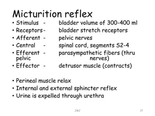
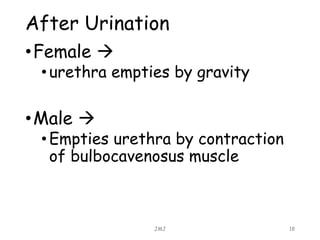
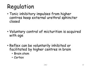
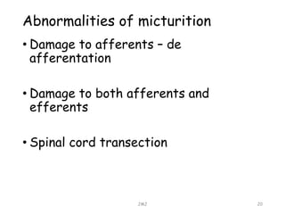
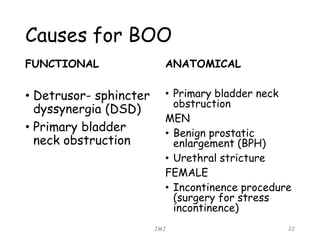
Tutorial on Bladder
Outflow Obstruction
K. Kavindya M. Fernando
JMJ 1
Anatomy of the bladder
• Made of whorls of smooth muscles, adapted for mass
contraction
• Distended bladder  globular (ovoid) shape
• Empty bladder  three sided pyramid
• Mean capacity of the bladder  220 ml
• Relations
• Apex
• Base (posterior surface)
• Inferolateral surface
• Superior surface
• Neck of the bladder
JMJ 2
JMJ 3
JMJ 4
JMJ 5
APEX
• Directed towards the top
of the pubic symphysis
• Attached to the uraches (
obliterated median
umbilican ligaments)
BASE (POSTERIOR SURFACE)
• Upper part  has peritoneal covering in males
(recto vesical pouch)
• Lower part  has no peritoneal coverings
• Males  seminal vesicles & ductus
differences are attached
• Uterers enter bladder at superolaretal
edge
• Females  firmly attached to the anterior
vaginal wall with connective tissue
INFERO-LATERAL SURFACES
• Slopes downwards and medially
• Lying against the pelvic diaphragm and obturator
internus
• Attached anteriorly to pubic bone (pubo vesical
ligament)
SUPERIOR SURFACE OF THE BLADDER
• Covered by peritoneum
• Distended bladder reaches the abdominal cavity,
while stripping peritoneum from anterior
abdominal wall
NECK OF THE BLADDER
• Base and inferior surface
meet together
• Pierced by urethra
• Attached to the base of
the prostate gland
• Most fixed part
• In females  it is related
to the pelvic fascia
Interior of the bladder
• Empty bladder
• Thick and folded mucus membrane
• Distended bladder
• Thin and stretched mucus membrane
• Trigone of bladder
• Lies in the posterior wall
• Bounded by 2 ureteric orifices and internal urethral
meatus
• Least mobile part
• Only slight increase in breath with distention of bladder
• Mucus membrane is smooth & firmly adhere to the
underlying mucus layer
• Ureters pierce bladder wall obliquely to lie at the lateral
extremity of trigone(flap valve to prevent reflux)
JMJ 6
Ligaments of the bladder
• True ligaments
• Condensation of pelvic fascia around the neck
and base of the bladder
• Lateral true ligament
• Lateral puboprostatic ligament
• Medial puboprostatic ligament ( females; pubovesical)
• Medial umbilical ligament
• Posterior ligament of bladder
JMJ 7
Ligaments of the bladder
• False ligaments
• These are peritoneal folds
• They do not form any support to the bladder
• Median umbilical fold
• Medial umbilical fod
• Lateral false ligament (formed by peritoneum of the
perivesical fossa)
• Posterior false ligament ( formed by peritoneum of
sacrogenital folds)
JMJ 8
Arterial supply to the bladder
• Mainly superior and inferior vesicle arteries
• Branches of internal iliac artery
• Small contribution from
• Obturator artery
• Inferior gluteal artery
• Uterine artery
• Vaginal artery
JMJ 9
Venous drainage of the bladder
• Do not follow arteries
• From a plexus and drain to vesico-prostatic
plexus
• Then to internal iliac vein
JMJ 10
Lymphatic drainage of the
bladder
• Runs along the arteries to
• Internal iliac nodes
• External iliac nodes
JMJ 11
Nervous supply to the bladder
JMJ 12
PARASYMPATHETIC
NERVES
• Main motor supply
• Via pelvic splanchnic
nerves
SYMPATHETIC NERVES
• Vasomotor and inhibitory to the bladder muscle
• From L1-2 via hypogastric and pelvic plexus
• Supply
• Superficial trigonal
• Internal urethral sphincter (prevent reflux
ejaculation of semen into bladder)
SENSORY NERVES
• Sensory nerves of bladder distention,
• (bladder stretch receptors),
• Travel along, Parasympathetic nerves to spinal cord (s1-2)
• Pain from bladder stone travel along sympathetic and parasympathetic fibers
to spinal cord
Normal
micturition
JMJ 13
Special features of bladder
that assist storage function
• Transitional epithelium
• Can be stretched without damage
• Impermeable to salt and water
• No exchange
• Bladder smooth muscle
• Plasticity
• Spherical structure
• Obeys laws of Laplace in filling
• P – 2T=r
JMJ 14
• First urge to void is felt at a bladder volume
of about 150ml
• Marked sense of fullness – 400 ml
• Micturition is a spinal autonomic neural
reflex
JMJ 15
JMJ 16
Micturition reflex
• Stimulus - bladder volume of 300-400 ml
• Receptors- bladder stretch receptors
• Afferent - pelvic nerves
• Central - spinal cord, segments S2-4
• Efferent - parasympathetic fibers (thru
pelvic nerves)
• Effector - detrusor muscle (contracts)
• Perineal muscle relax
• Internal and external sphincter reflex
• Urine is expelled through urethra
JMJ 17
After Urination
•Female 
•urethra empties by gravity
•Male 
•Empties urethra by contraction
of bulbocavenosus muscle
JMJ 18
Regulation
• Tonic inhibitory impulses from higher
centres keep external urethral sphincter
closed
• Voluntary control of micturition is acquired
with age
• Reflex can be voluntarily inhibited or
facilitated by higher centres in brain
• Brain stem
• Cortex
JMJ 19
Abnormalities of micturition
• Damage to afferents – de
afferentation
• Damage to both afferents and
efferents
• Spinal cord transection
JMJ 20
BLADDER OUTLET
OBSTRUCTION
JMJ 21
Causes for BOO
FUNCTIONAL
• Detrusor- sphincter
dyssynergia (DSD)
• Primary bladder
neck obstruction
ANATOMICAL
• Primary bladder neck
obstruction
MEN
• Benign prostatic
enlargement (BPH)
• Urethral stricture
FEMALE
• Incontinence procedure
(surgery for stress
incontinence)
JMJ 22
• Whatever the etiology, BOO produces
compression or resistance upon the bladder
outflow channel at any location from the
bladder neck to urethral meatus.
JMJ 23
Symptoms of BOO
• May be minimal
• Hesitancy, narrowing and diminished force
of the urinary stream, terminal dribbling –
typical features
• Infections commonly occur
• Increased frequency, urge incontinace, dysuria,
passage of foul smelly urine
• May precipitate acute retention
JMJ 24
Signs of BOO
• Loin tenderness may be present
• An enlarged hydrinephrotic kidney may
palpable
• In acute or chronic urine retention,
the enlarged bladder can be felt or
percussed
JMJ 25
Lower urinary tract
symptoms (LUTS)
OBSTRUCTIVE
• Hesitancy
• Sensation of
incomplete bladder
emptying
• Diminished urinary
stream
• Post voiding urinary
dribbling
IRRITATIVE
• Urinary urgency
• Frequency of
urination
• Occasional dysuria
• Nocturia
JMJ 26
Investigations
• Blood chemistries to look for signs of kidney
damage
• Cystoscopy and retrograde urethrogram (x-ray)
to look for narrowing of the urethra
• Tests to determine how fast urine flows out of
the body (uroflowmetry)
• Tests to see how much the urine flow is blocked
and how well the bladder contracts (urodynamic
testing)
• Ultrasound to locate the blockage of urine and
find out how well the bladder empties
• Urinalysis to look for blood or signs of infection
in the urine
• Urine culture to check for an infection
JMJ 27
Thank You!
JMJ 28

Bladder Anatomy and Bladder Outlet Obstruction

  • 1.
    Tutorial on Bladder OutflowObstruction K. Kavindya M. Fernando JMJ 1
  • 2.
    Anatomy of thebladder • Made of whorls of smooth muscles, adapted for mass contraction • Distended bladder  globular (ovoid) shape • Empty bladder  three sided pyramid • Mean capacity of the bladder  220 ml • Relations • Apex • Base (posterior surface) • Inferolateral surface • Superior surface • Neck of the bladder JMJ 2
  • 3.
  • 4.
  • 5.
    JMJ 5 APEX • Directedtowards the top of the pubic symphysis • Attached to the uraches ( obliterated median umbilican ligaments) BASE (POSTERIOR SURFACE) • Upper part  has peritoneal covering in males (recto vesical pouch) • Lower part  has no peritoneal coverings • Males  seminal vesicles & ductus differences are attached • Uterers enter bladder at superolaretal edge • Females  firmly attached to the anterior vaginal wall with connective tissue INFERO-LATERAL SURFACES • Slopes downwards and medially • Lying against the pelvic diaphragm and obturator internus • Attached anteriorly to pubic bone (pubo vesical ligament) SUPERIOR SURFACE OF THE BLADDER • Covered by peritoneum • Distended bladder reaches the abdominal cavity, while stripping peritoneum from anterior abdominal wall NECK OF THE BLADDER • Base and inferior surface meet together • Pierced by urethra • Attached to the base of the prostate gland • Most fixed part • In females  it is related to the pelvic fascia
  • 6.
    Interior of thebladder • Empty bladder • Thick and folded mucus membrane • Distended bladder • Thin and stretched mucus membrane • Trigone of bladder • Lies in the posterior wall • Bounded by 2 ureteric orifices and internal urethral meatus • Least mobile part • Only slight increase in breath with distention of bladder • Mucus membrane is smooth & firmly adhere to the underlying mucus layer • Ureters pierce bladder wall obliquely to lie at the lateral extremity of trigone(flap valve to prevent reflux) JMJ 6
  • 7.
    Ligaments of thebladder • True ligaments • Condensation of pelvic fascia around the neck and base of the bladder • Lateral true ligament • Lateral puboprostatic ligament • Medial puboprostatic ligament ( females; pubovesical) • Medial umbilical ligament • Posterior ligament of bladder JMJ 7
  • 8.
    Ligaments of thebladder • False ligaments • These are peritoneal folds • They do not form any support to the bladder • Median umbilical fold • Medial umbilical fod • Lateral false ligament (formed by peritoneum of the perivesical fossa) • Posterior false ligament ( formed by peritoneum of sacrogenital folds) JMJ 8
  • 9.
    Arterial supply tothe bladder • Mainly superior and inferior vesicle arteries • Branches of internal iliac artery • Small contribution from • Obturator artery • Inferior gluteal artery • Uterine artery • Vaginal artery JMJ 9
  • 10.
    Venous drainage ofthe bladder • Do not follow arteries • From a plexus and drain to vesico-prostatic plexus • Then to internal iliac vein JMJ 10
  • 11.
    Lymphatic drainage ofthe bladder • Runs along the arteries to • Internal iliac nodes • External iliac nodes JMJ 11
  • 12.
    Nervous supply tothe bladder JMJ 12 PARASYMPATHETIC NERVES • Main motor supply • Via pelvic splanchnic nerves SYMPATHETIC NERVES • Vasomotor and inhibitory to the bladder muscle • From L1-2 via hypogastric and pelvic plexus • Supply • Superficial trigonal • Internal urethral sphincter (prevent reflux ejaculation of semen into bladder) SENSORY NERVES • Sensory nerves of bladder distention, • (bladder stretch receptors), • Travel along, Parasympathetic nerves to spinal cord (s1-2) • Pain from bladder stone travel along sympathetic and parasympathetic fibers to spinal cord
  • 13.
  • 14.
    Special features ofbladder that assist storage function • Transitional epithelium • Can be stretched without damage • Impermeable to salt and water • No exchange • Bladder smooth muscle • Plasticity • Spherical structure • Obeys laws of Laplace in filling • P – 2T=r JMJ 14
  • 15.
    • First urgeto void is felt at a bladder volume of about 150ml • Marked sense of fullness – 400 ml • Micturition is a spinal autonomic neural reflex JMJ 15
  • 16.
  • 17.
    Micturition reflex • Stimulus- bladder volume of 300-400 ml • Receptors- bladder stretch receptors • Afferent - pelvic nerves • Central - spinal cord, segments S2-4 • Efferent - parasympathetic fibers (thru pelvic nerves) • Effector - detrusor muscle (contracts) • Perineal muscle relax • Internal and external sphincter reflex • Urine is expelled through urethra JMJ 17
  • 18.
    After Urination •Female  •urethraempties by gravity •Male  •Empties urethra by contraction of bulbocavenosus muscle JMJ 18
  • 19.
    Regulation • Tonic inhibitoryimpulses from higher centres keep external urethral sphincter closed • Voluntary control of micturition is acquired with age • Reflex can be voluntarily inhibited or facilitated by higher centres in brain • Brain stem • Cortex JMJ 19
  • 20.
    Abnormalities of micturition •Damage to afferents – de afferentation • Damage to both afferents and efferents • Spinal cord transection JMJ 20
  • 21.
  • 22.
    Causes for BOO FUNCTIONAL •Detrusor- sphincter dyssynergia (DSD) • Primary bladder neck obstruction ANATOMICAL • Primary bladder neck obstruction MEN • Benign prostatic enlargement (BPH) • Urethral stricture FEMALE • Incontinence procedure (surgery for stress incontinence) JMJ 22
  • 23.
    • Whatever theetiology, BOO produces compression or resistance upon the bladder outflow channel at any location from the bladder neck to urethral meatus. JMJ 23
  • 24.
    Symptoms of BOO •May be minimal • Hesitancy, narrowing and diminished force of the urinary stream, terminal dribbling – typical features • Infections commonly occur • Increased frequency, urge incontinace, dysuria, passage of foul smelly urine • May precipitate acute retention JMJ 24
  • 25.
    Signs of BOO •Loin tenderness may be present • An enlarged hydrinephrotic kidney may palpable • In acute or chronic urine retention, the enlarged bladder can be felt or percussed JMJ 25
  • 26.
    Lower urinary tract symptoms(LUTS) OBSTRUCTIVE • Hesitancy • Sensation of incomplete bladder emptying • Diminished urinary stream • Post voiding urinary dribbling IRRITATIVE • Urinary urgency • Frequency of urination • Occasional dysuria • Nocturia JMJ 26
  • 27.
    Investigations • Blood chemistriesto look for signs of kidney damage • Cystoscopy and retrograde urethrogram (x-ray) to look for narrowing of the urethra • Tests to determine how fast urine flows out of the body (uroflowmetry) • Tests to see how much the urine flow is blocked and how well the bladder contracts (urodynamic testing) • Ultrasound to locate the blockage of urine and find out how well the bladder empties • Urinalysis to look for blood or signs of infection in the urine • Urine culture to check for an infection JMJ 27
  • 28.