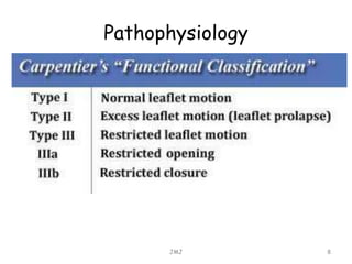
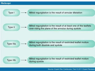
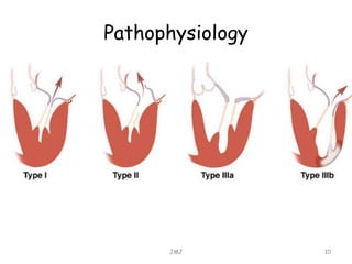
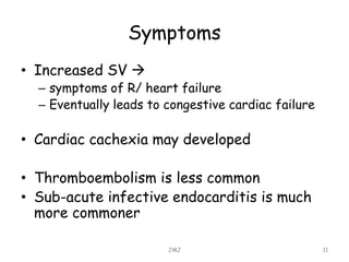
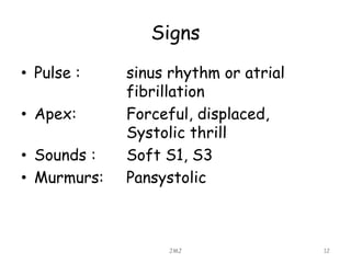
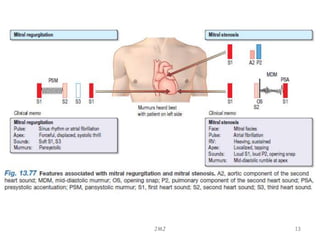
MITRAL REGERGITATION
K. Kavindya M. Fernando
JMJ 1
Mitral regergitation
• Mitral valve
– Consist of fibrous annulus,
– Anterior & posterior leaflets
– Chordae tendinae,
– Papillary muscle
JMJ 2
Causes
• Can be due to abnormalities of the
– valve leaflets
– Annulus
– Chordae tendineae
– Papillary muscle of the left ventricle
JMJ 3
Causes
• Most common causes
– Degenerative (myxomatous) disease
– Ischemic heart disease
– Rheumatic heart disease
– Infectious endocarditis
JMJ 4
Causes
• MR also seen in diseases of the
myocardium (dialated and hypertrophic
cardiomyopathy),
• Rheumatic autoimmune diseases
• Eg: SLE
• Collagen diseases
• Eg: Marfan’s syndrome
• Ehlers –Danlos syndrome
• Drugs inducing centrally acting appetite
suppressants, and dopamine agonists
JMJ 5
Pathophysiology
• Due to regurgitation into L atrium
– L/ atrium dilates
– Little increase in left atrial pressure if the
regurgitation is long standing
• In acute MR
– L/ atriam does not dilate
– So pressure inside it increases
JMJ 6
Pathophysiology
• Thus in acute MR
– Left arterial v-wave is greatly increased
– Pulmonary venous pressure rises to produce
pulmonary edema
• Proportion of the stroke volume
regurgitate
• Stroke volume increases to maintain CO
• So Left ventricle enlarges
JMJ 7
Pathophysiology
JMJ 8
Pathophysiology
JMJ 9
Pathophysiology
JMJ 10
Symptoms
• Increased SV 
– symptoms of R/ heart failure
– Eventually leads to congestive cardiac failure
• Cardiac cachexia may developed
• Thromboembolism is less common
• Sub-acute infective endocarditis is much
more commoner
JMJ 11
Signs
• Pulse : sinus rhythm or atrial
fibrillation
• Apex: Forceful, displaced,
Systolic thrill
• Sounds : Soft S1, S3
• Murmurs: Pansystolic
JMJ 12
Signs
JMJ 13
Signs of uncomplicated MR
• Laterally displaced diffuse apex beat and a
systolic thrill
• Soft 1st heart sound
• Pansystolic murmur
– Loudest at apex
– Radiating widely over the precordium & into
the axilla
• With a floppy mitral valve : there may be a
mild systolic click
• Prominent 3rd heart
JMJ 14
Investigation
• Chest X-ray
• Electrocardiogram
• Echocardiogram
• Cardiac catheterization
JMJ 15
Investigation – X-ray
• Left atrial, Left ventricular
enlargement
• Increase in CTR, and valve calcification
is seen
JMJ 16
Investigation – ECG
• Features of
– left atrial delay (bifid P wave)
– Left ventricular hypertrophy
• Tall R waves in left lateral leads (I & V6)
• Deep S wave in R sided precordial area ( V1, V2)
JMJ 17
Investigation – ECG
JMJ 18
Investigation – Echocardiogram
• Dilate left atrium and left ventricle
• May be specific features of chordal or
papillary muscle rupture
• TOE can be done
JMJ 19
Treatment
• Prophylaxis against endocarditis
• Any evedence of progressive cardiac
enlargement  surgical
– Mitral valve repair
– Mitral valve replacement
JMJ 20
Prolapsing (Billowing) mitral valve
• Barlow’s syndrome/ floppy mitral valve
• Due to excessively large mitral valve
leaflets,enlarged mitral annulus
• More common in young females
• Cause is unknown but associate with
Marfan’s syndrome, thyrotoxicosis,
rheumatic or ischemic heart disease
JMJ 21
JMJ 22

Mitral regurgitation

  • 1.
  • 2.
    Mitral regergitation • Mitralvalve – Consist of fibrous annulus, – Anterior & posterior leaflets – Chordae tendinae, – Papillary muscle JMJ 2
  • 3.
    Causes • Can bedue to abnormalities of the – valve leaflets – Annulus – Chordae tendineae – Papillary muscle of the left ventricle JMJ 3
  • 4.
    Causes • Most commoncauses – Degenerative (myxomatous) disease – Ischemic heart disease – Rheumatic heart disease – Infectious endocarditis JMJ 4
  • 5.
    Causes • MR alsoseen in diseases of the myocardium (dialated and hypertrophic cardiomyopathy), • Rheumatic autoimmune diseases • Eg: SLE • Collagen diseases • Eg: Marfan’s syndrome • Ehlers –Danlos syndrome • Drugs inducing centrally acting appetite suppressants, and dopamine agonists JMJ 5
  • 6.
    Pathophysiology • Due toregurgitation into L atrium – L/ atrium dilates – Little increase in left atrial pressure if the regurgitation is long standing • In acute MR – L/ atriam does not dilate – So pressure inside it increases JMJ 6
  • 7.
    Pathophysiology • Thus inacute MR – Left arterial v-wave is greatly increased – Pulmonary venous pressure rises to produce pulmonary edema • Proportion of the stroke volume regurgitate • Stroke volume increases to maintain CO • So Left ventricle enlarges JMJ 7
  • 8.
  • 9.
  • 10.
  • 11.
    Symptoms • Increased SV – symptoms of R/ heart failure – Eventually leads to congestive cardiac failure • Cardiac cachexia may developed • Thromboembolism is less common • Sub-acute infective endocarditis is much more commoner JMJ 11
  • 12.
    Signs • Pulse :sinus rhythm or atrial fibrillation • Apex: Forceful, displaced, Systolic thrill • Sounds : Soft S1, S3 • Murmurs: Pansystolic JMJ 12
  • 13.
  • 14.
    Signs of uncomplicatedMR • Laterally displaced diffuse apex beat and a systolic thrill • Soft 1st heart sound • Pansystolic murmur – Loudest at apex – Radiating widely over the precordium & into the axilla • With a floppy mitral valve : there may be a mild systolic click • Prominent 3rd heart JMJ 14
  • 15.
    Investigation • Chest X-ray •Electrocardiogram • Echocardiogram • Cardiac catheterization JMJ 15
  • 16.
    Investigation – X-ray •Left atrial, Left ventricular enlargement • Increase in CTR, and valve calcification is seen JMJ 16
  • 17.
    Investigation – ECG •Features of – left atrial delay (bifid P wave) – Left ventricular hypertrophy • Tall R waves in left lateral leads (I & V6) • Deep S wave in R sided precordial area ( V1, V2) JMJ 17
  • 18.
  • 19.
    Investigation – Echocardiogram •Dilate left atrium and left ventricle • May be specific features of chordal or papillary muscle rupture • TOE can be done JMJ 19
  • 20.
    Treatment • Prophylaxis againstendocarditis • Any evedence of progressive cardiac enlargement  surgical – Mitral valve repair – Mitral valve replacement JMJ 20
  • 21.
    Prolapsing (Billowing) mitralvalve • Barlow’s syndrome/ floppy mitral valve • Due to excessively large mitral valve leaflets,enlarged mitral annulus • More common in young females • Cause is unknown but associate with Marfan’s syndrome, thyrotoxicosis, rheumatic or ischemic heart disease JMJ 21
  • 22.