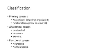
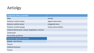
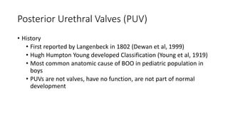
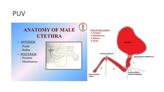
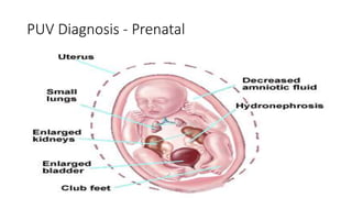
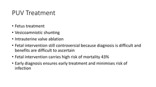
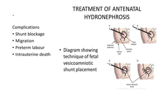
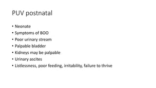
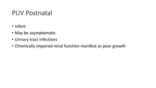
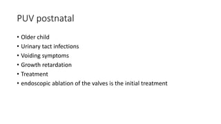
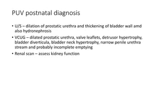
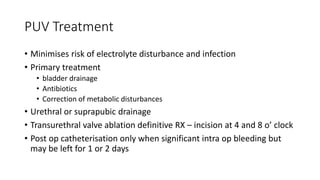
This document discusses bladder outlet obstruction (BOO). It begins by defining BOO and describing that it can be partial or complete. It then discusses the various causes of BOO including congenital and acquired factors. The pathophysiology of BOO is explained, noting how it can lead to renal damage over time if not treated. The document outlines the evaluation and management of BOO, including various imaging modalities and the importance of early surgical treatment to prevent long term renal issues. Posterior urethral valves are highlighted as a common cause of BOO in boys.
POSTERIOR URETHRAL VALVES- Pediatric Surgery
• Dear viewers,
• Greetings from “ Surgical Educator”
• Today I have uploaded one more video in Pediatric Surgery/Pediatric Urology- “ Posterior Urethral Valves”
• Posterior Urethral Valves is the congenital cause for Bladder Outlet Obstruction, resulting in abnormal development of the kidneys as well as the bladder.
• In this video, I talked about the learning outcomes, introduction, etiopathogenesis, clinical features, investigations, differential diagnosis, treatment, follow-up and prognosis of “ Posterior Urethral Valves”
• I hope you will enjoy the video for its educational value.
• You can watch all my teaching videos in the following links
• surgicaleducator.blogspot.com youtube.com/c/surgicaleducator
• Thank you for watching the video.
Ureteropelvic junction obstruction by\ Eman Salman
It was used for student presentation in Urology course rotation
I Hope you find what is helpful for your knowledge ♥
---------- Forwarded message ----------
From: cormac joyce <cjoyce78@hotmail.com>
Date: 2009/2/28
Subject: lecture
To: ucdgrad09@gmail.com
------------------------------
Join the all-new Windows Live Messenger family Click here!<http://get.live.com/>
HIRSCHSPRUNG DISEASE of neonate wrr.pptxShambelNegese
disease is a condition that affects the large intestine (colon) and causes problems with passing stool. The condition is present at birth (congenital) as a result of missing nerve cells in the muscles of the baby's colon.
micro teaching on communication m.sc nursing.pdfAnurag Sharma
Microteaching is a unique model of practice teaching. It is a viable instrument for the. desired change in the teaching behavior or the behavior potential which, in specified types of real. classroom situations, tends to facilitate the achievement of specified types of objectives.
Title: Sense of Smell
Presenter: Dr. Faiza, Assistant Professor of Physiology
Qualifications:
MBBS (Best Graduate, AIMC Lahore)
FCPS Physiology
ICMT, CHPE, DHPE (STMU)
MPH (GC University, Faisalabad)
MBA (Virtual University of Pakistan)
Learning Objectives:
Describe the primary categories of smells and the concept of odor blindness.
Explain the structure and location of the olfactory membrane and mucosa, including the types and roles of cells involved in olfaction.
Describe the pathway and mechanisms of olfactory signal transmission from the olfactory receptors to the brain.
Illustrate the biochemical cascade triggered by odorant binding to olfactory receptors, including the role of G-proteins and second messengers in generating an action potential.
Identify different types of olfactory disorders such as anosmia, hyposmia, hyperosmia, and dysosmia, including their potential causes.
Key Topics:
Olfactory Genes:
3% of the human genome accounts for olfactory genes.
400 genes for odorant receptors.
Olfactory Membrane:
Located in the superior part of the nasal cavity.
Medially: Folds downward along the superior septum.
Laterally: Folds over the superior turbinate and upper surface of the middle turbinate.
Total surface area: 5-10 square centimeters.
Olfactory Mucosa:
Olfactory Cells: Bipolar nerve cells derived from the CNS (100 million), with 4-25 olfactory cilia per cell.
Sustentacular Cells: Produce mucus and maintain ionic and molecular environment.
Basal Cells: Replace worn-out olfactory cells with an average lifespan of 1-2 months.
Bowman’s Gland: Secretes mucus.
Stimulation of Olfactory Cells:
Odorant dissolves in mucus and attaches to receptors on olfactory cilia.
Involves a cascade effect through G-proteins and second messengers, leading to depolarization and action potential generation in the olfactory nerve.
Quality of a Good Odorant:
Small (3-20 Carbon atoms), volatile, water-soluble, and lipid-soluble.
Facilitated by odorant-binding proteins in mucus.
Membrane Potential and Action Potential:
Resting membrane potential: -55mV.
Action potential frequency in the olfactory nerve increases with odorant strength.
Adaptation Towards the Sense of Smell:
Rapid adaptation within the first second, with further slow adaptation.
Psychological adaptation greater than receptor adaptation, involving feedback inhibition from the central nervous system.
Primary Sensations of Smell:
Camphoraceous, Musky, Floral, Pepperminty, Ethereal, Pungent, Putrid.
Odor Detection Threshold:
Examples: Hydrogen sulfide (0.0005 ppm), Methyl-mercaptan (0.002 ppm).
Some toxic substances are odorless at lethal concentrations.
Characteristics of Smell:
Odor blindness for single substances due to lack of appropriate receptor protein.
Behavioral and emotional influences of smell.
Transmission of Olfactory Signals:
From olfactory cells to glomeruli in the olfactory bulb, involving lateral inhibition.
Primitive, less old, and new olfactory systems with different path
Flu Vaccine Alert in Bangalore Karnatakaaddon Scans
As flu season approaches, health officials in Bangalore, Karnataka, are urging residents to get their flu vaccinations. The seasonal flu, while common, can lead to severe health complications, particularly for vulnerable populations such as young children, the elderly, and those with underlying health conditions.
Dr. Vidisha Kumari, a leading epidemiologist in Bangalore, emphasizes the importance of getting vaccinated. "The flu vaccine is our best defense against the influenza virus. It not only protects individuals but also helps prevent the spread of the virus in our communities," he says.
This year, the flu season is expected to coincide with a potential increase in other respiratory illnesses. The Karnataka Health Department has launched an awareness campaign highlighting the significance of flu vaccinations. They have set up multiple vaccination centers across Bangalore, making it convenient for residents to receive their shots.
To encourage widespread vaccination, the government is also collaborating with local schools, workplaces, and community centers to facilitate vaccination drives. Special attention is being given to ensuring that the vaccine is accessible to all, including marginalized communities who may have limited access to healthcare.
Residents are reminded that the flu vaccine is safe and effective. Common side effects are mild and may include soreness at the injection site, mild fever, or muscle aches. These side effects are generally short-lived and far less severe than the flu itself.
Healthcare providers are also stressing the importance of continuing COVID-19 precautions. Wearing masks, practicing good hand hygiene, and maintaining social distancing are still crucial, especially in crowded places.
Protect yourself and your loved ones by getting vaccinated. Together, we can help keep Bangalore healthy and safe this flu season. For more information on vaccination centers and schedules, residents can visit the Karnataka Health Department’s official website or follow their social media pages.
Stay informed, stay safe, and get your flu shot today!
The prostate is an exocrine gland of the male mammalian reproductive system
It is a walnut-sized gland that forms part of the male reproductive system and is located in front of the rectum and just below the urinary bladder
Function is to store and secrete a clear, slightly alkaline fluid that constitutes 10-30% of the volume of the seminal fluid that along with the spermatozoa, constitutes semen
A healthy human prostate measures (4cm-vertical, by 3cm-horizontal, 2cm ant-post ).
It surrounds the urethra just below the urinary bladder. It has anterior, median, posterior and two lateral lobes
It’s work is regulated by androgens which are responsible for male sex characteristics
Generalised disease of the prostate due to hormonal derangement which leads to non malignant enlargement of the gland (increase in the number of epithelial cells and stromal tissue)to cause compression of the urethra leading to symptoms (LUTS
Tom Selleck Health: A Comprehensive Look at the Iconic Actor’s Wellness Journeygreendigital
Tom Selleck, an enduring figure in Hollywood. has captivated audiences for decades with his rugged charm, iconic moustache. and memorable roles in television and film. From his breakout role as Thomas Magnum in Magnum P.I. to his current portrayal of Frank Reagan in Blue Bloods. Selleck's career has spanned over 50 years. But beyond his professional achievements. fans have often been curious about Tom Selleck Health. especially as he has aged in the public eye.
Follow us on: Pinterest
Introduction
Many have been interested in Tom Selleck health. not only because of his enduring presence on screen but also because of the challenges. and lifestyle choices he has faced and made over the years. This article delves into the various aspects of Tom Selleck health. exploring his fitness regimen, diet, mental health. and the challenges he has encountered as he ages. We'll look at how he maintains his well-being. the health issues he has faced, and his approach to ageing .
Early Life and Career
Childhood and Athletic Beginnings
Tom Selleck was born on January 29, 1945, in Detroit, Michigan, and grew up in Sherman Oaks, California. From an early age, he was involved in sports, particularly basketball. which played a significant role in his physical development. His athletic pursuits continued into college. where he attended the University of Southern California (USC) on a basketball scholarship. This early involvement in sports laid a strong foundation for his physical health and disciplined lifestyle.
Transition to Acting
Selleck's transition from an athlete to an actor came with its physical demands. His first significant role in "Magnum P.I." required him to perform various stunts and maintain a fit appearance. This role, which he played from 1980 to 1988. necessitated a rigorous fitness routine to meet the show's demands. setting the stage for his long-term commitment to health and wellness.
Fitness Regimen
Workout Routine
Tom Selleck health and fitness regimen has evolved. adapting to his changing roles and age. During his "Magnum, P.I." days. Selleck's workouts were intense and focused on building and maintaining muscle mass. His routine included weightlifting, cardiovascular exercises. and specific training for the stunts he performed on the show.
Selleck adjusted his fitness routine as he aged to suit his body's needs. Today, his workouts focus on maintaining flexibility, strength, and cardiovascular health. He incorporates low-impact exercises such as swimming, walking, and light weightlifting. This balanced approach helps him stay fit without putting undue strain on his joints and muscles.
Importance of Flexibility and Mobility
In recent years, Selleck has emphasized the importance of flexibility and mobility in his fitness regimen. Understanding the natural decline in muscle mass and joint flexibility with age. he includes stretching and yoga in his routine. These practices help prevent injuries, improve posture, and maintain mobilit
These lecture slides, by Dr Sidra Arshad, offer a quick overview of physiological basis of a normal electrocardiogram.
Learning objectives:
1. Define an electrocardiogram (ECG) and electrocardiography
2. Describe how dipoles generated by the heart produce the waveforms of the ECG
3. Describe the components of a normal electrocardiogram of a typical bipolar leads (limb II)
4. Differentiate between intervals and segments
5. Enlist some common indications for obtaining an ECG
Study Resources:
1. Chapter 11, Guyton and Hall Textbook of Medical Physiology, 14th edition
2. Chapter 9, Human Physiology - From Cells to Systems, Lauralee Sherwood, 9th edition
3. Chapter 29, Ganong’s Review of Medical Physiology, 26th edition
4. Electrocardiogram, StatPearls - https://www.ncbi.nlm.nih.gov/books/NBK549803/
5. ECG in Medical Practice by ABM Abdullah, 4th edition
6. ECG Basics, http://www.nataliescasebook.com/tag/e-c-g-basics
TEST BANK for Operations Management, 14th Edition by William J. Stevenson, Ve...kevinkariuki227
TEST BANK for Operations Management, 14th Edition by William J. Stevenson, Verified Chapters 1 - 19, Complete Newest Version.pdf
Ozempic: Preoperative Management of Patients on GLP-1 Receptor Agonists Saeid Safari
Preoperative Management of Patients on GLP-1 Receptor Agonists like Ozempic and Semiglutide
ASA GUIDELINE
NYSORA Guideline
2 Case Reports of Gastric Ultrasound
2. Introduction
• BOO describes blockage of urine outflow from the bladder and
through the urethra
• Obstruction may be partial or complete
• Childhood Urethral obstruction are mostly congenital
• Only posterior urethral valves commonly give rise to secondary
changes in the upper renal tracts
• Compared with the adult kidney, the developing kidney is highly
susceptible to injury from obstruction to urine flow
• The earlier the obstruction develops the worse the impact on the
upper tracts
3. Incidence
• Various causes of BOO and vary in incidence
• In Africa, reports on the incidence are scanty, probably
• Voiding dysfunction describe abnormalities in either the filling and/or
emptying of the bladder
• Constitutes 40% of paediatric urology clinic visits.
4. The Urinary system
• The urinary tract includes two kidneys, two
ureters, a bladder, and a urethra.
• Urine flows from the kidneys through the
ureters to the bladder which stores urine until
releasing it through the urethra by urination
• Lower urinary tract – bladder and urethra
• Obstruction may occur anywhere
• More common in males
5. Embryology
• Lower urinary tract development is closely interrelated with genital
tract and the hindgut.
• 3rd week of gestation, the cloaca meets the ectoderm of body wall at
cloacal membrane
• 5th week, cloaca divided by urorectal septum to form the primitive
rectum posteriorly and urogenital sinus anteriorly
• Allantois, bladder, pelvic, and phallic portions of the urogenital system
are recognisable in the 6th week.
7. Embryology
• 7th week gestation, the mesonephric ducts (vas deferens) shift
further caudal in the sinus and come to lie close to each other
• Metanephric buds (ureters) arise from mesonephric ducts
• root of the mesonephric duct exstrophies into the posterior wall of
the developing bladder
• This process brings the openings of the ureteric buds into the bladder
wall
• mesonephric duct is carried inferiorly to the level of the pelvic urethra
• The triangular region of exstrophied mesonephric duct incorporated
into the posterior bladder wall forms the trigone of the bladder
9. Embryology
• 9th week of gestation, the bladder cavity expands, urachus elongates
and continues with the allantoic stalk at the umbilicus.
• 12th week gestation, extra embryonic allantois degenerates, urachus
closes forming the median umbilical ligament.
• BOO manifest in some foetuses with closure of urachus, but delayed
closure may protect others.
10. Embryology
• The early bladder epithelium initially consists of a single cell layer, but
later become transitional
• Bladder epithelium or Chwalla’s membrane temporarily covers and
occludes the ureteral orifice
• Bladder muscle arises from mesenchyme as a longitudinal layer on
the dorsal surface and spreads cephalad to the intrarenal collecting
system
• 16th week of gestation, the bladder is completely muscularized and
the urachus is closed
11. Developmental Anomalies
• Bladder Agenesis : rare, is due to failure of allantoic stalk to develop
• Urachal anomalies (e.g., patent urachus) result from delayed closure
of the urachus, that arise from lower urinary tract obstruction at less
than 12 weeks gestation
• Some anomalies result from a general mesodermal failure (the
urachal diverticulum of the Prune belly syndrome).
• Intravesical (simple) ureterocoeles are thought to be due to the
persistence of Chwalla’s membrane beyond the time when urine flow
begins
12. • Bladder outlet obstruction caused by the urethral obstruction of
posterior urethral valves or urethral hypoplasia may be detected from
4 weeks gestation concurrent with the absorption of the mesonephric
duct and the resorption of the urogenital membrane.
• Abnormal dilatation of Cowper’s glands may give rise to an
obstructive urethral syringocele
13. Bladder Innervation
• Autonomic nervous system
Parasympathetic (S2–S4)
Sympathetic (T11–L2)
• Inhibit parasympathetic to allow detrusor relaxation and expansion
• Constrict internal sphincter
• Somatic nervous system (pudendal nerve(S1–S4)
• Control external sphincter
14. Physiology of voiding
• Normal voiding cycle is a two-phased process, consisting of
• low-pressure and adequate volume bladder filling
• complete evacuation of the bladder.
• Bladder filling is passive determined by
• Vesicoelastic properties of the bladder wall
• Inhibition of bladder contraction
• Increased urinary sphincter
• Urinary continence
• maintained by contraction of sphincter complex which keeps
urethral pressure above normal bladder pressure
15. Physiology of voiding
• Normal voiding is a spinal reflex modulated by the CNS (brain, pontine
micturition centre, and spinal cord), which coordinates the functions
of the bladder and urinary sphincter
• As the bladder fills, the pudendal nerve becomes excited, resulting in
contraction of the external urethral sphincter
• As the bladder approaches fullness, tension receptors in the bladder
wall trigger afferent nerves that send signals of the need to void.
• Voluntary voiding involves inhibition of sphincter contractions and
coordinated contraction of the bladder smooth muscle.
16. Physiology of voiding
• Bladder filling in neonates is followed by reflexive emptying
• This micturition cycle occurs hourly under sacral spinal cord control
• After 6 months of age, the urinary volumes increase, the frequency of
voiding decreases as the control of voiding shifts from the sacral cord
to the pontine voiding center
• Brain maturation and development, leads to bladder and urinary
sphincter control to inhibit involuntary voiding
• At 2 years the child develops conscious sensation of bladder fullness
and has urge incontinence
17. Physiology of voiding
• Between 2 and 4 years of age acquires voluntary control of bowel and
bladder function according to the following sequence:
• 1. nocturnal bowel control
• 2. daytime bowel control
• 3. daytime bladder control
• 4. nocturnal bladder control.
• By 4 years of age, most children demonstrate adult voiding pattern.
• The timing of this sequence is influenced by ethnic, cultural,
economic, and individual family differences.
18. Pathophysiology of BOO
• BOO results in an elevation of intravesical storage pressure
• Detrusor muscles generate excessive force against outlet resistance
• Massive hypertrophy of the detrusor muscles results
• Formation of sacculation, trabeculation, and ultimately diverticula
• The antireflux mechanism of the ureterovesical junction becomes
incompetent
• Ureteral peristalsis is overcome
• increased hydrostatic pressures are transmitted directly to the nephron
• Results in impairment of renal development and function
20. Pathophysiology of BOO
• GFR falls as pressure in the proximal tubule and bowman space increase
• After 12–24 hours of complete obstruction, intratubular pressure
decreases to preobstruction levels
• If not relieved, a depressed GFR is maintained by decreases in renal blood
flow mediated by thromboxane A2 and angiotensin II
• With continued obstruction, there is a progressive fall in renal blood flow,
ischaemia, and nephron damage.
• GFR falls, but tubular function is particularly severely affected, with a high
water and sodium loss resulting in the high output renal failure of
obstructive nephropathy.
21. Pathophysiology of BOO
• In the foetus, the placenta functions as the primary excretory organ in
place of the kidney throughout gestation.
• Hence, in the new-born with BOO, the renal function is usually similar
to the normal maternal levels initially because of the placental
function.
• The kidneys commence gradual glomerular filtration by the 11th to
12th weeks of gestation.
• About 90% of amniotic fluid is produced in the kidneys, and only 10%
comes from the GIT, lungs, and skin.
22. Pathophysiology of BOO
• Inadequate foetal urine production leads to oligohydramnios
• lung movement is therefore restricted
• Poor acinar growth and decreased surfactant production due to
decreased fluid in the bronchial tree
• The newborn may have compressed limbs, Potter’s facies, and
pulmonary hypoplasia presenting with respiratory distress and
pneumothorax
23. Pathophysiology of BOO
• The pathophysiology of this process is complex and incompletely
understood.
• Long-term bladder dysfunction may plague these patients in their
daily lives despite relieving the obstruction.
• Obstruction impacts renal concentrating ability, glomerular filtration
rate, and renal morphology.
26. Clinical Presentation
• History Elicit prenatal health, birth and development, perinatal
complication, and bowel and bladder habits.
• Many are asymptomatic for a long time or present with constitutional
features that often are misleading to an unsuspecting practitioner
• Acute or chronic urinary retention, overflow incontinence, UTI, or
renal failure.
27. Neonate
• Presentation is characterised by
• abdominal distention
• palpable suprapubic or flank masses (bladder and the kidneys);
• And/or urachal cyst, fistula, or abscess.
• Neonatal sepsis and respiratory distress may also be present.
• Parents may give a history of urine dribble, poor stream, and failure
to thrive in the young infant
28. Infants and children
• Enuresis, weight loss, vomiting, and diarrhoea.
• Constipation may be present in cases of dysfunctional voiding, faecal
impaction, Hirschsprung’s disease, or compression from pelvic
masses.
• Other features include urine dribbling, incontinence, straining,
frequency, and intermittency or “staccato” stream.
29. Physical Examination
• In the African setting, many children present with complications , due
to late presentation and lack of prenatal diagnosis
• small stature for age,
• anaemia,
• gross pitting pedal oedema,
• ascites, and
• abdominal masses
30. Physical examination
• Other features depend on primary or secondary causes.
• SPINE examine for defects, and the lower extremities for reflexes,
muscle mass and strength, sensation, and gait.
• PERINEUM check for gluteal fold symmetry, natal cleft depth, absent
coccyx, perineal sensation, tone and reflexes.
• Digital Rectal Examination check anal sphincteric tone, faecal
impaction, distended rectum, and presacral and pelvic masses.
31. Investigations
• Depend on availability and condition
• A variety of tests may be required to confirm the diagnosis and
determine the extent of damage
• Imaging may be sufficient to define:
• site and extent of an obstructing pathology;
• extent of urinary tract reaction to the BOO
• split and total renal function and scarring;
• presence and degree of reflux; and
• associated anomalies, where present.
32. Investigations
• Haemogram to quantify anaemia. The platelet count may also drop.
• Serum biochemistry to assess the electrolytes, urea, creatinine, and
acid-base balance
33. Investigations
• Plain radiography
• can identify radio-opaque stones.
• May suggest an enlarged or poorly emptying bladder,
• displacement of the bowel shadow from the hypochondrium by a
large fluid-filled kidney, and/or outline associated vertebral and
other bony anomalies.
34. Investigations
• Ultrasonography
• Demonstrate filling, dilated, or narrow posterior urethra or the
presence of intraluminal lesions.
• Size and shape of the kidneys
• bladder as well as dilated pelvicaliceal system and ureter.
• US should be performed with a moderately filled urinary bladder,
and if an anomaly is found, US should be repeated with the
bladder empty.
• It can be used to estimate the post void residual volume of urine,
and a spinal ultrasound in children younger than 3 months of age
is useful for spinal lesions (tethering, defects, or masses)
35. Investigations
• CT scan/MRI
• Done with or without contrast enhancement
• Provide precise anatomical details of the lesion
• Extent of damage
• Good-quality ultrasound with Doppler is a good substitute.
36. Investigations
• Micturating cystourethrography
• Essential for demonstration of bladder shape and capacity,
including diverticulum and vesicoureteric reflux and grading,
posterior urethral dilatation, and possible filling defects and
urinary incontinence.
• A filling phase is important to identify ureterocoele
37. Investigations
• Scintigraphy
• Dimercaptosuccinic acid (DMSA) scintigraphy is a radionuclide
technique that provides a functional cortical map of the kidney,
quantifying renal tubular cell mass.
• Useful for identifying scars of reflux nephropathy and estimating
differential function, provided there is no obstruction.
• Diethylenetriamine penta-acetic acid (DTPA) scintigram, the DTPA is
filtered by the glomerulus and gives a dynamic study similar to an IVU.
• It identifies dilatation and obstruction, and in the latter stages of the
study it can give information concerning reflux.
• Differential renal function can also be quantified.
38. Investigations
• Intravenous urogram
• An IVU study shows morphology of the urinary tract, provided the
kidney is working well enough.
• Is disappointing and sometimes dangerous in infants
• In the neonate, due to the low GFR, the urinary tract does not
opacify well.
• The x-ray exposure for IVU is relatively high, and US scans are
performed where possible
39. Investigations
• Urodynamic studies
• Urodynamic studies of the physiologic function of the bladder
mechanics during filling and voiding
• used to ascertain the aetiology and epidemiology of
nonneurogenic bladder sphincter dysfunction (NNBSD) with the
aid of x-ray screening
• Egs of such studies include uroflowmetry and cystometrogram.
40. Investigations
• Cystourethroscopy
• Approach of choice for identification and treatment of structural
abnormalities.
• Posterior urethral valves, ureterocoeles may be treated, and
Polyps May Be biopsied or resected.
• In the African setting, however, appropriate scopes may not be
readily available or accessible for either adults or paediatric
surgical services
41. Management
• BOO is a potentially curable form of lower urinary tract and renal
disease
• Determinants of bladder dysfunction - Degree and duration of
obstruction
• Early recognition and treatment are key to preventrenal function loss
• Definitive treatment of mechanical causes of BOO is mostly surgical
42. Posterior Urethral Valves (PUV)
• History
• First reported by Langenbeck in 1802 (Dewan et al, 1999)
• Hugh Humpton Young developed Classification (Young et al, 1919)
• Most common anatomic cause of BOO in pediatric population in
boys
• PUVs are not valves, have no function, are not part of normal
development
44. PUV classification
• Classification(Young)
• Type I – 95%
2 folds extend anteroinferiorly from caudal aspect of
verumontanum often fusing anteriorly at a lower level
• Type II - now discounted as non-obstructive folds of superficial
muscle and mucosa, between the verumontanum and the bladder
neck
• Type III – 5%
Circular diaphragm with a central or eccentric narrow aperture in
membranous urethra
46. • Dewan and Ransley’s challenged Youngs
classification; the types are secondary to urethral
instrumentation
• Characteristics of the urethral valve vary but include:
• attachment posteriorly to the distal part of the
verumontanum;
• a small hole adjacent to the verumontanum
• an oblique membrane with the distal attachment
lying anteriorly
• paramedian parallel reinforcements;
• Distal ballooning with suprapubic pressure;
• traversing the urethral sphincter
47. PUV Incidence
• Incidence is between 1 in 5000 - 8000 male births
• Few familial cases recorded, including in siblings, but no established
genetic predisposition
• Represent 10% of urological anomalies detected by prenatal U/S in Europe
• Accounts for less than 1% of antenatally diagnosed hydronephrosis
• Overall mortality is 25–50%.
• Many foetuses are lost antenatally, and 45% of survivors have renal failure
• Other associated anomalies = chromosomal abnormalities, and some
deformations related to the oligohydramnios.
48. PUV symptoms
• Severe obstruction leads to intrauterine renal failure and fetal death
in utero or soon after birth from potters syndrome.
• Less severe obstruction allows the fetus to survive, but if not detected
early leads to
• Septic complications from UTI and
• metabolic abnormalities caused by renal failure
49. PUV Diagnosis - Prenatal
Fetus
• Prenatal U/S detects 80% of cases, 50% between 16-20 weeks
• Obstruction develop at 7 wks GA and are detectable by U/S by 14 wks
50. PUV Diagnosis – Prenatal U/S features
• Bilateral upper tract dilatation
• Persistently distended full bladder
Predictors of poor functional outcome an early-onset renal failure
• Detection before 24 weeks’ gestation
• Bladder wall thickening
• Echo-bright kidneys (renal dysplasia)
• Oligohydramnios
52. PUV Treatment
• Fetus treatment
• Vesicoamniotic shunting
• Intrauterine valve ablation
• Fetal intervention still controversial because diagnosis is difficult and
benefits are difficult to ascertain
• Fetal intervention carries high risk of mortality 43%
• Early diagnosis ensures early treatment and minimises risk of
infection
54. PUV postnatal
• Neonate
• Symptoms of BOO
• Poor urinary stream
• Palpable bladder
• Kidneys may be palpable
• Urinary ascites
• Listlessness, poor feeding, irritability, failure to thrive
55. PUV Postnatal
• Infant
• May be asymptomatic
• Urinary tract infections
• Chronically impaired renal function manifest as poor growth
56. PUV postnatal
• Older child
• Urinary tact infections
• Voiding symptoms
• Growth retardation
• Treatment
• endoscopic ablation of the valves is the initial treatment
57. PUV postnatal diagnosis
• U/S – dilation of prostatic urethra and thickening of bladder wall amd
also hydronephrosis
• VCUG – dilated prostatic urethra, valve leaflets, detrusor hypertrophy,
bladder diverticula, bladder neck hypertrophy, narrow penile urethra
stream and probably incomplete emptying
• Renal scan – assess kidney function
58. PUV Treatment
• Minimises risk of electrolyte disturbance and infection
• Primary treatment
• bladder drainage
• Antibiotics
• Correction of metabolic disturbances
• Urethral or suprapubic drainage
• Transurethral valve ablation definitive RX – incision at 4 and 8 o’ clock
• Post op catheterisation only when significant intra op bleeding but
may be left for 1 or 2 days
59. • Vesicostomy and delayed valve ablation
• Indication
• markedly impaired renal function, especially
• deteriorating despite catheterisation,
• gross bilateral vesicoureteric reflux.
• Stoma created at the apex of the bladder to
minimise risk of prolapse
• Closure done after valve ablation, by 6–18
months, depending on the
• initial indication
• the child’s level of renal function
60. PUV treatment
• Urinary leaks due to high intravesical pressure
• Urinary ascites and perinephric collections or ‘urinomas’ usually
respond to a short period of bladder drainage.
• Large or persistent perirenal collections may require ultrasound-
guided percutaneous aspiration or open drainage (in combination
with open nephrostomy)
62. Patent Urachus
• Associated with drainage of urine from the umbilicus.
• Suspect Clear drainage from the umbilicus
• Investigate the urinary tract for bladder outlet obstruction in which the
urachus is functioning as a relief valve
• A patent urachus may be approached either through the umbilicus or
through an infraumbilical incision.
• It is important to identify all the umbilical structures for a definitive
diagnosis.
• The patent urachus is ligated and transected at the level of the bladder;
broad-based connections are closed in two layers with absorbable sutures
63. Urethral Polyps
• Urethral polyps are fibromuscular epithelial structures with
transitional epithelium covering the surface.
• They may occur either in the posterior or anterior urethra.
• In either position, they may cause hematuria, urgency, or obstructive
symptoms.
• Bladder ultrasound may show the polyps in the posterior urethra but
VCUG and cystoscopy are diagnostic and excision through the
cystoscope is usually curative.
• Polyps can present as congenital obstructing lesions with all of the
characteristics of bladder outlet obstruction.
64. References
• Coran Pediatric Surgery 7th Edition
• Essential of Pediatric Urology, 2nd Edition, Edited by David FM Thomas
• Clinical Pediatric Nephrology, 2nd Edition, Edited by Kanwal K Kher
MD
• Langmans medical embryology, 12th Edition