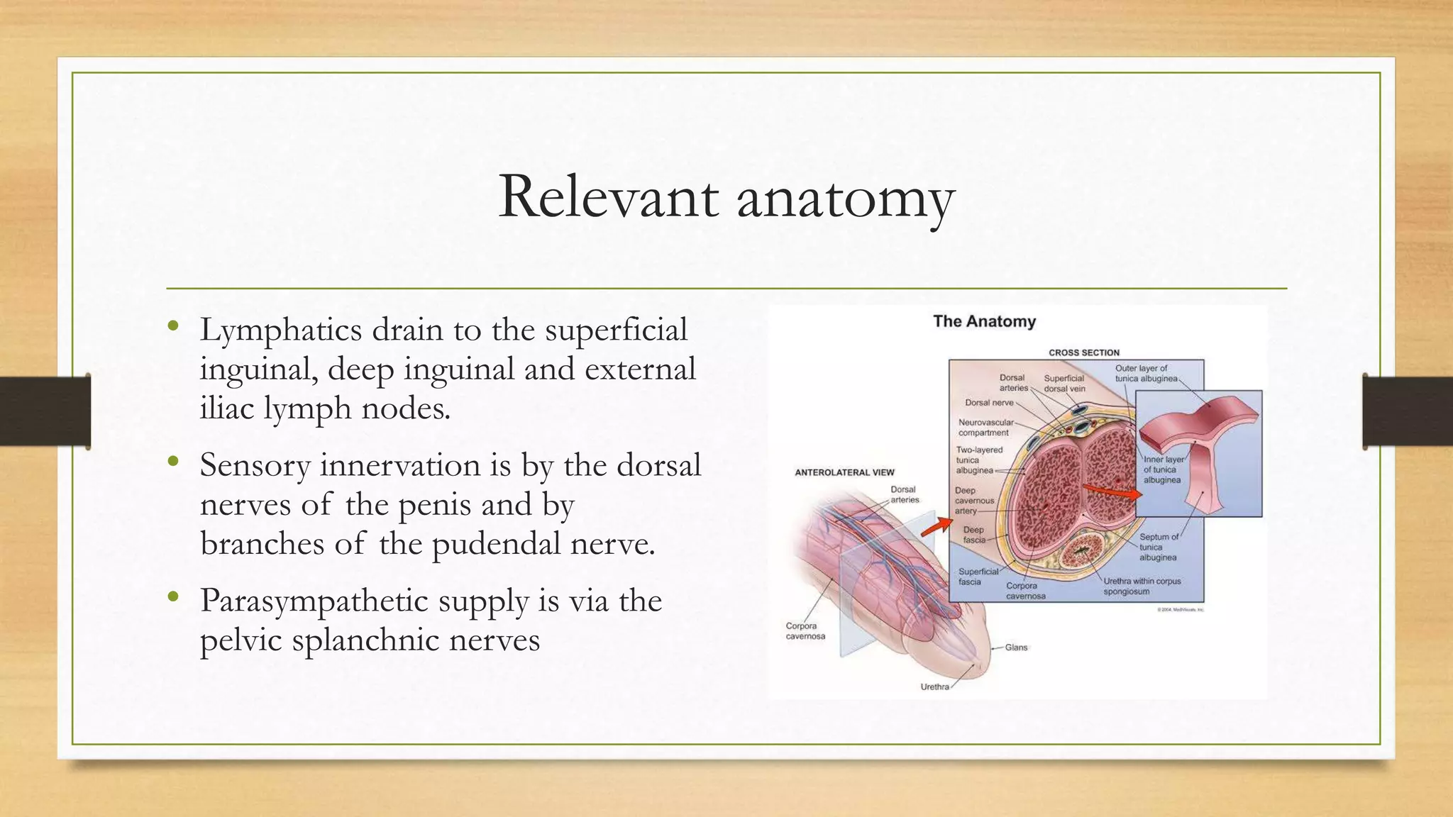
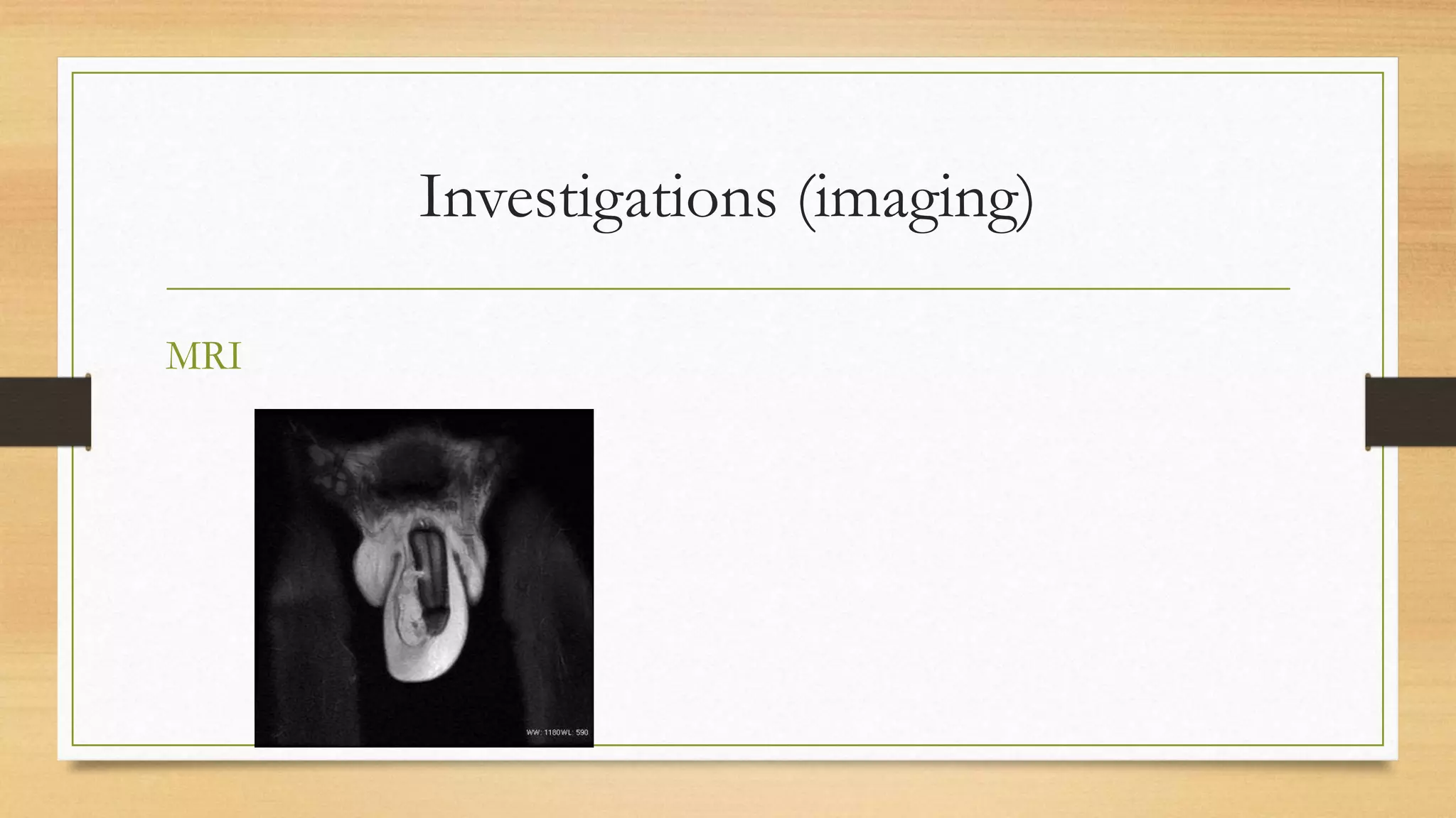
4. Introduction
• There are few emergencies that cause as much embarrassment to a man as
penile fracture.
• Its name is actually a misnomer as technically in the human penis, there is no
bone to be fractured.
• It is defined as the blunt traumatic rupture of the corpus cavernosum of an
erect penis, particularly the tunica albuginea surrounding it.
• This may be of one or both of the corpora cavernosa.
5. Relevant anatomy
• The layers of the penis include the
• Skin
• Dartos fascia
• Bucks fascia
• Tunica albuginea (covers the
cavernous bodies only)
6. Relevant anatomy
• The penis has 3 parts
• Root
• Body (Shaft)
• Glans
• Arterial supply is mostly via branches
of the internal pudendal artery
• Venous drainage is via the dorsal vein
and deep dorsal vein
7. Relevant anatomy
• Lymphatics drain to the superficial
inguinal, deep inguinal and external
iliac lymph nodes.
• Sensory innervation is by the dorsal
nerves of the penis and by
branches of the pudendal nerve.
• Parasympathetic supply is via the
pelvic splanchnic nerves
8. Epidemiology
• Most common in the 4th decade of life
• Mostly unilateral and mostly right sided (54%)
• Urethral injury seen in roughly 20 – 30%
10. Pathophysiology
• Injury to the flaccid penis is rare due to the flexibility and mobility of the
organ.
• Erection is achieved via engorgement of the corporal bodies especially the
sinusoids of the cavernous bodies.
• The erect penis is less mobile and the tunica albuginea reduces on thickness
from 2mm to 0.25mm.
• The differential between inflow and outflow is responsible for the
maintenance of tumescence.
11. Pathophysiology
• Sudden longitudinal trauma to the erect penis or a quick forceful lateral bending to
the erect penis can result in a tear or rent in the tunica albuginea.
• With tear of the cavernosa, there is an extra corporal loss of blood into the loose
fascia of the penis with sudden detumescence.
• There is a subsequent haematoma formation, abnormal shape of the penis and
pain.
• If the corpus spongiosum is involved with urethral injury, there may be bleeding per
urethra and depending on the severity of urethral injury, urinary retention.
12. Clinical presentation.
• Most cases present as emergencies and the complaints are mostly those of
• Pain
• Swelling
• Loss of erection
• Popping sound
• ± bleeding per urethra and inability to pass urine.
13. Clinical presentation
• It is important to ascertain the sexual position (most commonly the female
partner is on top straddling the phallus)
• Most are coincidental with sexual intercourse.
• The use of performance enhancing medication should also be obtained.
14. Examination
• Discomfort
• Anxious
• Egg plant deformity
• Lateral bend of the phallus if only one
side is involved
• Butterfly pattern ecchymoses
• Bleeding per urethra
• Tenderness
15. Diagnosis
• This is essentially clinical but there are some instances where investigations
may be required for confirmation.
• Differentials
• Rupture of the dorsal penile vessels
• Rupture of the suspensory ligament of the penis
16. Investigations (Imaging)
• Imaging modalities are not essential to make a diagnosis as they usually just
increase cost and do not necessarily change the treatment plan.
• Supportive investigations
• Penile ultrasound
• Magnetic Resonance imaging
• Penile Cavernosography
• Retrograde urethrography
21. Management
• At the emergency room,
• Adequate analgesia
• Evaluate state of the urethra by asking the patient to void. Urinary diversion to be
performed if there is urinary retention
• Counselling
• Consent for surgery
• Anaesthetic evaluation
22. Treatment
• Surgical
• Conservative.
• Rarely performed.
• Higher rates of complications
• May be attempted if the history is suggestive but the examination findings are not in
line with penile fracture.
• Investigations do not show any tear in the tunica albuginea ot the tear is < 0.5cm with
minimal haematoma formation.
23. Surgical principles
• Adequate exposure
• Haematoma evacuation
• Identification and repair of the tear in the tunica albuginea
• Repair of urethral injury if any.
24. Surgical approaches
• Circumferential incision and degloving
• Most common approach
• Gives good exposure
• Excellent results
• Inguinoscrotal
• Usually for injuries at or close t the base of the phallus
• Incision over defect.
25. Intra-op
• Anaesthesia
• General or Spinal
• Supine position
• Cleaning and draping.
• Application of tourniquet
• Circumferential incision through to Bucks fascia and then degloving
• Evacuation of the haematoma
26. Surgery
• Thorough inspection both corpora cavernosa and the corpus spongiosum for tears
• Injection of contrast to identify the tear in the tunica albuginea.
• Freshen the edges of the tear and repair with small absorbable sutures.
• Test the repair by inducing tumescence with the aid of normal saline injected into
the cavernous body.
• If there is a urethral injury, a catheter is passed under direct vision and a primary
repair of the tear or an anastomosis in the event of a transection is performed.
28. Surgery
• Repair the fascial layers and then the skin with absorbable sutures ensuring
the proper orientation and length of penis is maintained.
• In the event of a urethral repair, catheter is left for a period of 7 – 14 days.
29. Post operative care
• Analgesics
• Antibiotics
• Erection suppression
• Stilboestrol
• Chlorpromazine
• Diazepam
• Abstinence from sexual activity for 6 – 8 weeks.
31. Management of chronic penile haematomas
• Patients may have a much delayed presentation following penile fracture
• In these patients, the haematoma may have become organised with resultant
fibrosis and curvature of the penis.
• The treatment is a modified Nesbit procedure.
32. Follow-up
• Discharge is usually within 5 days of the procedure and follow-up is on an
outpatient basis
• Most patients make a full recovery with restoration of normal sexual
function and micturition.
• The catheter in patients that had a urethral repair is removed after about 2
weeks.
33. Conclusion
• Penile fracture as an emergency is under reported due to the embarrassment
the patients feel
• It is a clinical diagnosis that’s easy to identify but may have some false
negatives.
• Surgical intervention is preferable as it has much better outcomes.
34. References
• Imtiaz Wani. Management of Penile Fractures. Oman Med J. 2008 Jul; 23(3): 162 –
165.
• Gregory S Jack et al. Current treatment options for penile fractures. Rev Urol. 2004
Summer; 6(3): 114 – 120.
• Galanakis I, Adamos K, Spyropoulos E, Mavrikos S. Delayed successful repair of a
penile fracture: A case report. Curr Urol. 2018; 12: 111 -112
• Richard A Santucci. Penile fracture and trauma. 2019 Jan. accessed via the Medscape
app
• Images obtained via a google search.
The corpora are incompletely separated by the septum in the midline for most of their length except at the root of the penis where they are separated into the crura.
Root – It is deep to the pubis to which it is attached by a ligament. Provides stability to the erect penis. It consists of the crura, bulb, ischiocavernosus and bulbospongiosus muscles.
Body – Makes up the bulk of the penis. Has the sponge-like erectile tissue, the corpora cavernosa and the corpus spongiosum through which the urethra passes.
Glans is an extension of the corpus spongiosum.
Arterial supply – Deep artery of the penis (paired) end as the cavernosal arteries, Bulbar artery, and Urethral artery
Omisanjo et al (LASUTH 2015) 5 year period
Ligament – floppy penis
Cavernosography; nvolves intracorporal injection of a contrast. Done under fluoroscopic guidance. There is a risk of fibrosis post procedure. There are also lots of false negatives. Allergy, priapism. Early films, 10 minute delayed films. Contrast (diatrizoate meglumine, diatrizoate sodium)
Urethrogram: A 12 – 14F urethral catheter is used. It is for extravasation of contrast from the injured urethra. Oblique film shot