Downloaded 923 times


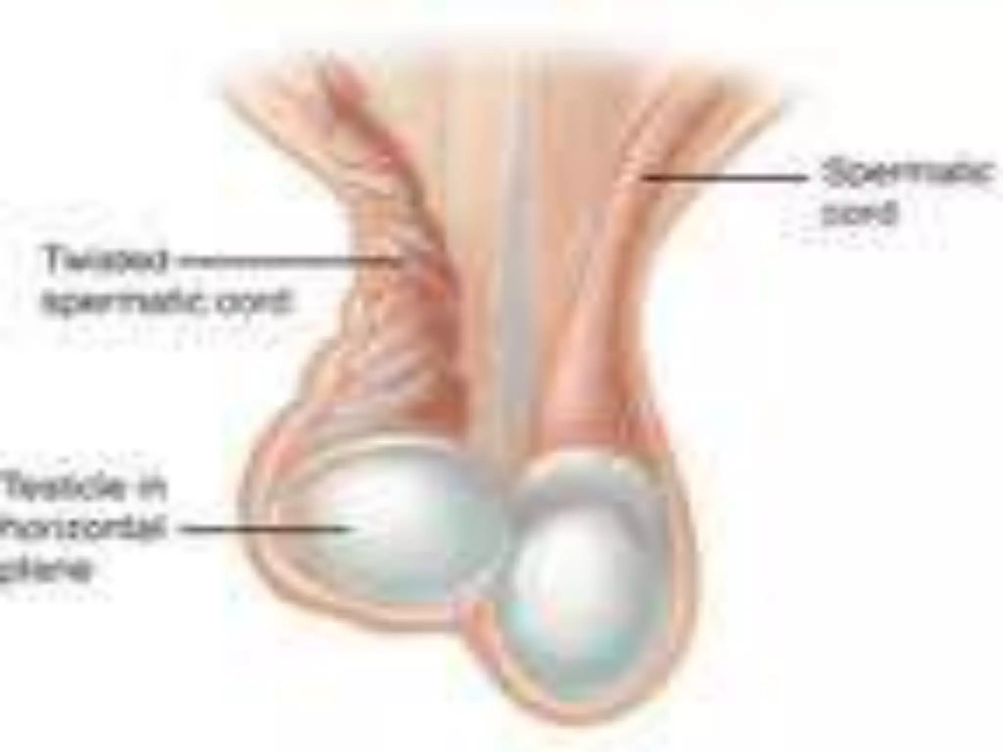
![Testicular torsion
Testicular torsion is a urologic emergency that is more
common in neonates and postpubertal boys, although it can
occur at any age [2].
The prevalence of testicular torsion in adult patients
hospitalized with acute scrotal pain is approximately 25 to 50
percent [2,4-7].](https://image.slidesharecdn.com/0xlzppquk91t8cm07qqg-signature-c8cfb560114f98eb613090c2eb289dfa9ca4e7cd7f74674ec5352d8caa8acbae-poli-150420204928-conversion-gate02/75/The-acute-scrotum-6-2048.jpg)
![ Testicular torsion results from inadequate fixation of the testis
to the tunica vaginalis producing ischemia from reduced
arterial inflow and venous outflow obstruction.
Testicular torsion may occur after an incidental event (eg,
trauma) or spontaneously [10].](https://image.slidesharecdn.com/0xlzppquk91t8cm07qqg-signature-c8cfb560114f98eb613090c2eb289dfa9ca4e7cd7f74674ec5352d8caa8acbae-poli-150420204928-conversion-gate02/75/The-acute-scrotum-7-2048.jpg)
![ It is generally felt that the testis suffers irreversible damage
after 12 hours of ischemia due to testicular torsion [8,9].
Infertility may result, even with a normal contralateral testis,
because the disruption of the immunologic "blood-testis"
barrier may expose antigens from germ cells and sperm to the
general circulation and lead to the development of anti-sperm
antibodies.](https://image.slidesharecdn.com/0xlzppquk91t8cm07qqg-signature-c8cfb560114f98eb613090c2eb289dfa9ca4e7cd7f74674ec5352d8caa8acbae-poli-150420204928-conversion-gate02/75/The-acute-scrotum-8-2048.jpg)
![Clinical features and diagnosis
The diagnosis of testicular torsion is usually determined by
acute onset of severe symptoms and characteristic physical
findings, although ultrasound may be needed in equivocal
cases.
The onset of pain in testicular torsion is usually sudden and
often occurs several hours after vigorous physical activity or
minor trauma to the testicles [11].
There may be associated nausea and vomiting.](https://image.slidesharecdn.com/0xlzppquk91t8cm07qqg-signature-c8cfb560114f98eb613090c2eb289dfa9ca4e7cd7f74674ec5352d8caa8acbae-poli-150420204928-conversion-gate02/75/The-acute-scrotum-9-2048.jpg)


![The cremasteric reflex
A normal response is cremasteric contraction with elevation of
the testis.
The reflex is usually absent in patients with testicular torsion .
This helps distinguish testicular torsion from epididymitis
and other causes of scrotal pain, in which the reflex is
typically intact [1].](https://image.slidesharecdn.com/0xlzppquk91t8cm07qqg-signature-c8cfb560114f98eb613090c2eb289dfa9ca4e7cd7f74674ec5352d8caa8acbae-poli-150420204928-conversion-gate02/75/The-acute-scrotum-15-2048.jpg)

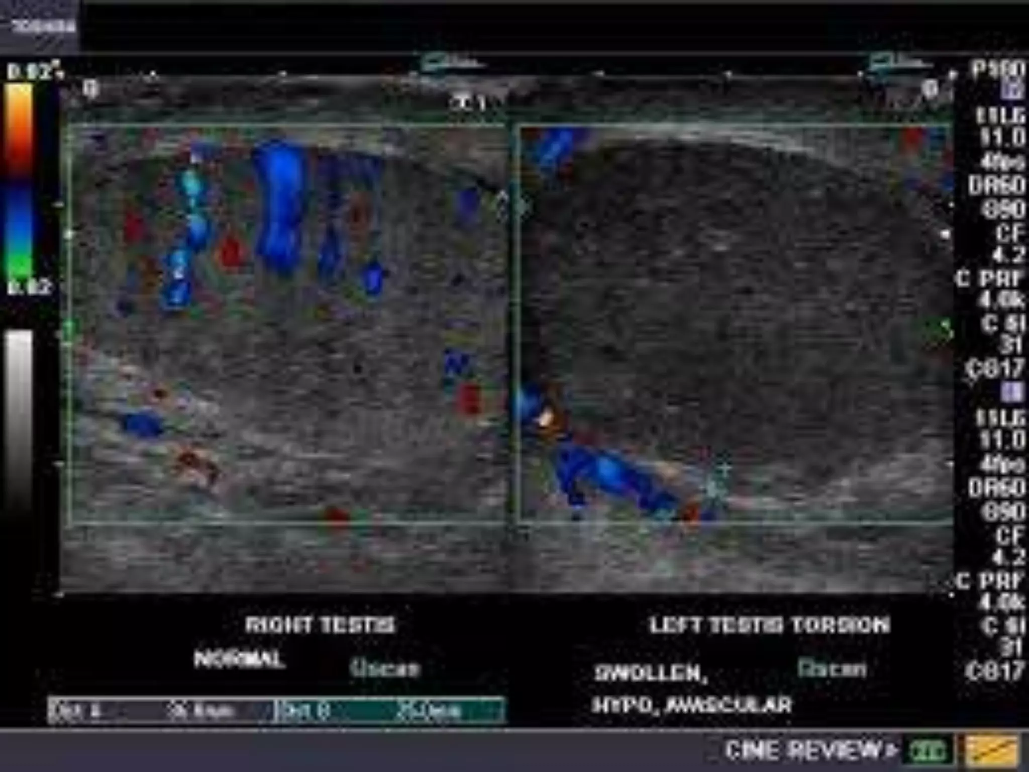
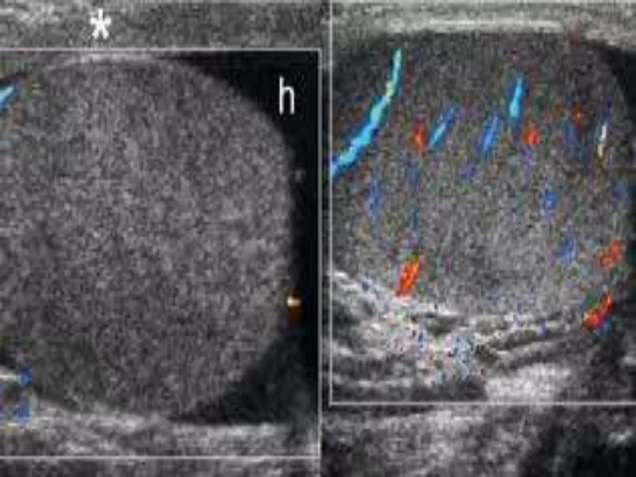
![ In a study of 56 patients who underwent surgical exploration
for acute scrotal pain and had Doppler ultrasound
examinations performed preoperatively [4] (sensitivity 100
percent and specificity 97 percent).](https://image.slidesharecdn.com/0xlzppquk91t8cm07qqg-signature-c8cfb560114f98eb613090c2eb289dfa9ca4e7cd7f74674ec5352d8caa8acbae-poli-150420204928-conversion-gate02/75/The-acute-scrotum-18-2048.jpg)


![Manual detorsion
If surgery is not immediately available (within two hours), it is
reasonable to attempt to manually detorse the testicle [16].
The classic teaching is that the testis usually rotates medially
during torsion and can be detorsed by rotating it outward
toward the thigh.](https://image.slidesharecdn.com/0xlzppquk91t8cm07qqg-signature-c8cfb560114f98eb613090c2eb289dfa9ca4e7cd7f74674ec5352d8caa8acbae-poli-150420204928-conversion-gate02/75/The-acute-scrotum-25-2048.jpg)
![ However, in a retrospective analysis of 200 consecutive males
age 18 months to 20 years who underwent surgical
exploration for testicular torsion, lateral rotation was present in
one-third of cases [17].](https://image.slidesharecdn.com/0xlzppquk91t8cm07qqg-signature-c8cfb560114f98eb613090c2eb289dfa9ca4e7cd7f74674ec5352d8caa8acbae-poli-150420204928-conversion-gate02/75/The-acute-scrotum-26-2048.jpg)
![successful detorsion is suggested by [18]:
Relief of pain
Resolution of the transverse lie of the testis to a longitudinal
orientation
Lower position of the testis in the scrotum
Return of normal arterial pulsations detected with a color
Doppler study](https://image.slidesharecdn.com/0xlzppquk91t8cm07qqg-signature-c8cfb560114f98eb613090c2eb289dfa9ca4e7cd7f74674ec5352d8caa8acbae-poli-150420204928-conversion-gate02/75/The-acute-scrotum-27-2048.jpg)
![ Surgical exploration is necessary even after clinically
successful manual detorsion because orchiopexy (securing
the testicle to the scrotal wall) must be performed to prevent
recurrence, and residual torsion may be present that can be
further relieved [17].](https://image.slidesharecdn.com/0xlzppquk91t8cm07qqg-signature-c8cfb560114f98eb613090c2eb289dfa9ca4e7cd7f74674ec5352d8caa8acbae-poli-150420204928-conversion-gate02/75/The-acute-scrotum-28-2048.jpg)
![Epididymitis
Epididymitis is the most common cause of scrotal pain in
adults in the outpatient setting [19].
Epididymitis is most commonly infectious in etiology, but can
also be due to noninfectious causes (eg, trauma, autoimmune
disease) [22].](https://image.slidesharecdn.com/0xlzppquk91t8cm07qqg-signature-c8cfb560114f98eb613090c2eb289dfa9ca4e7cd7f74674ec5352d8caa8acbae-poli-150420204928-conversion-gate02/75/The-acute-scrotum-29-2048.jpg)

![Investigations
A urinalysis and urine culture should be performed in all
patients suspected of epididymitis, although urine studies are
often negative in patients without urinary complaints [8].
A urethral swab should be obtained in patients with urethral
discharge and sent for culture
should be performed in patients with acute onset of
testiculUltrasound ar pain to assess for testicular torsion.](https://image.slidesharecdn.com/0xlzppquk91t8cm07qqg-signature-c8cfb560114f98eb613090c2eb289dfa9ca4e7cd7f74674ec5352d8caa8acbae-poli-150420204928-conversion-gate02/75/The-acute-scrotum-31-2048.jpg)


![Torsion of the appendix testis
Testicular pain from torsion of the appendix testis is usually more
gradual than with testicular torsioIt is the leading cause of acute
scrotal pathology in childhood. Torsion of the appendix testis
rarely occurs in adults [29].
It is not uncommon for patients to have several days of scrotal
discomfort before they present for evaluation. Pain ranges widely
from mild to severe.
Careful inspection of the scrotal wall at this location may detect the
classic "blue dot" sign caused by infarction and necrosis of the
appendix testis .](https://image.slidesharecdn.com/0xlzppquk91t8cm07qqg-signature-c8cfb560114f98eb613090c2eb289dfa9ca4e7cd7f74674ec5352d8caa8acbae-poli-150420204928-conversion-gate02/75/The-acute-scrotum-39-2048.jpg)


![Referred pain
Men who have the acute onset of scrotal pain without local
inflammatory signs or a scrotal mass on examination may be
suffering from referred pain to the scrotum.
The diseases that may cause referred scrotal pain are diverse,
reflecting the anatomy of the three somatic nerves that travel
to the scrotum: the genitofemoral, ilioinguinal, and posterior
scrotal nerves [31].](https://image.slidesharecdn.com/0xlzppquk91t8cm07qqg-signature-c8cfb560114f98eb613090c2eb289dfa9ca4e7cd7f74674ec5352d8caa8acbae-poli-150420204928-conversion-gate02/75/The-acute-scrotum-44-2048.jpg)


This document discusses the differential diagnosis and management of acute scrotal pathology. Testicular torsion and epididymitis are the most common causes of acute scrotal pain in adults. Testicular torsion is a surgical emergency that requires detorsion and fixation to prevent tissue damage from lack of blood flow. Epididymitis is usually infectious and treated with antibiotics, anti-inflammatories, and scrotal elevation. Other potential causes include Fournier's gangrene, trauma, testicular cancer, and referred pain from conditions like kidney stones. Physical exam, ultrasound, and surgical exploration can help determine the appropriate treatment.


