
Recommended
More Related Content
What's hot
What's hot (20)
Viewers also liked
Viewers also liked (20)
Similar to Horseshoe kidney & PCNL
Similar to Horseshoe kidney & PCNL (20)
More from GAURAV NAHAR
More from GAURAV NAHAR (20)
Recently uploaded
Recently uploaded (20)
Horseshoe kidney & PCNL
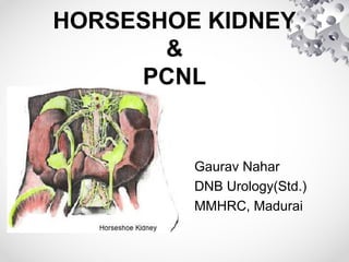
- 1. HORSESHOE KIDNEY & PCNL Gaurav Nahar DNB Urology(Std.) MMHRC, Madurai
- 2. HORSESHOE KIDNEY • Most common congenital renal anomaly. • Two distinct renal masses lying vertically on either side of the midline & connected at their respective lower poles by a parenchymatous or fibrous isthmus that crosses the midplane of the body.
- 3. • Result of a median fusion of metanephric tissue during early gestation. • Subsequent entrapment of fused lower pole isthmus by IMA results in an incomplete cephalad migration and an associated malrotation of the kidney.
- 4. HK: INCIDENCE • Occurs in 0.25% of the population, or about 1 in 400 persons. • More common in males; M:F~2:1. • discovered in all age groups, ranging from fetal life to 80 years. • In autopsy series, more prevalent in children.
- 6. HK: EMBYROLOGY Normal Embryology • Pronephros, Mesonephros, Metanephros • Metanephros → Adult Kidney • Formed from condensing blastema of Metanephric Mesenchyme and Ureteric Bud from Wolffian(Mesonephric) duct.
- 7. • Kidney advances superiorly(6th -9th week) as fetus grows caudally. • Kidney rotates 90° • Hilum migrates anterior to medial. • Kidneys land at ~L1 by week 9. • Blood supply changes throughout migration
- 10. • Shh(Sonic Hedgehog gene) in notochord and floor plate specifically inactivated. • Depleting axial source of Shh→kidney fusion (even in presence of notochord). • Thus notochord is not necessary for nephrogenesis but is required for correct positioning of the metanephric kidney, while axial Shh signal is critical for kidney positioning along the mediolateral axis.
- 11. HK: VARIATIONS • Lower pole fusion(95%). Upper pole/Interpolar region fusion(rare). • Generally, Isthmus is bulky and consists of parenchymatous tissue with its own blood supply. Occasionally, it is flimsy fibrous tissue band. • Isthmus adjacent to L3 or L4 vertebra just below the origin of IMA from aorta, resulting in paired kidneys lying lower than normal.
- 12. • Calyces- normal number, but atypical orientation(point posteriorly due to failure of rotation of kidneys.) • Ureter- inserts high & laterally into the pelvis and has a characteristic bend as it crosses over & anterior to the isthmus(predisposition to UPJO).
- 13. • Blood supply: One renal artery to each kidney. Duplicate or triplicate renal arteries to 1 or both kidneys. Isthmus may receive a branch from each main renal artery, or separate supply from aorta, or branches from inferior mesenteric, common or external iliac or sacral arteries.
- 15. HK: ASSOCIATED ANOMALIES EXTRA G.U. ANOMALIES: • Most commonly affected organ systems: Skeletal, CVS(ventriculoseptal defects), & CNS. • Found in 3% of NTDs. • ARMs. • 20% of Trisomy 18 (Edward syndrome). • 60% of Turner syndrome. • Townes-Brock syndrome.
- 16. G.U. ANOMALIES: • Hypospadias & UdT(4%) - males. • Bicornuate uterus or septate vagina or both(7%) - females. • Ureteral duplication(10%). • Ectopic ureterocoele. • VUR(50%). • UPJO(20%). • Multicystic dysplasia, & ADPKD.
- 17. HK: SYMPTOMS • Asymptomatic in 50%. • Symptoms typically related to hydronephrosis, infection, or calculus formation. • MC symptom is vague abdominal pain that may radiate to lower lumbar region. • GI complaints may also be present: nausea, vomiting, abdominal pain on hyperextension of spine.
- 18. • UTIs occur in 30% of patients, and • Calculi noted in 20% to 80%.
- 19. HK: DIAGNOSIS & RADIOGRAPHIC APPEARANCE PLAIN ABDOMINAL RADIOGRAPH: • Kidneys somewhat low lying & close to vertebral column; have a vertical or outward axis with lower poles being more medial than in normal kidney. USG: • Prenatal USG detects most HKs. • USG detects the isthmus joining two lower poles of kidneys in midline. • scanning horizontally along midline in a craniocaudal direction.
- 20. HK ON RADIOGRAPH
- 21. HK ON USG
- 22. RADIONUCLIDE SCAN: • demonstrates abnormal axis of a horseshoe kidney. • A continuous band across midline is observed if isthmus contains functioning parenchyma. CT & MRU: • Both characterize the isthmus. MR ANGIOGRAPHY: • accurately delineate vascular anatomy for preoperative planning.
- 23. HK ON CT
- 24. HK ON BONE SCINTIGRAM
- 25. HK ON RADIONUCLIDE SCAN
- 26. HK: GADOLINIUM ENHANCED MR ANGIOGRAM
- 27. CALCULI IN HORSESHOE KIDNEY • MC complication of HK is calculi formation. • Earlier hypothesis: calculi formation due to higher rate of infection, stasis, & obstruction because of abnormal position of pelvis & ureter. • Recent review suggest combined anatomic & metabolic causes. • Hypovolemia, hypercalciuria & hypocitraturia: most common metabolic defects. • MC type of calculi: Ca oxalate.
- 28. KUB showing stones in a horseshoe kidney with stones extending in the isthmus.
- 29. CT scan showing isthmic stone in a horseshoe kidney
- 30. Preoperative IVU showing Calculi in Hoseshoe kidney.
- 31. ESWL TO TREAT CALCULI IN HK • Favorable factors for SWL of HK: (i) a stone burden <1.5 cm, & (ii) nonobstructed collecting system drainage. • Anomalous kidney orientation makes localization of calculi more difficult, especially for stones in anteromedial calyces. • Prone position may facilitate localization of stone. • Alternatively, a “blast path” technique may be employed. • Also it may interfere with fragment passage after SWL.
- 32. • Clearance rate for lower calyceal stones was inferior to that of middle and upper calyceal stones. • A higher number of shockwaves per treatment required and a higher re- treatment rate observed. • High recurrence rate in the presence of persistent fragments after ESWL.
- 33. RIRS TO TREAT CALCULI IN HK • Ureteroscopy may be an effective treatment modality for a stone burden < 1.5 cm in HK. • Flexible ureteroscopes, and devices such as Ho:YAG laser, nitinol graspers, and ureteral access sheaths used.
- 34. PCNL TO TREAT CALCULI IN HK • PCNL is ToC for calculi in HK > 1.5-2.0 cm, or when SWL fails. • Percutaneous access to a HK is more favorable than in normal kidneys. • Abnormal anatomic position(lower & incomplete/non rotation of kidneys, calyceal orientation) causes PCNL to be easier and more safe.
- 35. • Preoperative CT or MRI: to assess for the possibility of retro-renal colon, and to assess the vasculature and relationship of the calyces to the anticipated puncture site.
- 36. ANATOMIC IMPLICATIONS OF HK IN PCNL • The anteroposterior tilt of kidney is prominent, which makes upper pole the most superficial and posterior aspect of HK. • Upper pole calyces are more posterior & lateral and often subcostal. • Convenient & relatively safe route for PCNL access. • Lower pole calyces are anterior, lie in a coronal plane, angled medially and inaccessible percutaneously.
- 38. • In horseshoe kidney, the frontal plane lies more or less in the sagittal plane of the body. • Consequently, posterior row of calyces point dorsomedially and ventral row dorsolaterally, and the renal pelvis is in a ventral position.
- 39. • In normal kidney anatomy, upper pole calyxes are in front of 11 & 12 ribs; entering them needs a supracostal approach that may cause thoracic complications such as pneumothorax. • However, in a HK, since kidney is placed lower than normal position, there is usually no need for supracostal approach.
- 41. • The optimal point of entry is through a posterior calyx, which is typically more medial than in normal kidney because of altered renal axis & rotation associated with midline fusion.
- 43. • Initial entry into a horseshoe kidney is more medial than in normal kidneys and can pass through the paraspinous musculature. • Slightly more difficult to dilate, because they traverse through erector spinae and quadrates lumborum muscles.
- 44. • (a) Retrograde pyelogram of a horseshoe kidney with intrarenal calculi. • (b) Fluoroscopically guided percutaneous access to a horseshoe kidney via upper pole access
- 45. • The vasculature of horseshoe kidneys is aberrant, but vessels enter and exit the kidney in an antero-medial location (except for some at the isthmus), so direct vessel injury is rare with well-planned access as puncture of the dorsal or dorsolateral aspect of kidney will be well away from major renal vessels. • However, direct access to isthmus calyxes are not suggested because aberrant vessels often enter the kidney in a dorsomedial direction.
- 46. • Standard site for PCNL puncture: along the posterior axillary line just caudad to 12th rib, but angle caudad rather than cephalad. • This provides percutaneous access to Upper pole posterior calyx: useful in HK because this is the easiest calyx to enter, puncture is subcostal, and it provides excellent access to most of the kidney and ureter owing to the alignment of long axis of the moiety.
- 47. • Entering kidney via upper pole facilitates access to upper pole calyxes, pelvis, lower pole calyces, PUJ and proximal ureter. • Additionally, because longitudinal axis of nephroscope is along the longitudinal axis of kidneys, pressure on kidney tissue by nephroscope and subsequent bleeding reduces (minimizing nephroscope torque on renal tissue during manipulation).
- 50. • The tracts are usually longer because of low-lying kidneys especially in obese or muscular patients; hence length of standard nephroscope may fall short. • Renal pelvis may be more anteriorly located. • In such cases flexible nephroscopes, longer rigid nephroscopes, and multiple access attempts are necessary to increase the possibility of achieving a stone-free outcome. • Flexible nephroscopy may help gain access to lower medial calyces, where stones are often found.
- 51. Intraoperative fluoroscopic image showing access to the lower pole.
- 52. • After dilatation of the tract, calculi localised & fragmented using rigid nephroscope & pneumatic/ballistic or ultrasonic lithotripsy. • For calculi inaccessible by rigid nephroscopy, flexible nephroscope & Ho:YAG laser lithotripsy done. • Complete stone clearance should be achieved as far as possible.
- 53. • Hence, PCNL offers a superior stone free rate as compared to ESWL or RIRS in the treatment of renal calculi in HK. • However, for stones in an isthmic location availability of flexible nephroscope might achieve a better clearance.
- 55. THANK YOU !!!