Downloaded 495 times


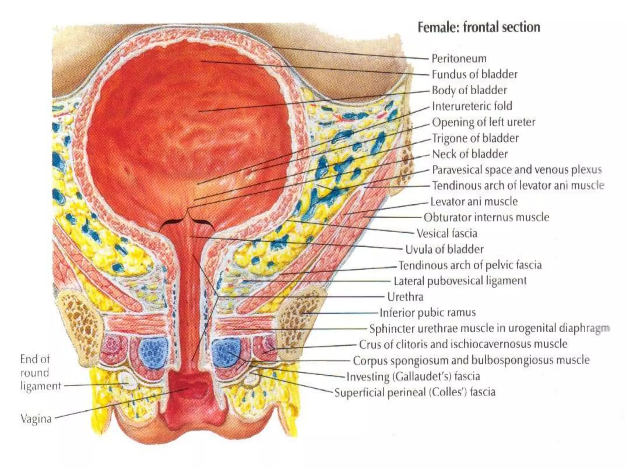
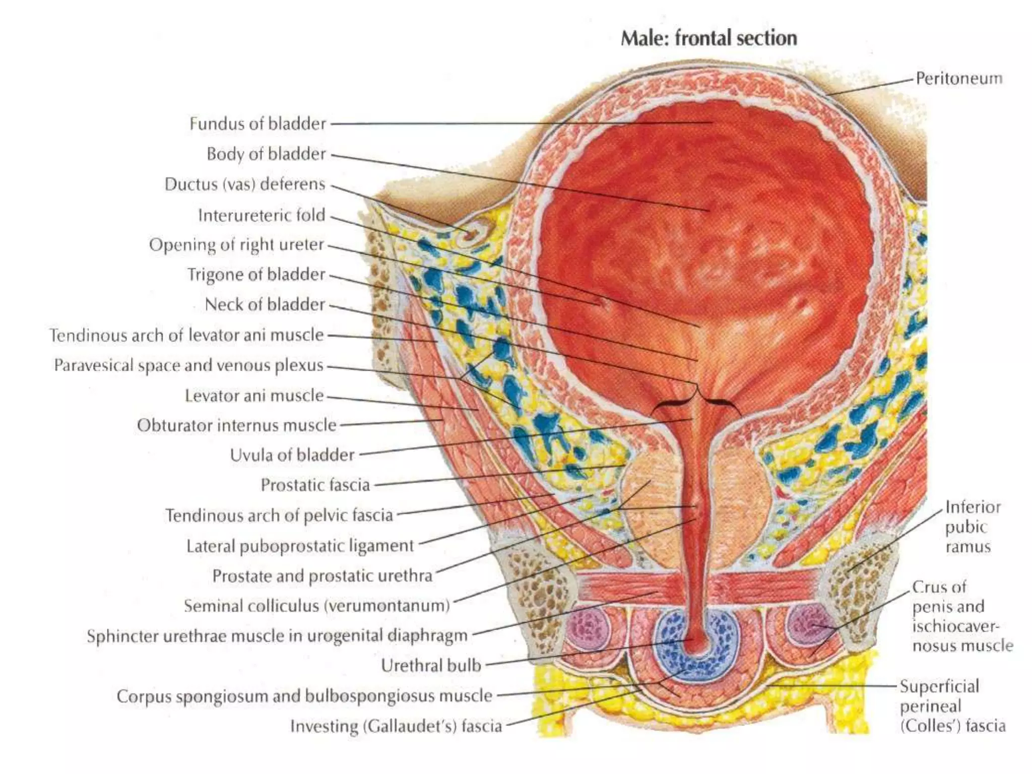


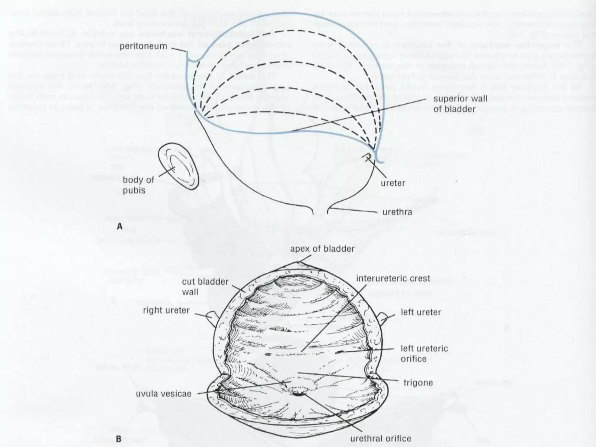


The document summarizes the anatomy and physiology of the urinary bladder. It describes the bladder as a hollow muscular organ that acts as a reservoir for urine. It notes that in adults the bladder normally holds 400-500 ml of urine. The document discusses the location of the bladder, ureter openings, blood supply, innervation, and functions of storage and emptying.