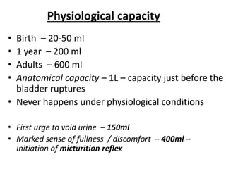
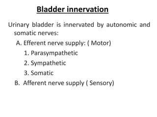
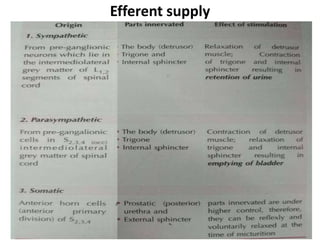
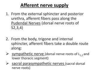
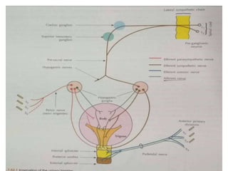
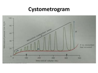
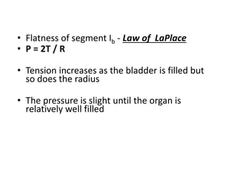
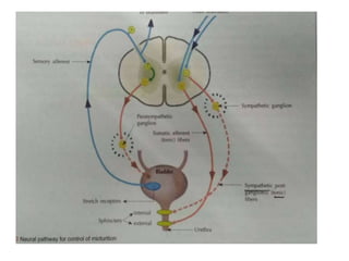
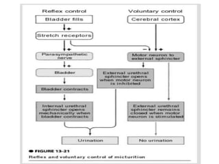
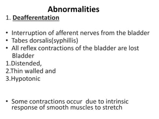
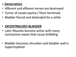
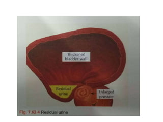
Physiology of urinary bladder
and Micturition
Functional anatomy of urinary
bladder
• Hollow organ
• lined by transitional epithelium
• stretched enormously
• Mucosa is thrown into folds called rugae
• except triangular area at the base - Trigone
• Smooth muscle – Detrusor
• Interlacing of fibres
• Internal sphincter – sphinteric mechanism
• Function – maintain tonic closure of urethral
opening
• prevent reflux of semen into urethra during
ejaculation
• External sphinter – voluntary skeletal m.
• can be reflexely/ voluntarily relaxed at the time
of micturition
• Upper end – Ureteric opening – ureters enter
obliquely
Physiological capacity
• Birth – 20-50 ml
• 1 year – 200 ml
• Adults – 600 ml
• Anatomical capacity – 1L – capacity just before the
bladder ruptures
• Never happens under physiological conditions
• First urge to void urine – 150ml
• Marked sense of fullness / discomfort – 400ml –
Initiation of micturition reflex
Bladder innervation
Urinary bladder is innervated by autonomic and
somatic nerves:
A. Efferent nerve supply: ( Motor)
1. Parasympathetic
2. Sympathetic
3. Somatic
B. Afferent nerve supply ( Sensory)
Efferent supply
Afferent nerve supply
1. From the external sphincter and posterior
urethra, afferent fibers pass along the
Pudendal Nerves (dorsal nerve roots of
S2,3,4)
2. From the body, trigone and internal
sphincter, afferent fibers take a double route
along:
 sympathetic nerve (dorsal nerve roots of L1,2 and
lower thoracic segment)
 sacral parasympathetic nerves (sacral dorsal
nerve roots)
Functions
1. Indicates the degree of distension of the
urinary bladder. (afferent nerves along sacral
parasympathetic)
2. To convey pain sensibility. (afferent nerves
along sympathetic)
Mechanics of micturition
• 2phases – Emptying and filling – interplay of
pressures in the bladder and internal sphincter
• Spinal reflex – facilitated and inhibited by higher
brain centres
• Plasticity – when it is stretched tension is not
maintained for long
Cystometrogram
• Plot of intravesical pressure against volume of
fluid in the bladder – Cystometrogram
1.Initial slight rise of pressure – Ia
2.Long nearly flat segment – Ib
3.Sudden sharp rise of pressure when the
micturition reflex is triggered – II
Cystometrogram
• Flatness of segment Ib - Law of LaPlace
• P = 2T / R
• Tension increases as the bladder is filled but
so does the radius
• The pressure is slight until the organ is
relatively well filled
Voluntary micturition-Afferent
pathway
Bladder fills with urine –stretch receptors++
↓
Sensory signals to the dorsal nerve root S2,3,4
segments of the spinal cord (pelvic nerves)
↓
Back to the bladder by the parasympathetic fibres
in the same nerve
↓
Micturition reflex
• Sympathetic efferents are reflexely
inhibited
• Reflex is integrated in the sacral segment of
spinal cord
• Volume of urine that initiates reflex
contraction – 300-400 ml
Higher control of micturition reflex
Threshold for voiding reflex is adjusted by :
Facilitatory areas
 Pontine
 Post. Hypothalamus
Inhibitory areas
 midbrain
• Voluntary contraction of abdominal muscles
aids in expulsion of urine
Mechanism of micturition
• During micturition – perineal muscle and
external urethral sphincters are relaxed
↓
Detrusor muscles contract
↓
Urine passes out through the urethra
• Voluntary contraction – relaxation of muscles of
pelvic floor
↓
Downward tug on detrusor muscles
• Perineal muscles and external urethra can be
contracted voluntarily – preventing urine from
passing down or interruption of flow
Abnormalities
1. Deafferentation
• Interruption of afferent nerves from the bladder
• Tabes dorsalis(syphillis)
• All reflex contractions of the bladder are lost
Bladder
1.Distended,
2.Thin walled and
3.Hypotonic
• Some contractions occur due to intrinsic
response of smooth muscles to stretch
2. Denervation
• Afferent and efferent nerves are destroyed
• Tumor of cauda equina / filum terminale
• Bladder flaccid and distended for a while
• DECENTRALISED BLADDER
• Later Muscles become active with many
contraction waves that cause dribbling
• Bladder becomes shrunken and bladder wall is
hypertrophied
3. Spinal cord transection
• During spinal shock – bladder is flaccid and
unresponsive
• Sphinter tone returns first
• Overfilled and urine dribbles out through the
sphincter (Overflow incontinence )
• After spinal shock has passed,
• voiding reflex returns
• no voluntary control and
• no facilitation/ inhibition from higher centres
• Paraplegic – Initiate voiding – mild mass reflex
• Sometimes voiding reflex becomes
hyperactive – capacity is reduced – walls
become hypertrophied (Spastic neurogenic
bladder )
• infection of the urinary bladder
• In all these defects the bladder contracts but
these contractions are not sufficient to empty
the viscus completely
• Residual urine is left in the bladder

Physiology of urinary bladder.pptx

  • 1.
    Physiology of urinarybladder and Micturition
  • 2.
    Functional anatomy ofurinary bladder • Hollow organ • lined by transitional epithelium • stretched enormously • Mucosa is thrown into folds called rugae • except triangular area at the base - Trigone • Smooth muscle – Detrusor • Interlacing of fibres
  • 4.
    • Internal sphincter– sphinteric mechanism • Function – maintain tonic closure of urethral opening • prevent reflux of semen into urethra during ejaculation • External sphinter – voluntary skeletal m. • can be reflexely/ voluntarily relaxed at the time of micturition • Upper end – Ureteric opening – ureters enter obliquely
  • 6.
    Physiological capacity • Birth– 20-50 ml • 1 year – 200 ml • Adults – 600 ml • Anatomical capacity – 1L – capacity just before the bladder ruptures • Never happens under physiological conditions • First urge to void urine – 150ml • Marked sense of fullness / discomfort – 400ml – Initiation of micturition reflex
  • 7.
    Bladder innervation Urinary bladderis innervated by autonomic and somatic nerves: A. Efferent nerve supply: ( Motor) 1. Parasympathetic 2. Sympathetic 3. Somatic B. Afferent nerve supply ( Sensory)
  • 8.
  • 9.
    Afferent nerve supply 1.From the external sphincter and posterior urethra, afferent fibers pass along the Pudendal Nerves (dorsal nerve roots of S2,3,4) 2. From the body, trigone and internal sphincter, afferent fibers take a double route along:  sympathetic nerve (dorsal nerve roots of L1,2 and lower thoracic segment)  sacral parasympathetic nerves (sacral dorsal nerve roots)
  • 11.
    Functions 1. Indicates thedegree of distension of the urinary bladder. (afferent nerves along sacral parasympathetic) 2. To convey pain sensibility. (afferent nerves along sympathetic)
  • 12.
    Mechanics of micturition •2phases – Emptying and filling – interplay of pressures in the bladder and internal sphincter • Spinal reflex – facilitated and inhibited by higher brain centres • Plasticity – when it is stretched tension is not maintained for long
  • 13.
    Cystometrogram • Plot ofintravesical pressure against volume of fluid in the bladder – Cystometrogram 1.Initial slight rise of pressure – Ia 2.Long nearly flat segment – Ib 3.Sudden sharp rise of pressure when the micturition reflex is triggered – II
  • 14.
  • 15.
    • Flatness ofsegment Ib - Law of LaPlace • P = 2T / R • Tension increases as the bladder is filled but so does the radius • The pressure is slight until the organ is relatively well filled
  • 16.
    Voluntary micturition-Afferent pathway Bladder fillswith urine –stretch receptors++ ↓ Sensory signals to the dorsal nerve root S2,3,4 segments of the spinal cord (pelvic nerves) ↓ Back to the bladder by the parasympathetic fibres in the same nerve ↓ Micturition reflex
  • 18.
    • Sympathetic efferentsare reflexely inhibited • Reflex is integrated in the sacral segment of spinal cord • Volume of urine that initiates reflex contraction – 300-400 ml
  • 19.
    Higher control ofmicturition reflex Threshold for voiding reflex is adjusted by : Facilitatory areas  Pontine  Post. Hypothalamus Inhibitory areas  midbrain • Voluntary contraction of abdominal muscles aids in expulsion of urine
  • 20.
    Mechanism of micturition •During micturition – perineal muscle and external urethral sphincters are relaxed ↓ Detrusor muscles contract ↓ Urine passes out through the urethra
  • 21.
    • Voluntary contraction– relaxation of muscles of pelvic floor ↓ Downward tug on detrusor muscles • Perineal muscles and external urethra can be contracted voluntarily – preventing urine from passing down or interruption of flow
  • 23.
    Abnormalities 1. Deafferentation • Interruptionof afferent nerves from the bladder • Tabes dorsalis(syphillis) • All reflex contractions of the bladder are lost Bladder 1.Distended, 2.Thin walled and 3.Hypotonic • Some contractions occur due to intrinsic response of smooth muscles to stretch
  • 24.
    2. Denervation • Afferentand efferent nerves are destroyed • Tumor of cauda equina / filum terminale • Bladder flaccid and distended for a while • DECENTRALISED BLADDER • Later Muscles become active with many contraction waves that cause dribbling • Bladder becomes shrunken and bladder wall is hypertrophied
  • 26.
    3. Spinal cordtransection • During spinal shock – bladder is flaccid and unresponsive • Sphinter tone returns first • Overfilled and urine dribbles out through the sphincter (Overflow incontinence )
  • 27.
    • After spinalshock has passed, • voiding reflex returns • no voluntary control and • no facilitation/ inhibition from higher centres • Paraplegic – Initiate voiding – mild mass reflex
  • 28.
    • Sometimes voidingreflex becomes hyperactive – capacity is reduced – walls become hypertrophied (Spastic neurogenic bladder ) • infection of the urinary bladder
  • 30.
    • In allthese defects the bladder contracts but these contractions are not sufficient to empty the viscus completely • Residual urine is left in the bladder

Editor's Notes

  • #13 Read about plasticity and la place law
  • #27 After complete transection of the spinal cord when reflex movements return , a mild noxious stimulus on the lower limb can cause withdrawl reflex and autonomic changes like urination, defecation , sweating and fluctuation in B.P