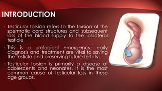
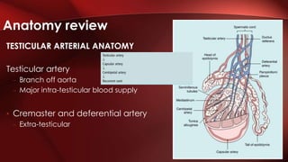
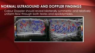
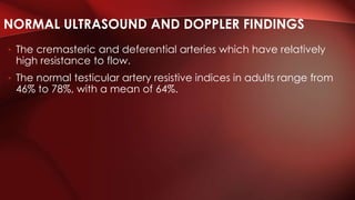
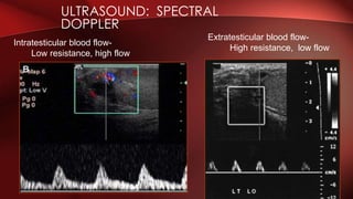
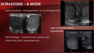
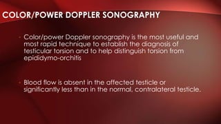
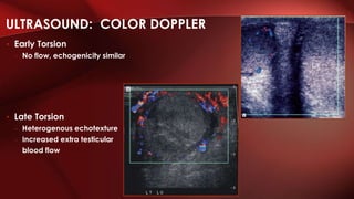
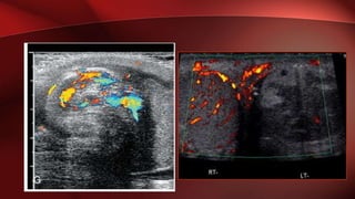
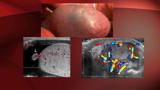
Testicular torsion refers to twisting of the spermatic cord and loss of blood supply to the testicle. It is a urological emergency as early diagnosis and treatment are needed to save the testicle. Ultrasound with Doppler is the primary imaging method and shows absent or decreased blood flow in the affected testicle compared to the normal side. Prompt surgical detorsion and orchioplexy are the definitive treatments.
Disclaimer: A lot from this slides were taken also from https://www.slideshare.net/babysurgeon/scrotal-swellings-1 (Dr Selvaraj Balasubramani)
This covers only :
ANATOMY
CAUSES
TORSION OF TESTIS
EPIDIDYMO-ORCHITIS
HYDROCELE
EPIDIDYMAL CYST
VARICOCELE
Testicular torsion refers to the torsion of the spermatic cord structures and subsequent loss of the blood supply to the ipsilateral testicle.
Urological emergency; early diagnosis and treatment are vital.
Mainly disease of Neonates, Adolescents.
The rate of testicular viability decreases significantly after 6 hours from onset of symptoms.
it is painful condition for boys , coming in emergency, ultrasound is basic imaging .it is to see testes and accordingly guide the surgeon whether testes could be saved
Flu Vaccine Alert in Bangalore Karnatakaaddon Scans
As flu season approaches, health officials in Bangalore, Karnataka, are urging residents to get their flu vaccinations. The seasonal flu, while common, can lead to severe health complications, particularly for vulnerable populations such as young children, the elderly, and those with underlying health conditions.
Dr. Vidisha Kumari, a leading epidemiologist in Bangalore, emphasizes the importance of getting vaccinated. "The flu vaccine is our best defense against the influenza virus. It not only protects individuals but also helps prevent the spread of the virus in our communities," he says.
This year, the flu season is expected to coincide with a potential increase in other respiratory illnesses. The Karnataka Health Department has launched an awareness campaign highlighting the significance of flu vaccinations. They have set up multiple vaccination centers across Bangalore, making it convenient for residents to receive their shots.
To encourage widespread vaccination, the government is also collaborating with local schools, workplaces, and community centers to facilitate vaccination drives. Special attention is being given to ensuring that the vaccine is accessible to all, including marginalized communities who may have limited access to healthcare.
Residents are reminded that the flu vaccine is safe and effective. Common side effects are mild and may include soreness at the injection site, mild fever, or muscle aches. These side effects are generally short-lived and far less severe than the flu itself.
Healthcare providers are also stressing the importance of continuing COVID-19 precautions. Wearing masks, practicing good hand hygiene, and maintaining social distancing are still crucial, especially in crowded places.
Protect yourself and your loved ones by getting vaccinated. Together, we can help keep Bangalore healthy and safe this flu season. For more information on vaccination centers and schedules, residents can visit the Karnataka Health Department’s official website or follow their social media pages.
Stay informed, stay safe, and get your flu shot today!
ARTIFICIAL INTELLIGENCE IN HEALTHCARE.pdfAnujkumaranit
Artificial intelligence (AI) refers to the simulation of human intelligence processes by machines, especially computer systems. It encompasses tasks such as learning, reasoning, problem-solving, perception, and language understanding. AI technologies are revolutionizing various fields, from healthcare to finance, by enabling machines to perform tasks that typically require human intelligence.
Tom Selleck Health: A Comprehensive Look at the Iconic Actor’s Wellness Journeygreendigital
Tom Selleck, an enduring figure in Hollywood. has captivated audiences for decades with his rugged charm, iconic moustache. and memorable roles in television and film. From his breakout role as Thomas Magnum in Magnum P.I. to his current portrayal of Frank Reagan in Blue Bloods. Selleck's career has spanned over 50 years. But beyond his professional achievements. fans have often been curious about Tom Selleck Health. especially as he has aged in the public eye.
Follow us on: Pinterest
Introduction
Many have been interested in Tom Selleck health. not only because of his enduring presence on screen but also because of the challenges. and lifestyle choices he has faced and made over the years. This article delves into the various aspects of Tom Selleck health. exploring his fitness regimen, diet, mental health. and the challenges he has encountered as he ages. We'll look at how he maintains his well-being. the health issues he has faced, and his approach to ageing .
Early Life and Career
Childhood and Athletic Beginnings
Tom Selleck was born on January 29, 1945, in Detroit, Michigan, and grew up in Sherman Oaks, California. From an early age, he was involved in sports, particularly basketball. which played a significant role in his physical development. His athletic pursuits continued into college. where he attended the University of Southern California (USC) on a basketball scholarship. This early involvement in sports laid a strong foundation for his physical health and disciplined lifestyle.
Transition to Acting
Selleck's transition from an athlete to an actor came with its physical demands. His first significant role in "Magnum P.I." required him to perform various stunts and maintain a fit appearance. This role, which he played from 1980 to 1988. necessitated a rigorous fitness routine to meet the show's demands. setting the stage for his long-term commitment to health and wellness.
Fitness Regimen
Workout Routine
Tom Selleck health and fitness regimen has evolved. adapting to his changing roles and age. During his "Magnum, P.I." days. Selleck's workouts were intense and focused on building and maintaining muscle mass. His routine included weightlifting, cardiovascular exercises. and specific training for the stunts he performed on the show.
Selleck adjusted his fitness routine as he aged to suit his body's needs. Today, his workouts focus on maintaining flexibility, strength, and cardiovascular health. He incorporates low-impact exercises such as swimming, walking, and light weightlifting. This balanced approach helps him stay fit without putting undue strain on his joints and muscles.
Importance of Flexibility and Mobility
In recent years, Selleck has emphasized the importance of flexibility and mobility in his fitness regimen. Understanding the natural decline in muscle mass and joint flexibility with age. he includes stretching and yoga in his routine. These practices help prevent injuries, improve posture, and maintain mobilit
Ozempic: Preoperative Management of Patients on GLP-1 Receptor Agonists Saeid Safari
Preoperative Management of Patients on GLP-1 Receptor Agonists like Ozempic and Semiglutide
ASA GUIDELINE
NYSORA Guideline
2 Case Reports of Gastric Ultrasound
micro teaching on communication m.sc nursing.pdfAnurag Sharma
Microteaching is a unique model of practice teaching. It is a viable instrument for the. desired change in the teaching behavior or the behavior potential which, in specified types of real. classroom situations, tends to facilitate the achievement of specified types of objectives.
- Video recording of this lecture in English language: https://youtu.be/lK81BzxMqdo
- Video recording of this lecture in Arabic language: https://youtu.be/Ve4P0COk9OI
- Link to download the book free: https://nephrotube.blogspot.com/p/nephrotube-nephrology-books.html
- Link to NephroTube website: www.NephroTube.com
- Link to NephroTube social media accounts: https://nephrotube.blogspot.com/p/join-nephrotube-on-social-media.html
Lung Cancer: Artificial Intelligence, Synergetics, Complex System Analysis, S...Oleg Kshivets
RESULTS: Overall life span (LS) was 2252.1±1742.5 days and cumulative 5-year survival (5YS) reached 73.2%, 10 years – 64.8%, 20 years – 42.5%. 513 LCP lived more than 5 years (LS=3124.6±1525.6 days), 148 LCP – more than 10 years (LS=5054.4±1504.1 days).199 LCP died because of LC (LS=562.7±374.5 days). 5YS of LCP after bi/lobectomies was significantly superior in comparison with LCP after pneumonectomies (78.1% vs.63.7%, P=0.00001 by log-rank test). AT significantly improved 5YS (66.3% vs. 34.8%) (P=0.00000 by log-rank test) only for LCP with N1-2. Cox modeling displayed that 5YS of LCP significantly depended on: phase transition (PT) early-invasive LC in terms of synergetics, PT N0—N12, cell ratio factors (ratio between cancer cells- CC and blood cells subpopulations), G1-3, histology, glucose, AT, blood cell circuit, prothrombin index, heparin tolerance, recalcification time (P=0.000-0.038). Neural networks, genetic algorithm selection and bootstrap simulation revealed relationships between 5YS and PT early-invasive LC (rank=1), PT N0—N12 (rank=2), thrombocytes/CC (3), erythrocytes/CC (4), eosinophils/CC (5), healthy cells/CC (6), lymphocytes/CC (7), segmented neutrophils/CC (8), stick neutrophils/CC (9), monocytes/CC (10); leucocytes/CC (11). Correct prediction of 5YS was 100% by neural networks computing (area under ROC curve=1.0; error=0.0).
CONCLUSIONS: 5YS of LCP after radical procedures significantly depended on: 1) PT early-invasive cancer; 2) PT N0--N12; 3) cell ratio factors; 4) blood cell circuit; 5) biochemical factors; 6) hemostasis system; 7) AT; 8) LC characteristics; 9) LC cell dynamics; 10) surgery type: lobectomy/pneumonectomy; 11) anthropometric data. Optimal diagnosis and treatment strategies for LC are: 1) screening and early detection of LC; 2) availability of experienced thoracic surgeons because of complexity of radical procedures; 3) aggressive en block surgery and adequate lymph node dissection for completeness; 4) precise prediction; 5) adjuvant chemoimmunoradiotherapy for LCP with unfavorable prognosis.
Pulmonary Thromboembolism - etilogy, types, medical- Surgical and nursing man...VarunMahajani
Disruption of blood supply to lung alveoli due to blockage of one or more pulmonary blood vessels is called as Pulmonary thromboembolism. In this presentation we will discuss its causes, types and its management in depth.
HOT NEW PRODUCT! BIG SALES FAST SHIPPING NOW FROM CHINA!! EU KU DB BK substit...GL Anaacs
Contact us if you are interested:
Email / Skype : kefaya1771@gmail.com
Threema: PXHY5PDH
New BATCH Ku !!! MUCH IN DEMAND FAST SALE EVERY BATCH HAPPY GOOD EFFECT BIG BATCH !
Contact me on Threema or skype to start big business!!
Hot-sale products:
NEW HOT EUTYLONE WHITE CRYSTAL!!
5cl-adba precursor (semi finished )
5cl-adba raw materials
ADBB precursor (semi finished )
ADBB raw materials
APVP powder
5fadb/4f-adb
Jwh018 / Jwh210
Eutylone crystal
Protonitazene (hydrochloride) CAS: 119276-01-6
Flubrotizolam CAS: 57801-95-3
Metonitazene CAS: 14680-51-4
Payment terms: Western Union,MoneyGram,Bitcoin or USDT.
Deliver Time: Usually 7-15days
Shipping method: FedEx, TNT, DHL,UPS etc.Our deliveries are 100% safe, fast, reliable and discreet.
Samples will be sent for your evaluation!If you are interested in, please contact me, let's talk details.
We specializes in exporting high quality Research chemical, medical intermediate, Pharmaceutical chemicals and so on. Products are exported to USA, Canada, France, Korea, Japan,Russia, Southeast Asia and other countries.
2. • Testicular torsion refers to the torsion of the
spermatic cord structures and subsequent
loss of the blood supply to the ipsilateral
testicle.
• This is a urological emergency; early
diagnosis and treatment are vital to saving
the testicle and preserving future fertility.
• Testicular torsion is primarily a disease of
adolescents and neonates. It is the most
common cause of testicular loss in these
age groups.
INTRODUCTION
3. Normal anatomy
• The tunica vaginalis does not
completely surround the testis and
epididymis, which are attached to the
posterior scrotal wall.
Anatomy review
4. • Bell-clapper anomaly.
The tunica vaginalis completely surrounds
the testis, epididymis, and part of the
spermatic cord, predisposing to torsion.
Anatomy review
5. TESTICULAR ARTERIAL ANATOMY
Testicular artery
– Branch off aorta
– Major intra-testicular blood supply
• Cremaster and deferential artery
– Extra-testicular
Anatomy review
6. • Colour Doppler should reveal bilaterally symmetric and relatively
uniform flow through both testes and epididymides.
NORMAL ULTRASOUND AND DOPPLER FINDINGS
7. • Spectral Doppler tracings of testicular arterial inflow demonstrate
relatively low resistance
NORMAL ULTRASOUND AND DOPPLER FINDINGS
8. • The cremasteric and deferential arteries which have relatively
high resistance to flow.
• The normal testicular artery resistive indices in adults range from
46% to 78%, with a mean of 64%.
NORMAL ULTRASOUND AND DOPPLER FINDINGS
10. • Torsion occurs as the testicle rotates between 90° and 180°,
compromising blood flow to and from the testicle.
• Complete torsion usually occurs when the testicle twists 360° or more;
incomplete or partial torsion occurs with lesser degrees of rotation.
The degree of torsion may extend to 720°.
• The twisting of the testicle causes venous occlusion and engorgement
as well as arterial ischemia and infarction of the testicle.
• The degree of torsion the testicle endures may play a role in the
viability of the testicle over time.
• In addition to the extent of torsion, the duration of torsion prominently
influences the rates of both immediate salvage and late testicular
atrophy. Testicular salvage is most likely if the duration of torsion is less
than 6-8 hours.
• If 24 hours or more elapse, testicular necrosis develops in most
patients.
PATHOPHYSIOLOGY
11. Intravaginal torsion
Is the more common type, occurring
most frequently at puberty. It results
from anomalous suspension of the testis
by a long stalk of spermatic cord,
resulting in complete investment of the
testis and epididymis by the tunica
vaginalis.
• This anomaly has been likened to a
bell-clapper
TWO TYPES OF TESTICULAR
TORSION
12. Extravaginal torsion
• Most often occurs in newborns
without the “bell clapper”
deformity.
• It is thought to result from a poor or
absent attachment of the testis to
the scrotal wall, allowing rotation of
the testis, epididymis, and tunica
vaginalis as a unit and causing
torsion of the cord at the level of
the external ring
TWO TYPES OF TESTICULAR
TORSION
14. • Severe unilateral scrotal pain
• Previous episodes, spontaneous resolution
• Related to activity, trauma, during sleep
• Nausea, vomiting, abdominal pain, fever
HISTORY
15. • Prenatal torsion, firm, hard, scrotal
mass, which does not transilluminate
in an otherwise asymptomatic
newborn male. The scrotal skin
characteristically fixes to the
necrotic gonad.
• Older patient, swollen, tender, high-
riding testis with abnormal transverse
lie and loss of the cremasteric reflex
PHYSICAL EXAMINATION
17. • Sensitivity 86%, specificity 100% experienced provider using color /
power doppler1
• Gray-scale findings on ultrasound depend on how much time has
passed since the torsion occurred.
• The gray-scale findings of acute and subacute torsion are not
specific and may be seen in testicular infarction caused by
epididymitis, epididymo-orchitis, and traumatic testicular rupture or
infarction.
ULTRASOUND FOR TESTICULAR TORSION
18. • Early stages, scrotal contents may have a normal sonographic
appearance.
• After 4 to 6 hours, the testis becomes swollen and hypoechoic,
• After 24 hours, the testis becomes heterogeneous as a result of
hemorrhage, infarction, necrosis, and vascular congestion
• The epididymal head appears enlarged and may have
decreased echogenicity or may become heterogeneous.
• The spermatic cord immediately cranial to the testis and
epididymis is twisted, causing a characteristic torsion knot or
“whirlpool pattern” of concentric layers
ULTRASOUND FOR TESTICULAR TORSION
19. • A reactive hydrocele and scrotal skin thickening are often seen
with torsion.
Large, echogenic or complex extratesticular masses caused by
hemorrhage in the tunica vaginalis or epididymis may be seen in
patients with undiagnosed torsion.
ULTRASOUND FOR TESTICULAR TORSION
20. ULTRASOUND – B-MODE
– Early ischemia: enlargement, no Δ echogenicity
– Hemorrhage: hyperechoic areas in an
infarcted testis, heterogenous
• Late ischemia/infarct:
hypoechoic
21. • Color/power Doppler sonography is the most useful and
most rapid technique to establish the diagnosis of
testicular torsion and to help distinguish torsion from
epididymo-orchitis
• Blood flow is absent in the affected testicle or
significantly less than in the normal, contralateral testicle.
COLOR/POWER DOPPLER SONOGRAPHY
22. ULTRASOUND: COLOR DOPPLER
• Early Torsion
– No flow, echogenicity similar
• Late Torsion
– Heterogenous echotexture
– Increased extra testicular
blood flow
23.
24. Meticulous scanning of the testicular parenchyma with
the use of low-flow detection Doppler settings
(low pulse repetition frequency, low wall filter, high
Doppler gain)
is important because testicular vessels are small and
have low flow velocities, especially in prepubertal
boys.
25. DIFFERENTIAL DIAGNOSIS OF ACUTE SCROTUM
• Epididymitis
• Scrotal abscess
• Torsion of epididymal appendage
• Intratesticular hematoma
26. • Is a common cause of acute scrotal pain and may mimic
testicular torsion clinically.
• Patients are rarely referred for imaging because the pain is
usually not severe, and the twisted appendage may be evident
clinically as the “blue dot” sign.
• The sonographic appearance of the twisted testicular
appendage has been described as an avascular hypoechoic
mass adjacent to a normally perfused testis and surrounded by
an area of increased color Doppler perfusion.
• However, the twisted appendage may appear as an echogenic
extratesticular mass situated between the head of the
epididymis and the upper pole of the testis.
TORSION OF THE TESTICULAR APPENDAGE
27.
28. • Technetium-99m pertechnetate is the agent of choice.
• Immediate radionuclide angiograms are obtained, with
subsequent static images as well.
• In the healthy patient, images show symmetric flow to the
testes, and delayed images show uniformly symmetric
activity.
NUCLEAR IMAGING
29. • Static images demonstrate a photopenic area in the
involved testis.
• In the subacute and late phases of torsion (missed torsion),
there is often increased flow to the affected hemiscrotum
via the pudendal artery with a photopenic testis and a rim
of surrounding increased activity on static images. This has
been called a rim, doughnut, or bull's-eye sign.
NUCLEAR IMAGING
30.
31.
32. • Near-infrared spectroscopy (NIRS) is an emerging tool to assess
testicular torsion.
• It can measure oxygen saturation 3-4 cm deep in the skin, is rapid
(lasting 20 seconds), and is noninvasive.
• Aydogdu et al performed a small prospective study evaluating 16
adult patients with testicular torsion and found NIRS to be 100%
sensitive and specific for torsion when compared with the
contralateral testis.
NEAR-INFRARED SPECTROSCOPY
33. TREATMENT
• Definitive treatment: surgical
detorsion and orchioplexy
• Manual detorsion: medial to
lateral; “opening a book”
– May need to rotate 2-3 times for
complete detorsion
Roberts: Clinical Procedures in Emergency Medicine
Editor's Notes
The right and left testicular arteries originate from the aorta just below the renal arteries.
They course through the deep inguinal ring to enter the spermatic cord, accompanied by the cremasteric and deferentialarteries, which supply the soft tissues of the scrotum , epididymis and vas deferens.
The testicular artery penetrates the tunica albuginea along the posterior aspect of the testis and gives off capsular branches which course through the tunic a vasculosa. The se capsularbranches then give rise to the centripetal arteries which carry blood from the capsular surface , centrally towards the mediastinum along the septula (Figs 13-5 and 13-6). Branches of the centripetal arteries then course backward towards the capsular surface; theseare known as recurrent rami.,
Transverse colour Doppler ultrasound image of both testicles using a split screen format. Note the symmetry of echotexture and theuniformity and symmetry of colour flow.
Spectral Doppler waveform of a normal left testicular artery. The resistive index is normal (approx. 60%).
Intrastesticular supply- TESTICULAR a off aorta, low resistance with high flow
Extratesticular supply- DEFERENTIAL and CREMASTERIC a, high resistance with low flow
Flow during diastole
Looking at echotexture
Straddle view to see portions of each in same image.
Enlarged to engorged blood
Epidid: enlarge hyperemic epydidymis
Abscess: comples fluid collection
Appendix: mass next to epididymal head with absence of flow
Hematoma has surrounding blood flow.