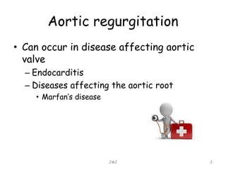
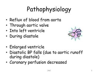
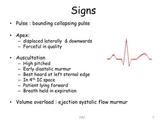
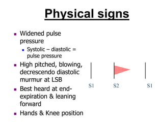
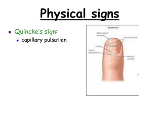
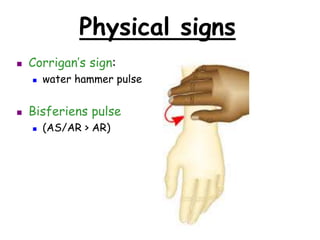
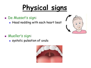
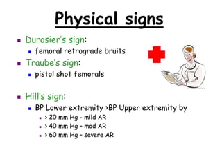
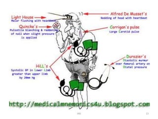
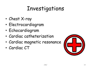
Atrial Regurgitation
K. Kavindya M. Fernando
JMJ 1
Aortic regurgitation
• Can occur in disease affecting aortic
valve
– Endocarditis
– Diseases affecting the aortic root
• Marfan’s disease
JMJ 2
Pathophysiology
• Reflux of blood from aorta
• Through aortic valve
• Into left ventricle
• During diastole
• Enlarged ventricle
• Diastolic BP falls (due to aortic runoff
during diastole)
• Coronary perfusion decreased
JMJ 3
Aeitiology
 Any conditions resulting in incompetent
aortic leaflets
 Congenital
 Bicuspid valve
 Aortopathy
 Collagen disorders (e.g. Marfan’s)
 Ehler-Danlos
 Osteogenesis imperfecta
 Pseudoxanthoma elasticum
Aeitiology
 Acquired
 Rheumatic heart disease
 Dilated aorta (e.g. hypertension..)
 Degenerative
 Connective tissue disorders
 E.g. ankylosing spondylitis, rheumatoid arthritis,
Reiter’s syndrome, Giant-cell arteritis )
 Syphilis (chronic aortitis)
 Acute AI: aortic dissection, infective
endocarditis, trauma
Symptoms
• Symptoms occur late
• They do not develop until L ventricular
failure
• Angina pectoris is a frequent complaint
• Dyspnea
• Arrhythmias are relatively uncommon
JMJ 6
Signs
• Pulse : bounding collapsing pulse
• Apex:
– displaced laterally & downwards
– Forceful in quality
• Auscultation
– High pitched
– Early diastolic murmur
– Best heard at left sternal edge
– In 4th IC space
– Patient lying forward
– Breath held in expiration
• Volume overload : ejection systolic flow murmur
JMJ 7
Physical signs
 Widened pulse
pressure
 Systolic – diastolic =
pulse pressure
 High pitched, blowing,
decrescendo diastolic
murmur at LSB
 Best heard at end-
expiration & leaning
forward
 Hands & Knee position
S1 S2 S1
Physical signs
 Quincke’s sign:
 capillary pulsation
Physical signs
 Corrigan’s sign:
 water hammer pulse
 Bisferiens pulse
 (AS/AR > AR)
Physical signs
 De Musset’s sign:
 Head nodding with each heart beat
 Mueller’s sign:
 systolic pulsation of uvula
Physical signs
 Durosier’s sign:
 femoral retrograde bruits
 Traube’s sign:
 pistol shot femorals
 Hill’s sign:
 BP Lower extremity >BP Upper extremity by
 > 20 mm Hg - mild AR
 > 40 mm Hg – mod AR
 > 60 mm Hg – severe AR
JMJ 13
Investigations
• Chest X-ray
• Electrocardiogram
• Echocardiogram
• Cardiac catheterization
• Cardiac magnetic resonance
• Cardiac CT
JMJ 14
Investigations
• Chest X-ray
– Features of left ventricular failure
– Dilation of ascending aorta
– Ascending aorta may be calcified : syphilis
JMJ 15
Investigations
• ECG
– Features of left ventricular falilure
– Tall R waves
– Deeply inverted T waves in the left side
chest leads
– Deep S waves in the R side chest leasds
– Normally sinus rhythm is present
JMJ 16
Treatment
• Underlying cause may need treatment
– Syphilitic aortitis
– Infective endocarditis
• Acute aortic regurgitation
– Treatment with vasodilators & inotropes
• Patients with left ventricular dysfunctions
– ACE inhibitors
– Beta blockers (may slow aortic dilatation in
Marfan’s patients)
JMJ 17
Treatment
• Aortic surgery indicated
– In acute severe aortic regurgitation
(endocarditis)
– In symptomatic (dyspnea, angina) patients
with chronic severe aortic regurgitation
– Ehen symptomatic with L ventricular
ejection fraction is < 50%
JMJ 18
Treatment
• Mechanical prostheses & tissue valves
• Antibiotic prophylaxis against infective
endocarditis is not recommended
JMJ 19
JMJ 20

Atrial regurgitation

  • 1.
  • 2.
    Aortic regurgitation • Canoccur in disease affecting aortic valve – Endocarditis – Diseases affecting the aortic root • Marfan’s disease JMJ 2
  • 3.
    Pathophysiology • Reflux ofblood from aorta • Through aortic valve • Into left ventricle • During diastole • Enlarged ventricle • Diastolic BP falls (due to aortic runoff during diastole) • Coronary perfusion decreased JMJ 3
  • 4.
    Aeitiology  Any conditionsresulting in incompetent aortic leaflets  Congenital  Bicuspid valve  Aortopathy  Collagen disorders (e.g. Marfan’s)  Ehler-Danlos  Osteogenesis imperfecta  Pseudoxanthoma elasticum
  • 5.
    Aeitiology  Acquired  Rheumaticheart disease  Dilated aorta (e.g. hypertension..)  Degenerative  Connective tissue disorders  E.g. ankylosing spondylitis, rheumatoid arthritis, Reiter’s syndrome, Giant-cell arteritis )  Syphilis (chronic aortitis)  Acute AI: aortic dissection, infective endocarditis, trauma
  • 6.
    Symptoms • Symptoms occurlate • They do not develop until L ventricular failure • Angina pectoris is a frequent complaint • Dyspnea • Arrhythmias are relatively uncommon JMJ 6
  • 7.
    Signs • Pulse :bounding collapsing pulse • Apex: – displaced laterally & downwards – Forceful in quality • Auscultation – High pitched – Early diastolic murmur – Best heard at left sternal edge – In 4th IC space – Patient lying forward – Breath held in expiration • Volume overload : ejection systolic flow murmur JMJ 7
  • 8.
    Physical signs  Widenedpulse pressure  Systolic – diastolic = pulse pressure  High pitched, blowing, decrescendo diastolic murmur at LSB  Best heard at end- expiration & leaning forward  Hands & Knee position S1 S2 S1
  • 9.
    Physical signs  Quincke’ssign:  capillary pulsation
  • 10.
    Physical signs  Corrigan’ssign:  water hammer pulse  Bisferiens pulse  (AS/AR > AR)
  • 11.
    Physical signs  DeMusset’s sign:  Head nodding with each heart beat  Mueller’s sign:  systolic pulsation of uvula
  • 12.
    Physical signs  Durosier’ssign:  femoral retrograde bruits  Traube’s sign:  pistol shot femorals  Hill’s sign:  BP Lower extremity >BP Upper extremity by  > 20 mm Hg - mild AR  > 40 mm Hg – mod AR  > 60 mm Hg – severe AR
  • 13.
  • 14.
    Investigations • Chest X-ray •Electrocardiogram • Echocardiogram • Cardiac catheterization • Cardiac magnetic resonance • Cardiac CT JMJ 14
  • 15.
    Investigations • Chest X-ray –Features of left ventricular failure – Dilation of ascending aorta – Ascending aorta may be calcified : syphilis JMJ 15
  • 16.
    Investigations • ECG – Featuresof left ventricular falilure – Tall R waves – Deeply inverted T waves in the left side chest leads – Deep S waves in the R side chest leasds – Normally sinus rhythm is present JMJ 16
  • 17.
    Treatment • Underlying causemay need treatment – Syphilitic aortitis – Infective endocarditis • Acute aortic regurgitation – Treatment with vasodilators & inotropes • Patients with left ventricular dysfunctions – ACE inhibitors – Beta blockers (may slow aortic dilatation in Marfan’s patients) JMJ 17
  • 18.
    Treatment • Aortic surgeryindicated – In acute severe aortic regurgitation (endocarditis) – In symptomatic (dyspnea, angina) patients with chronic severe aortic regurgitation – Ehen symptomatic with L ventricular ejection fraction is < 50% JMJ 18
  • 19.
    Treatment • Mechanical prostheses& tissue valves • Antibiotic prophylaxis against infective endocarditis is not recommended JMJ 19
  • 20.