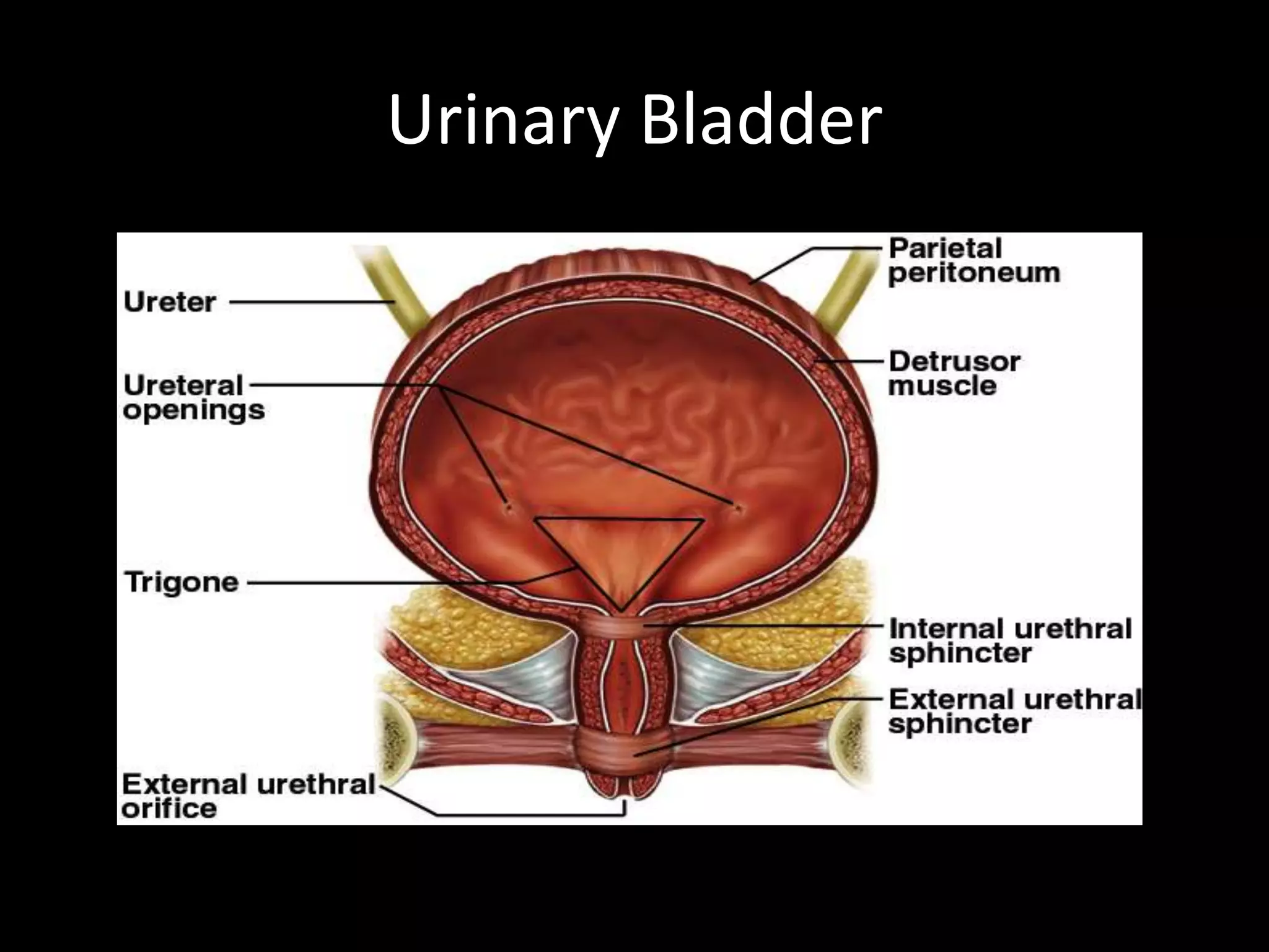
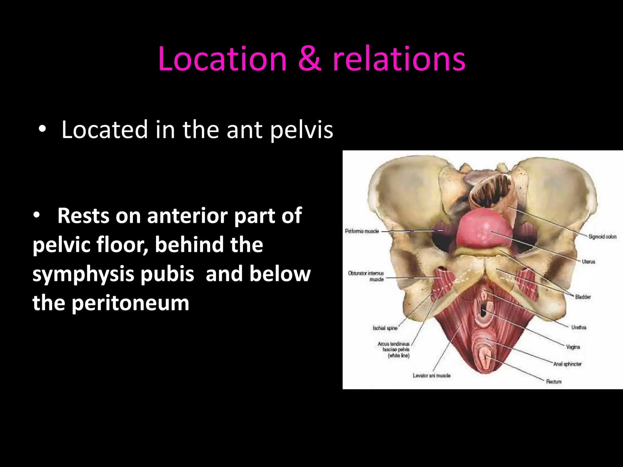
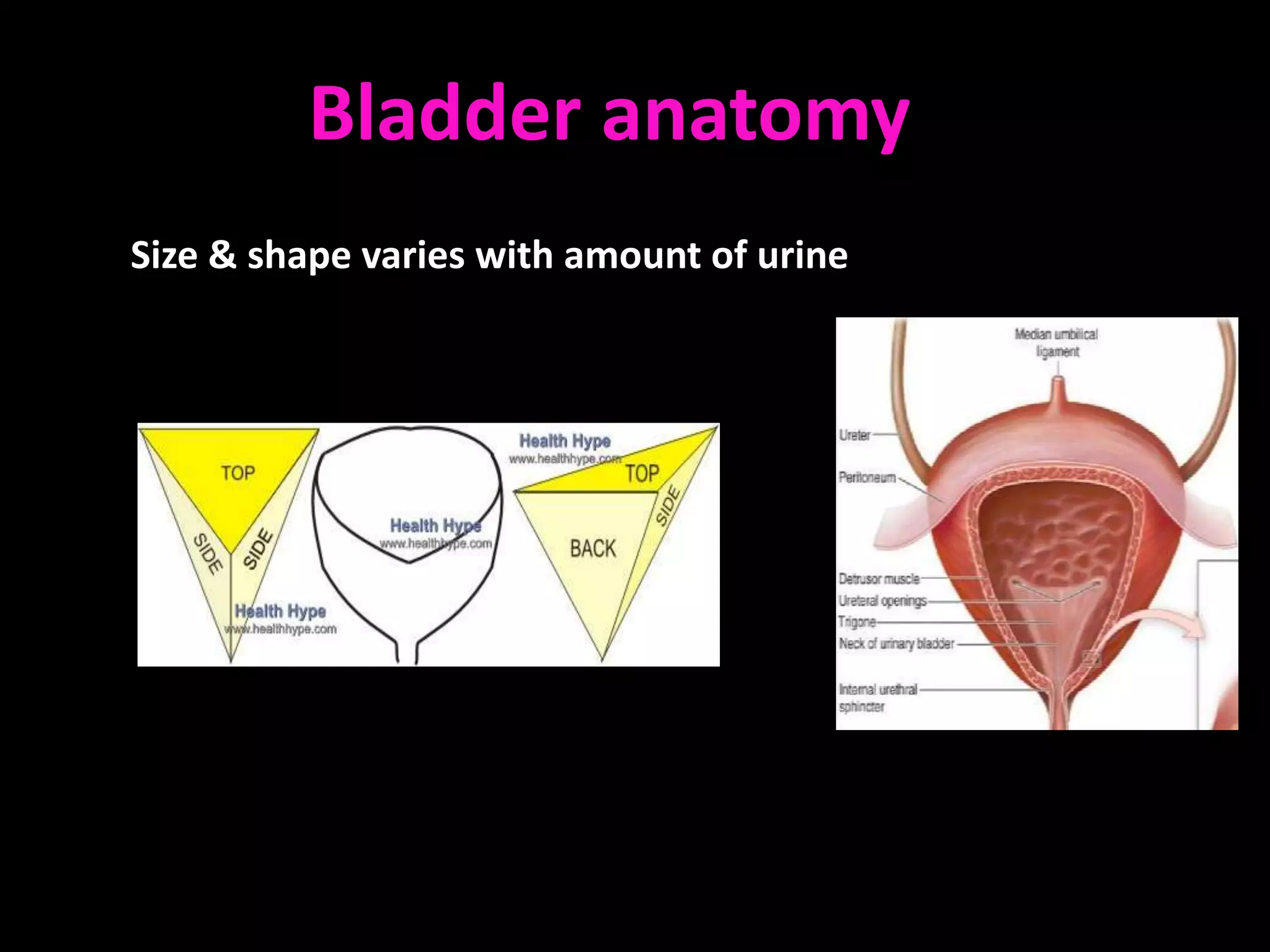
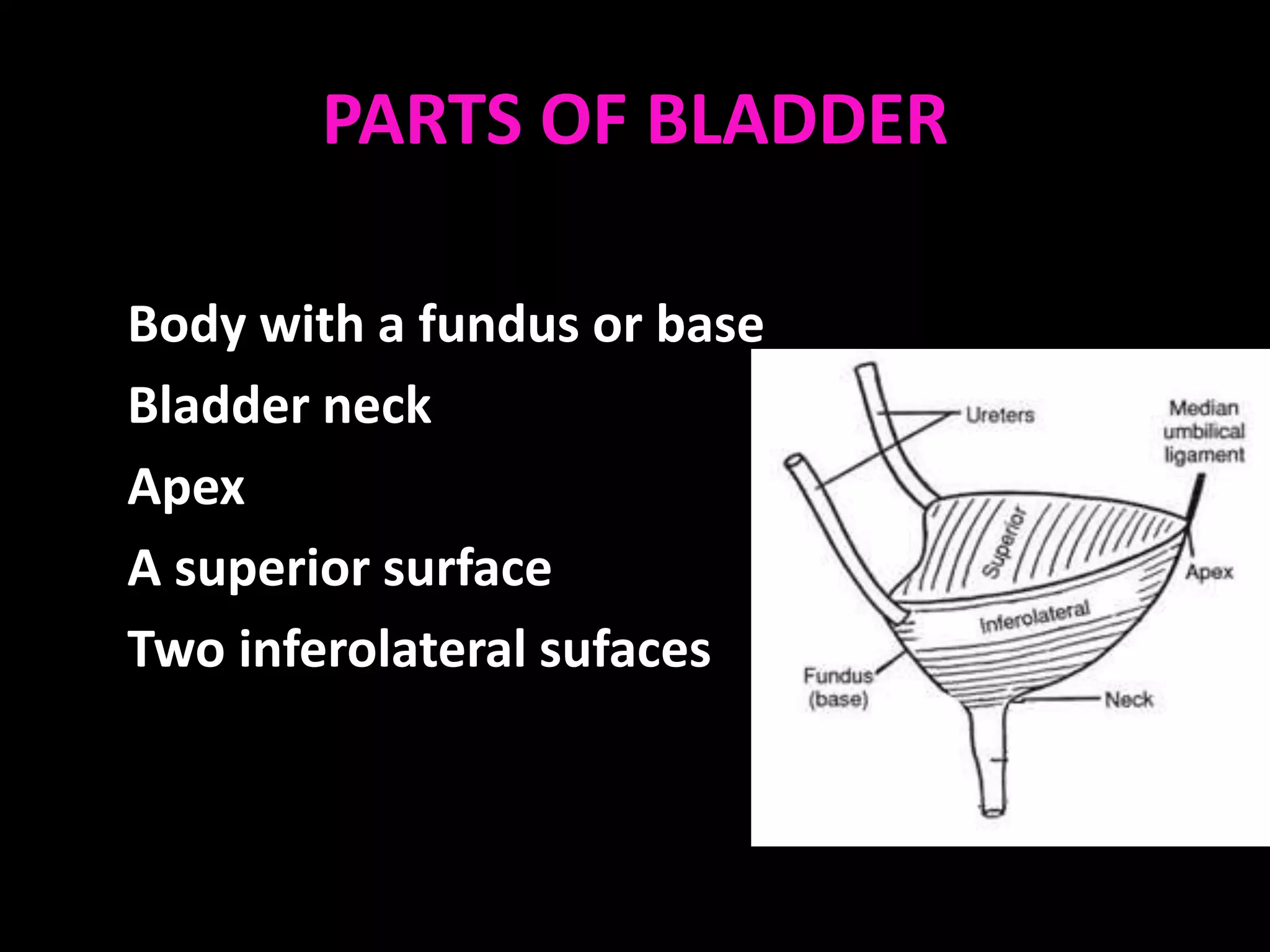
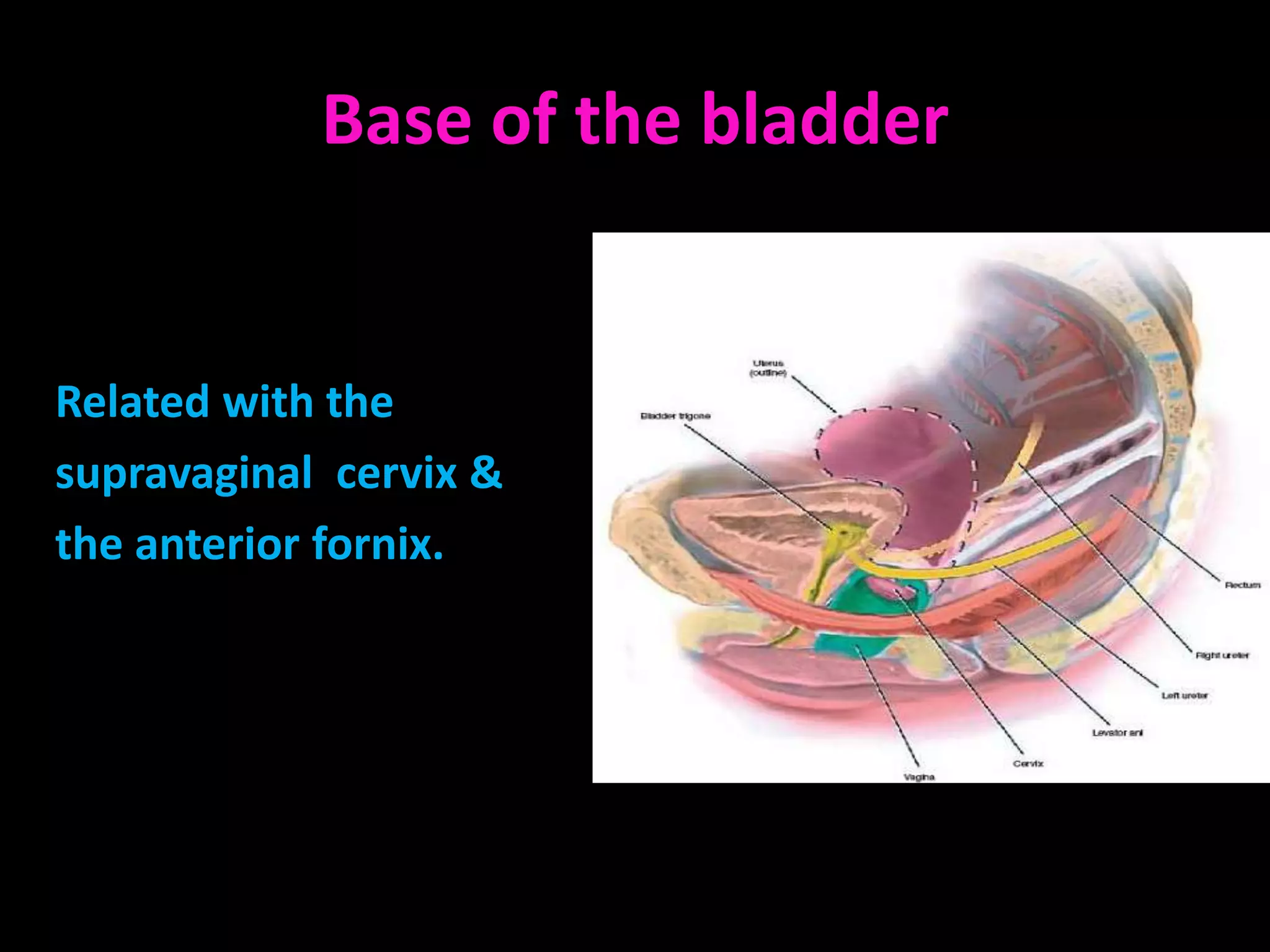
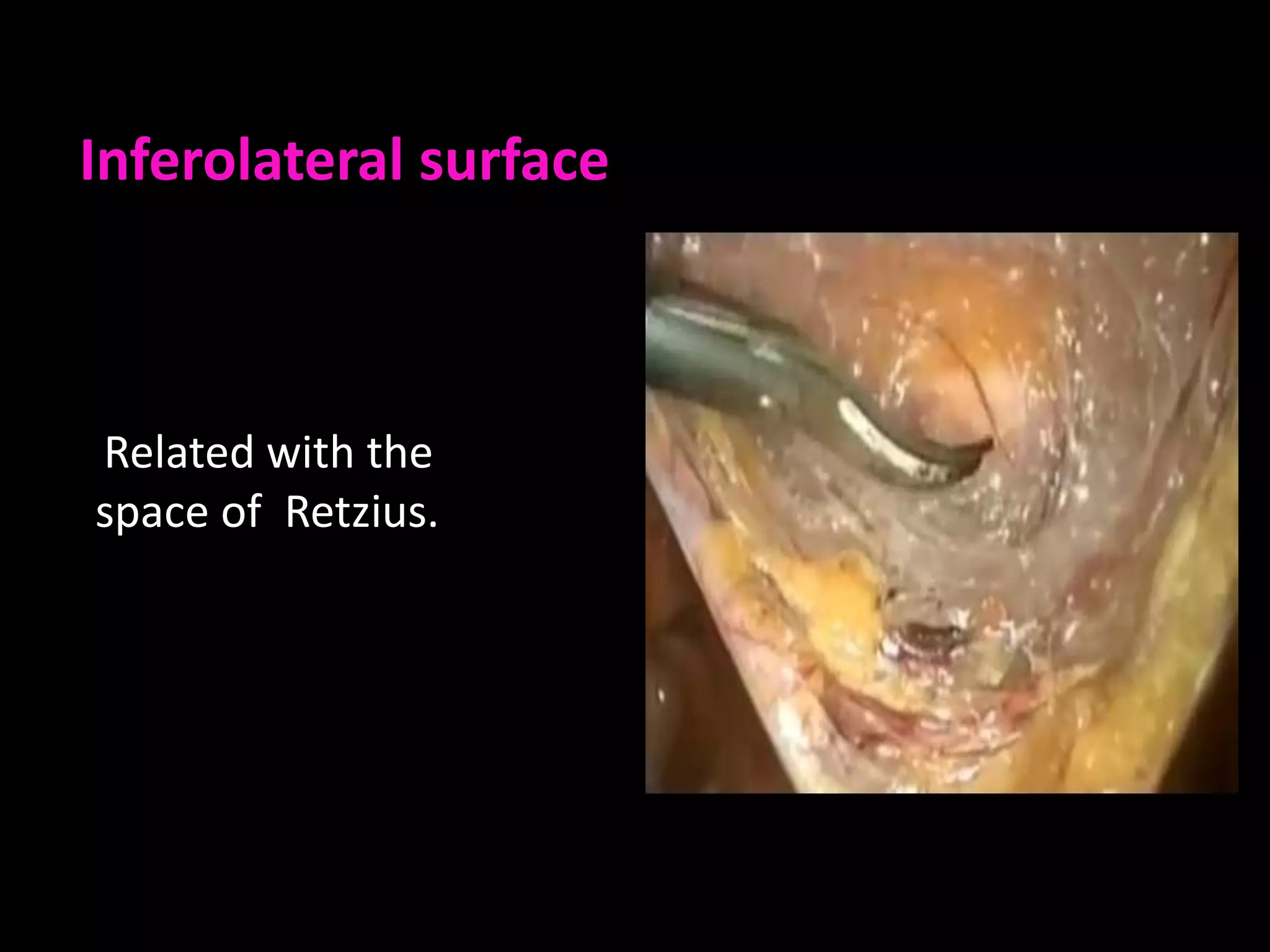
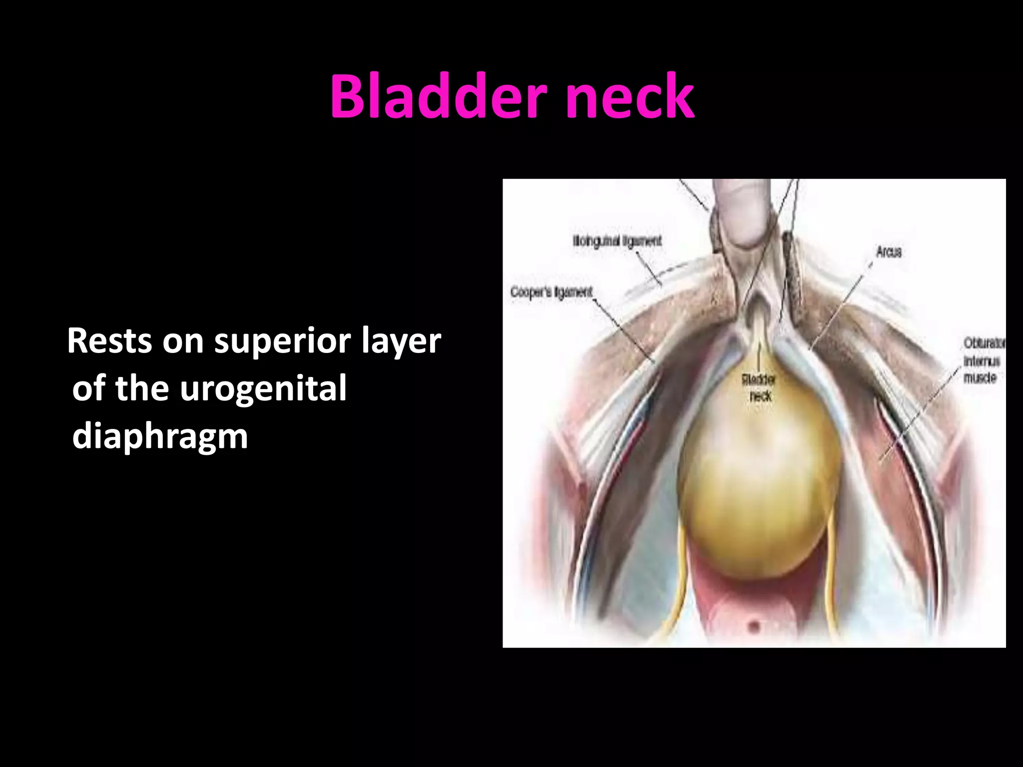
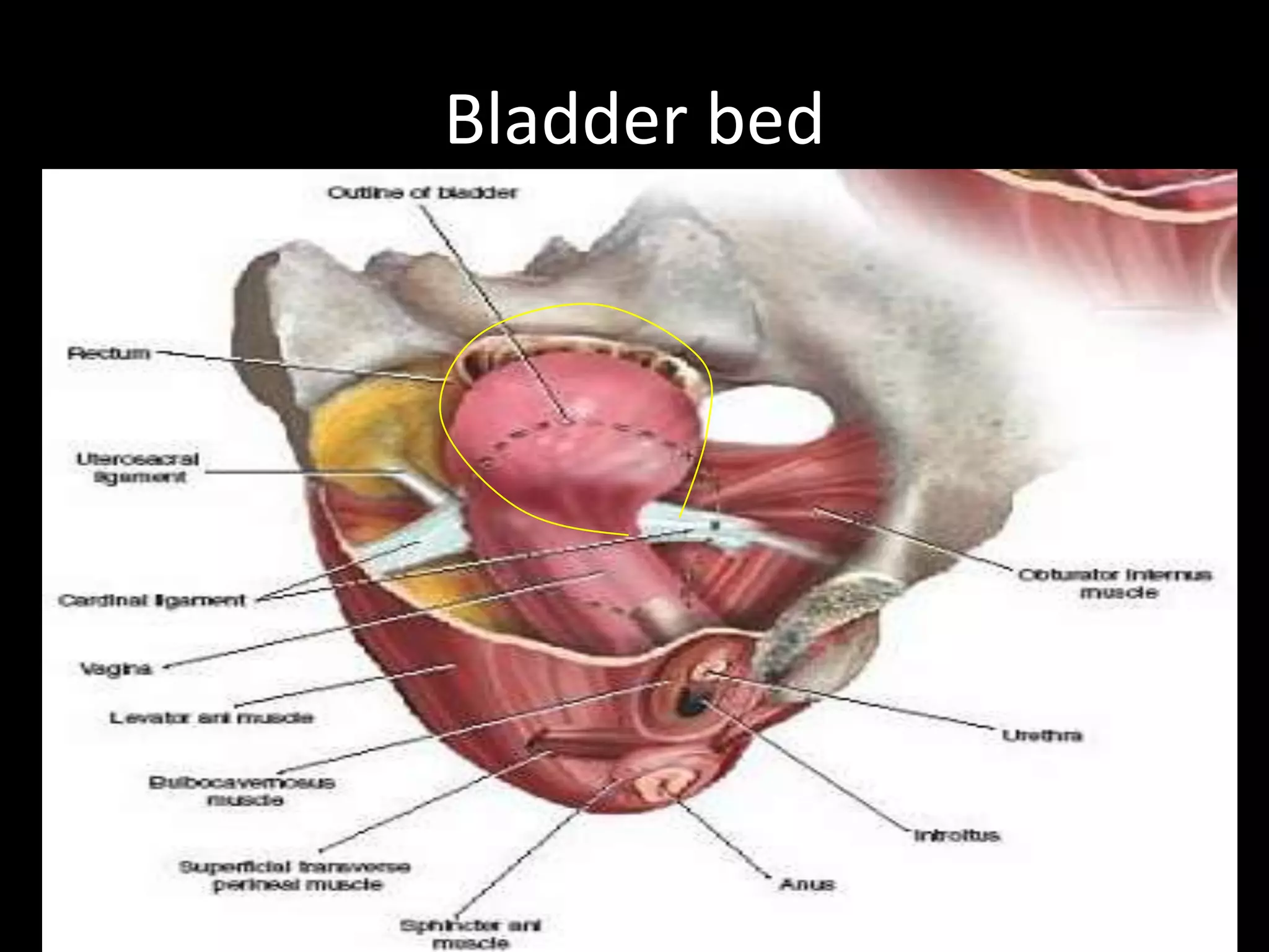
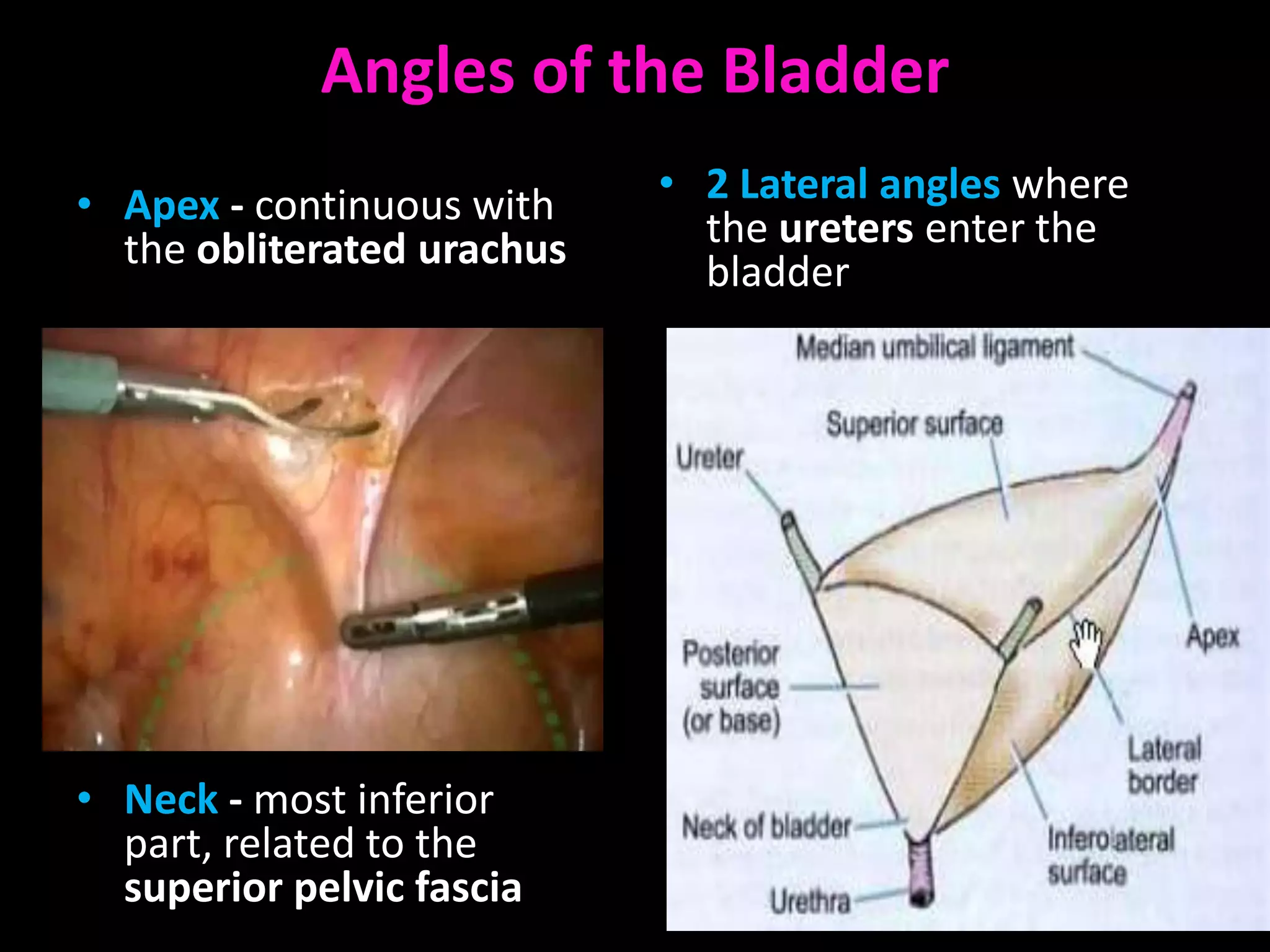
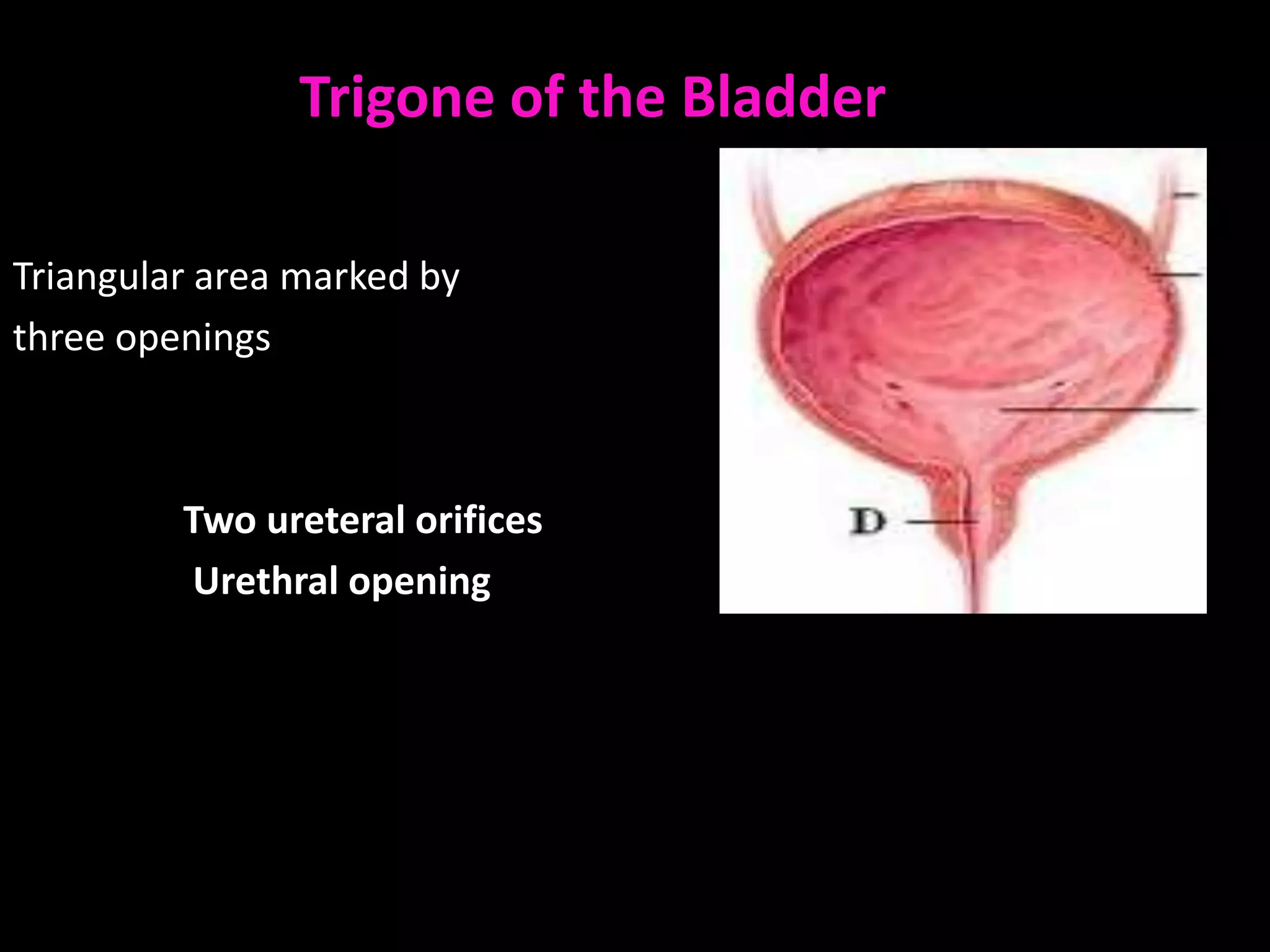
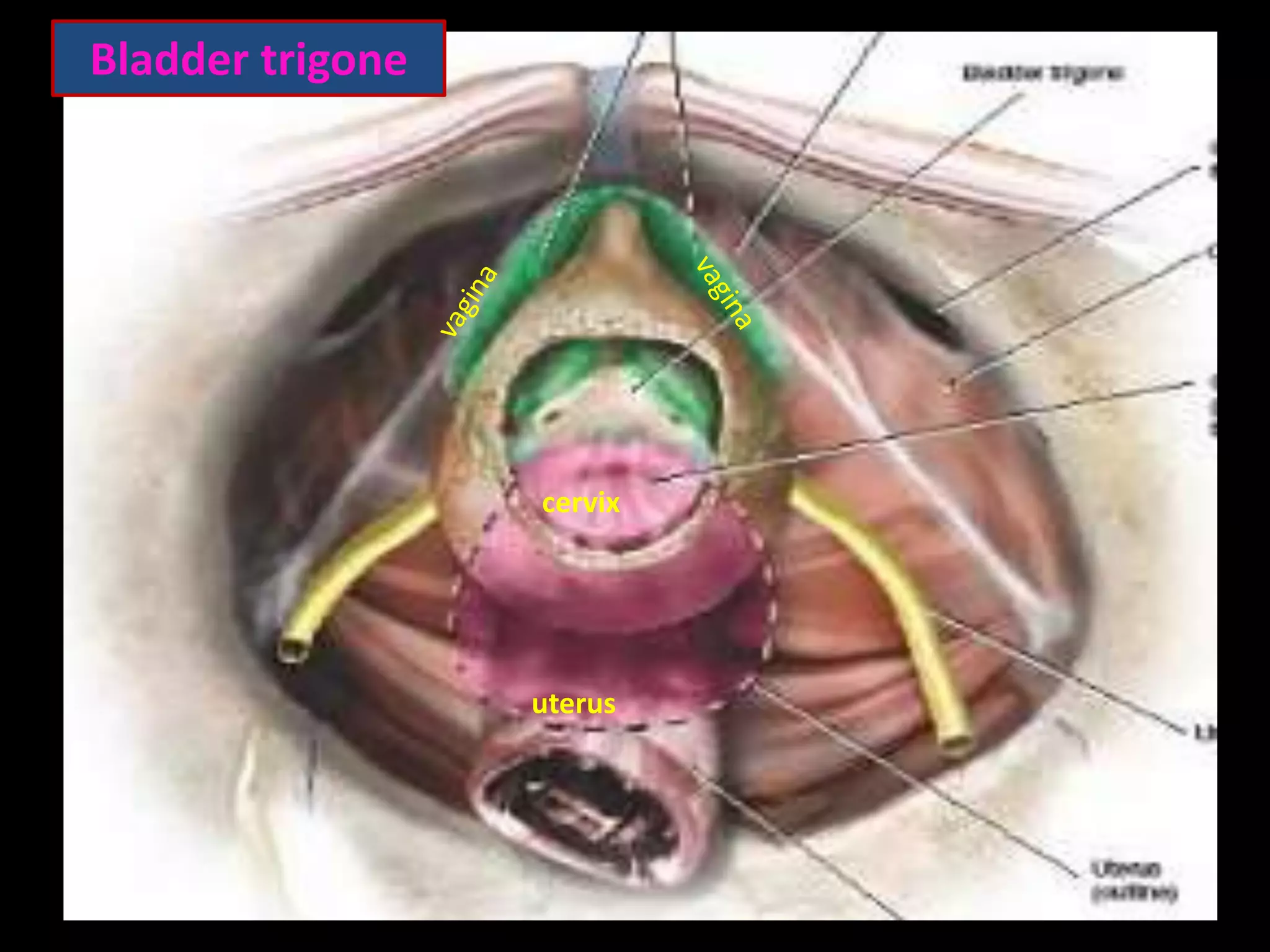
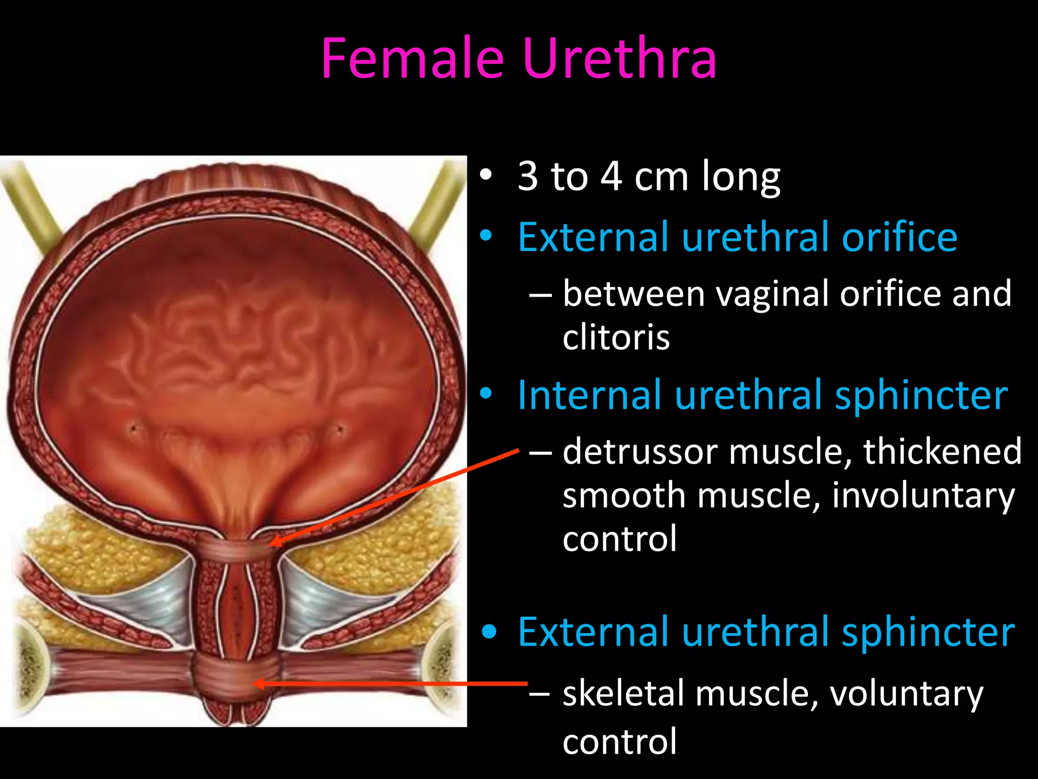
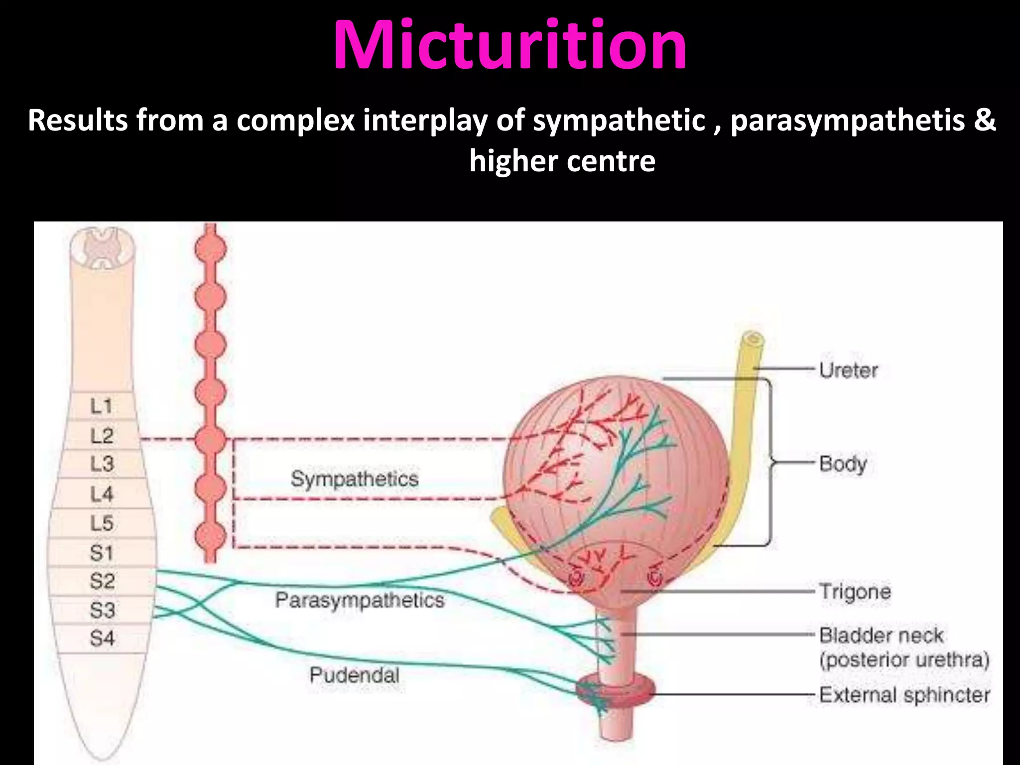
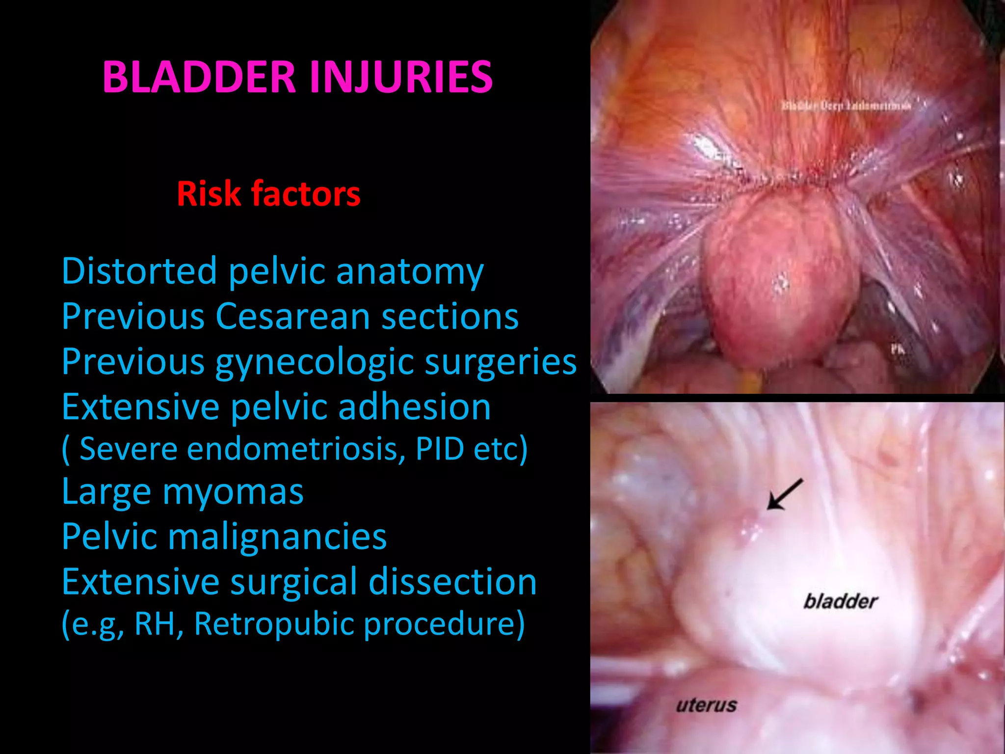
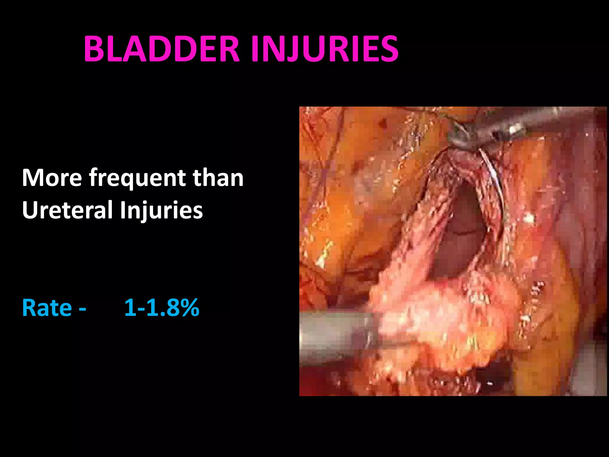
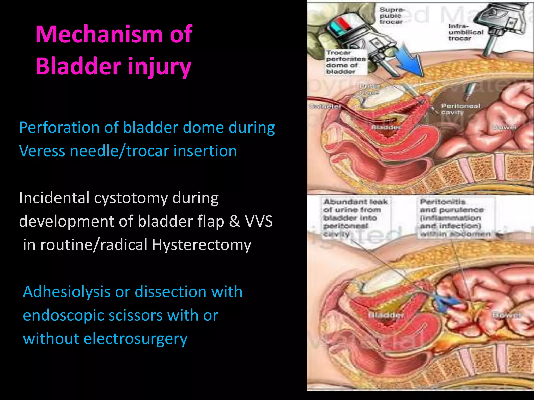
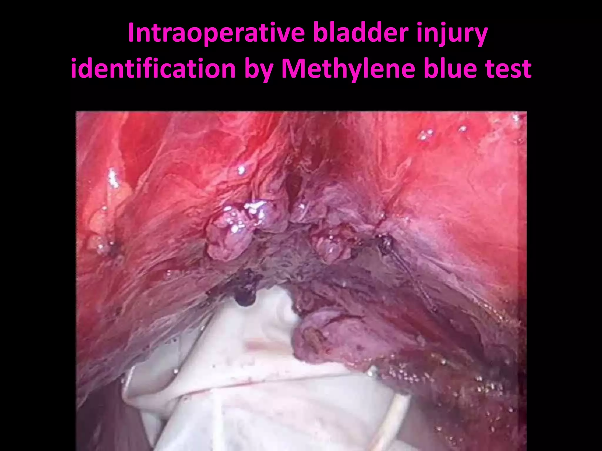
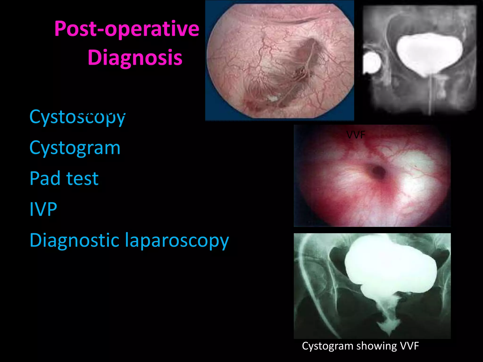
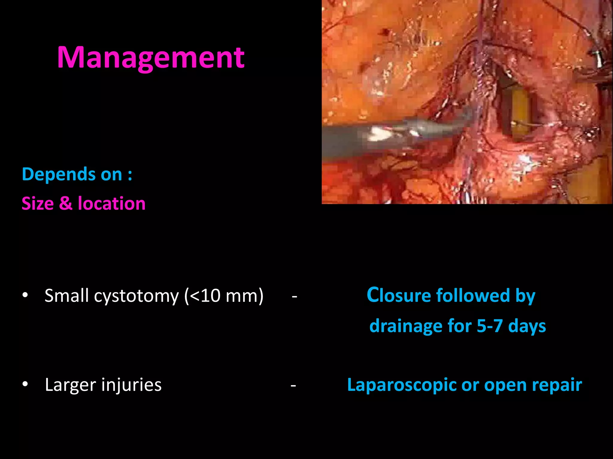
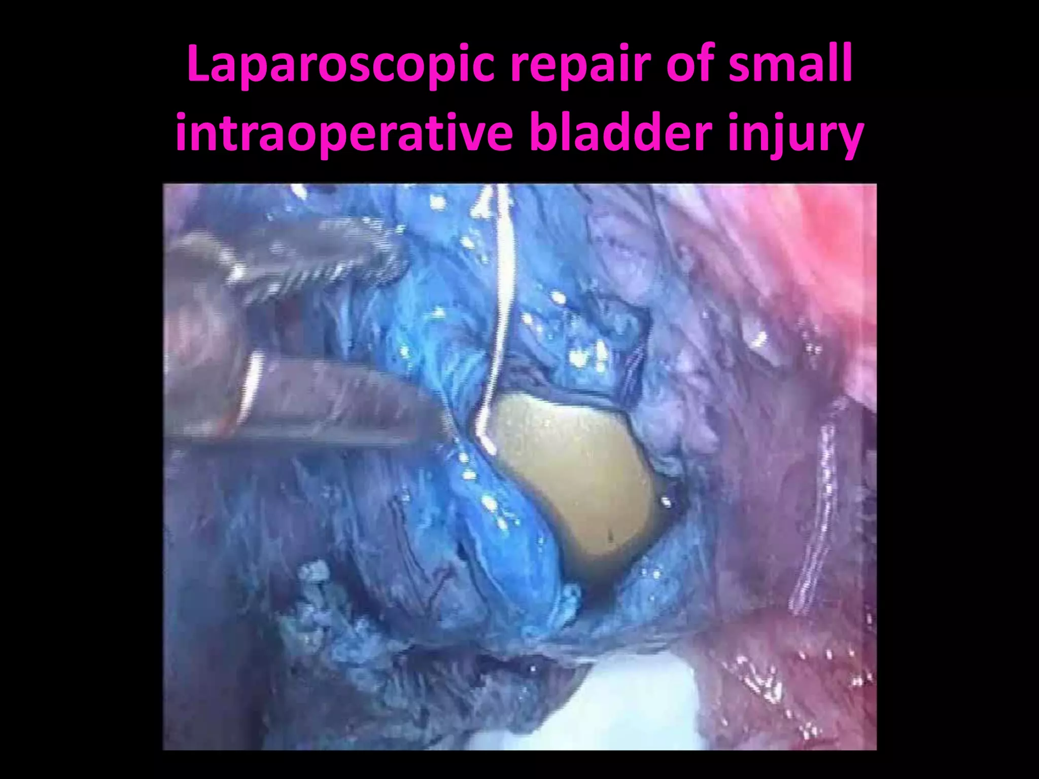
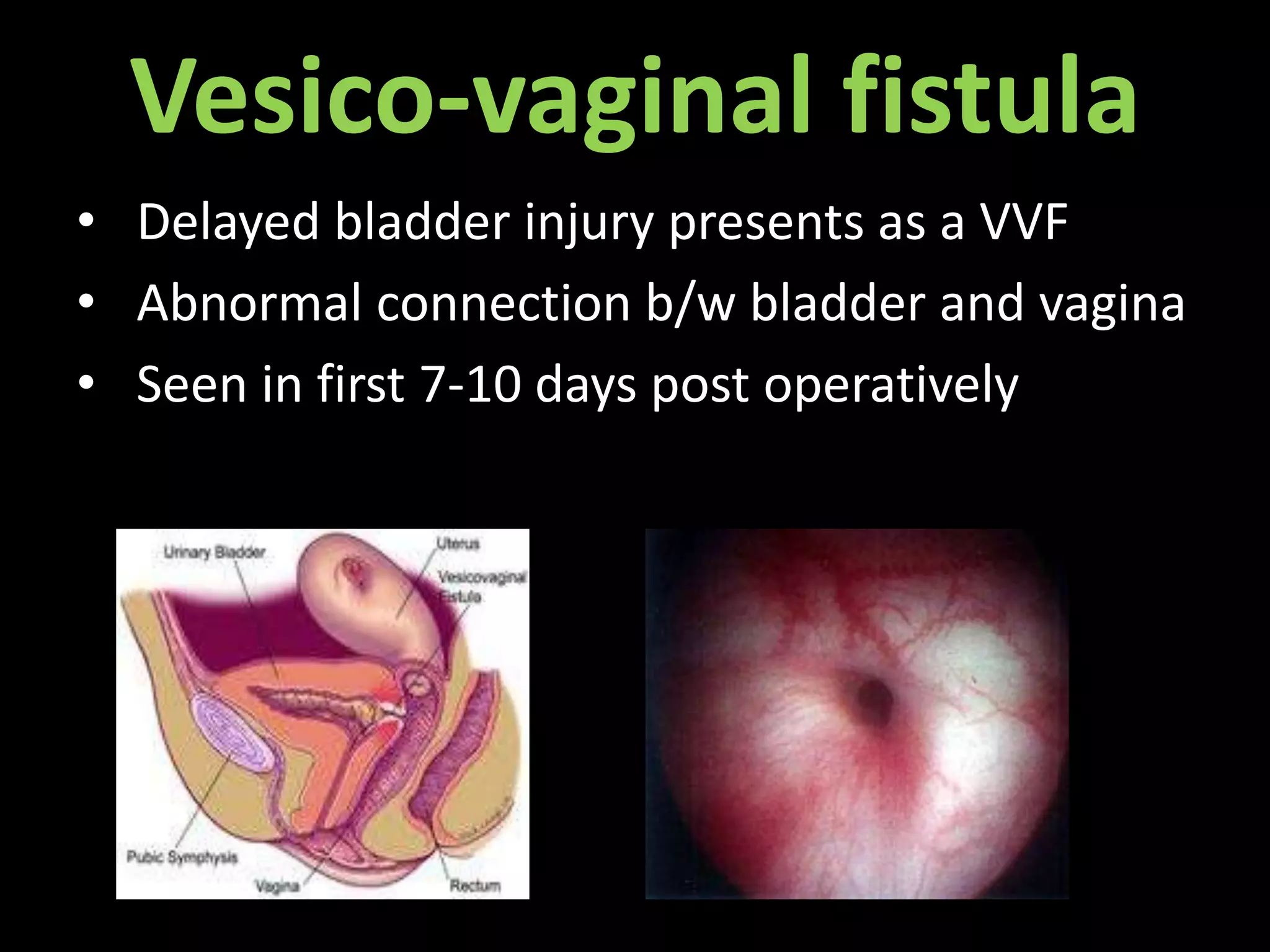
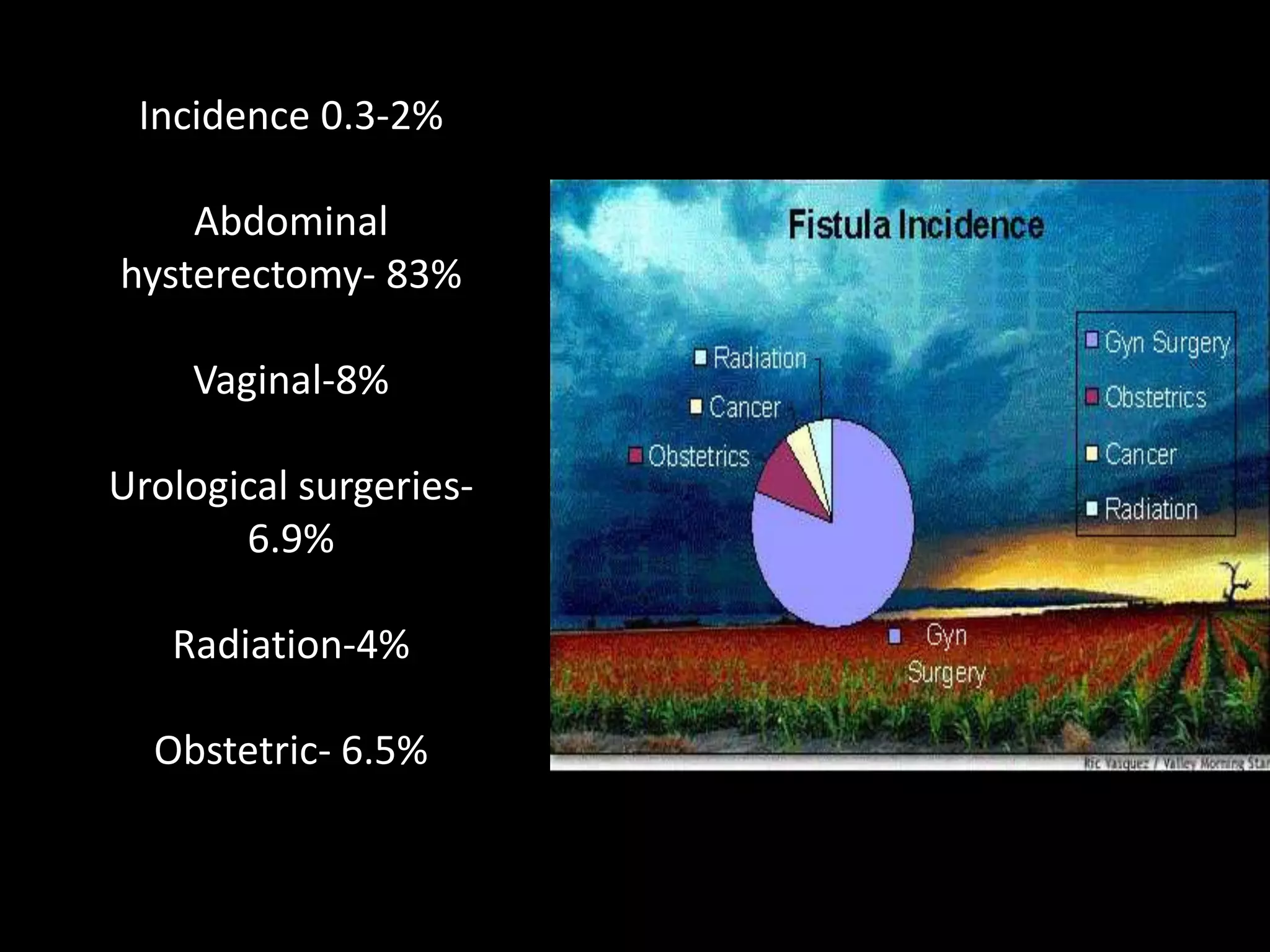
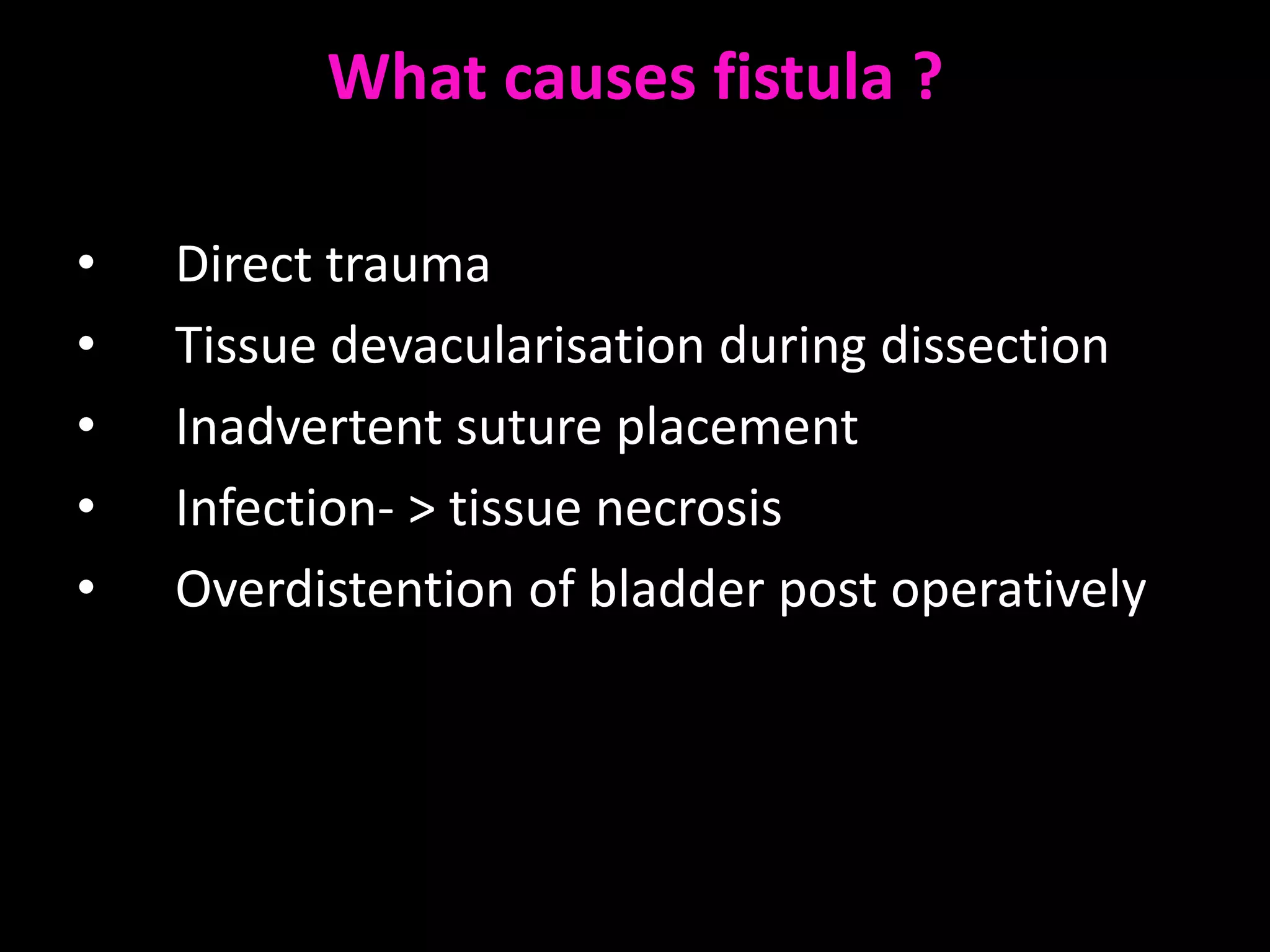
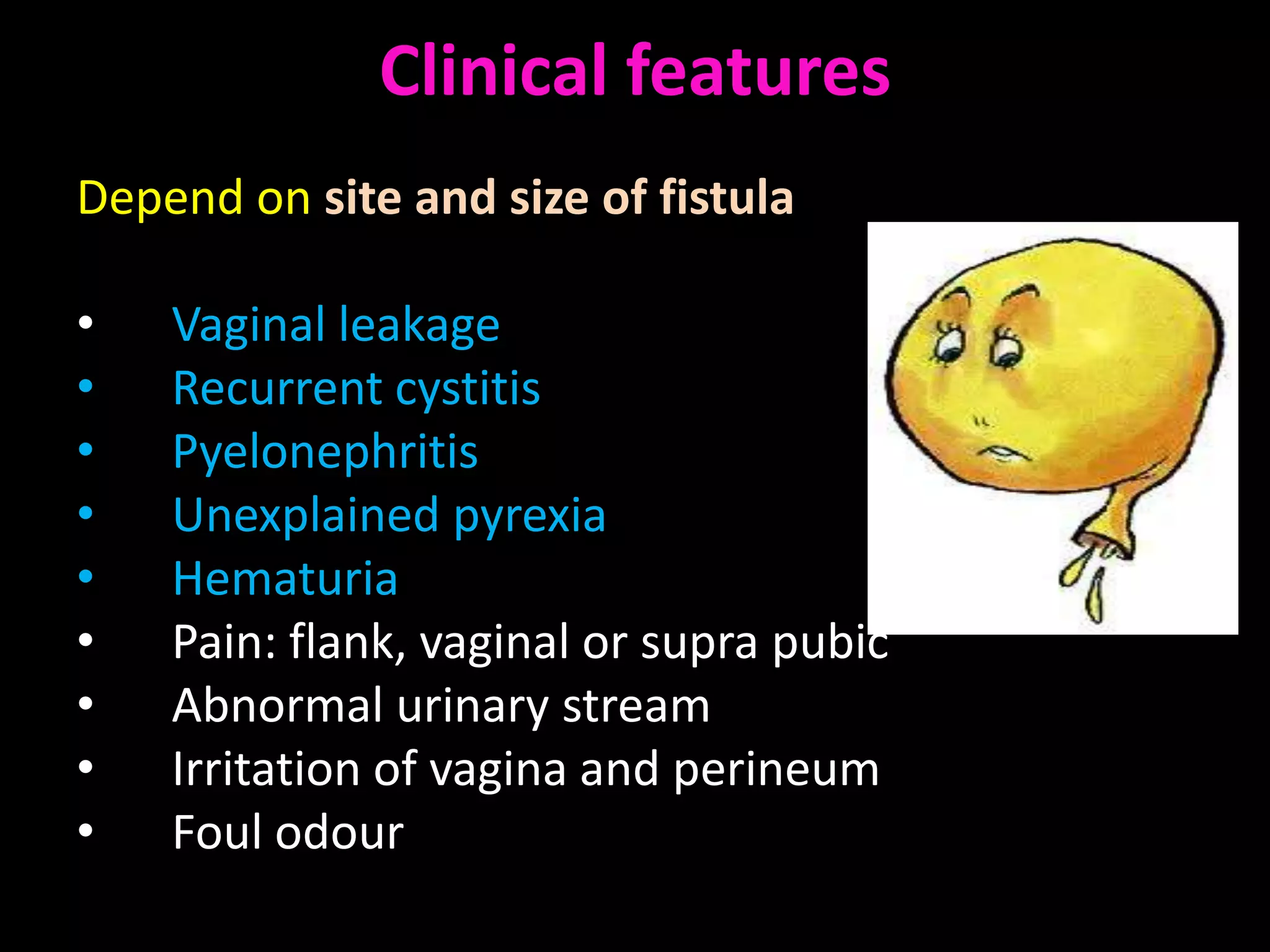
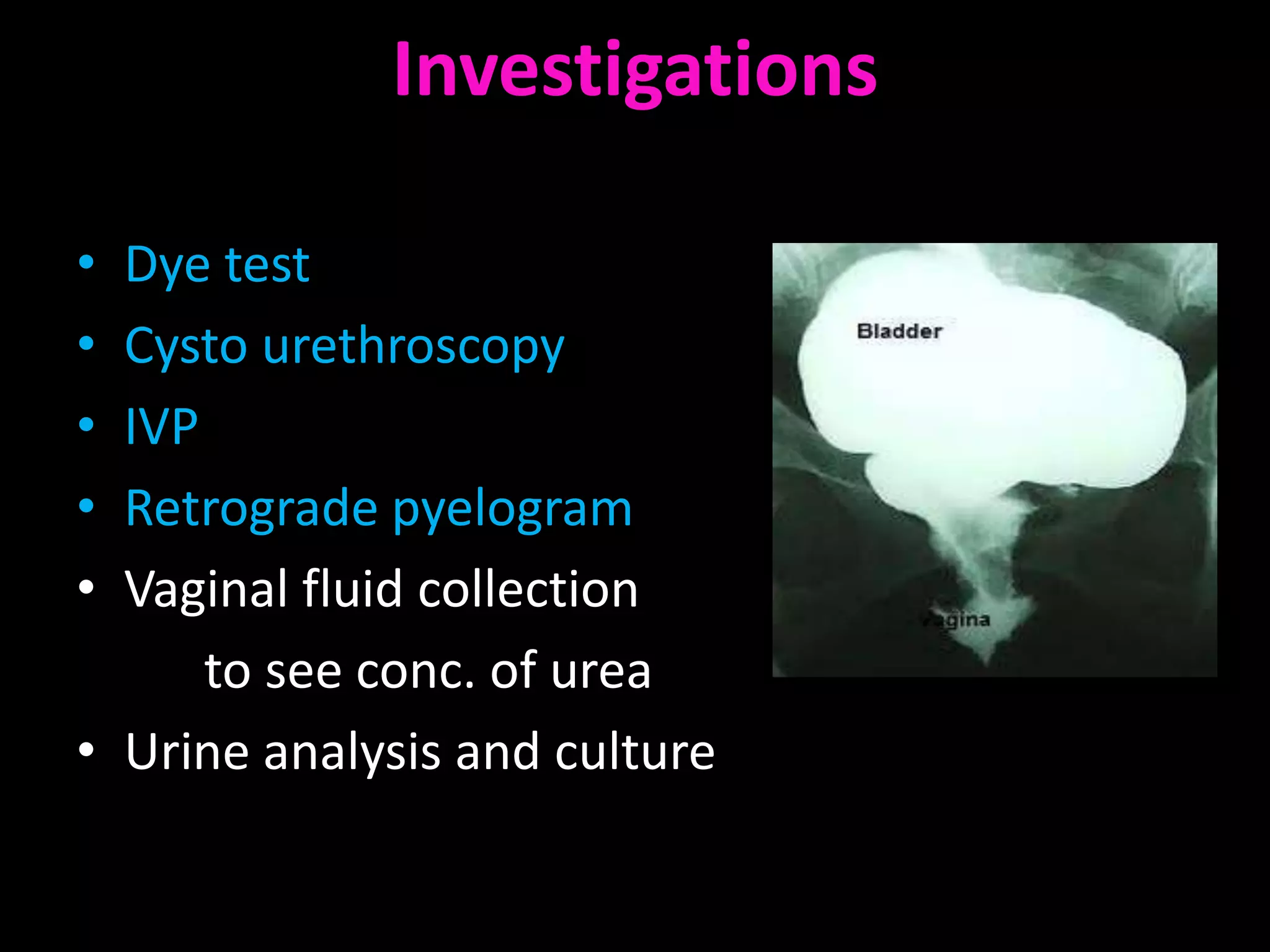
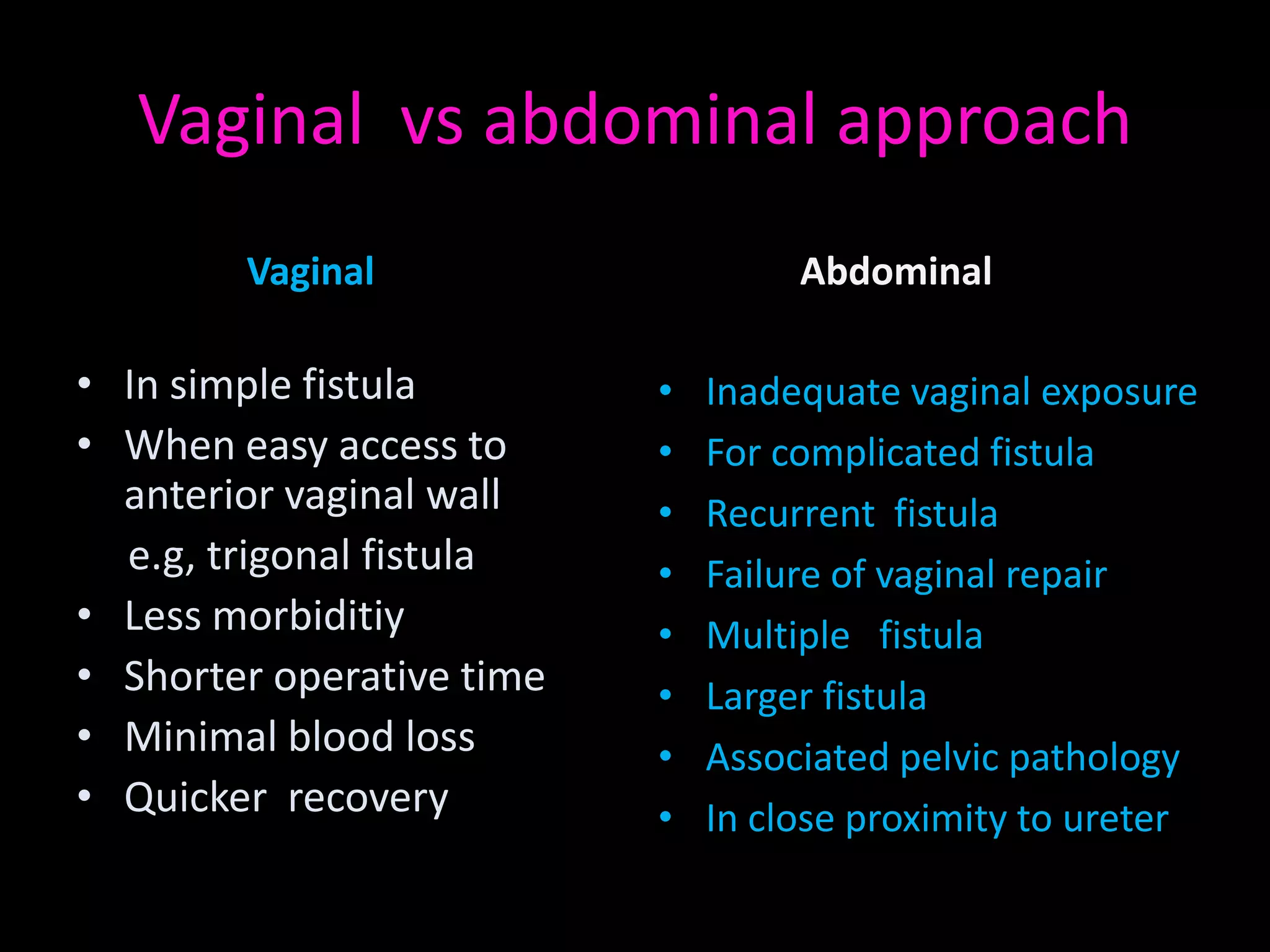
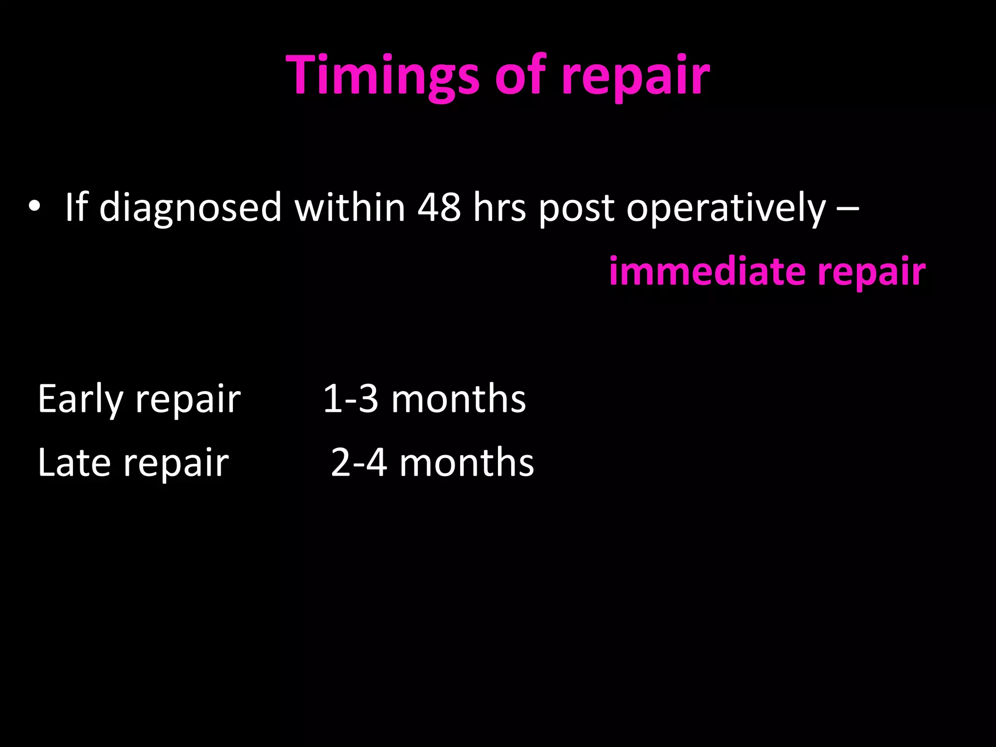
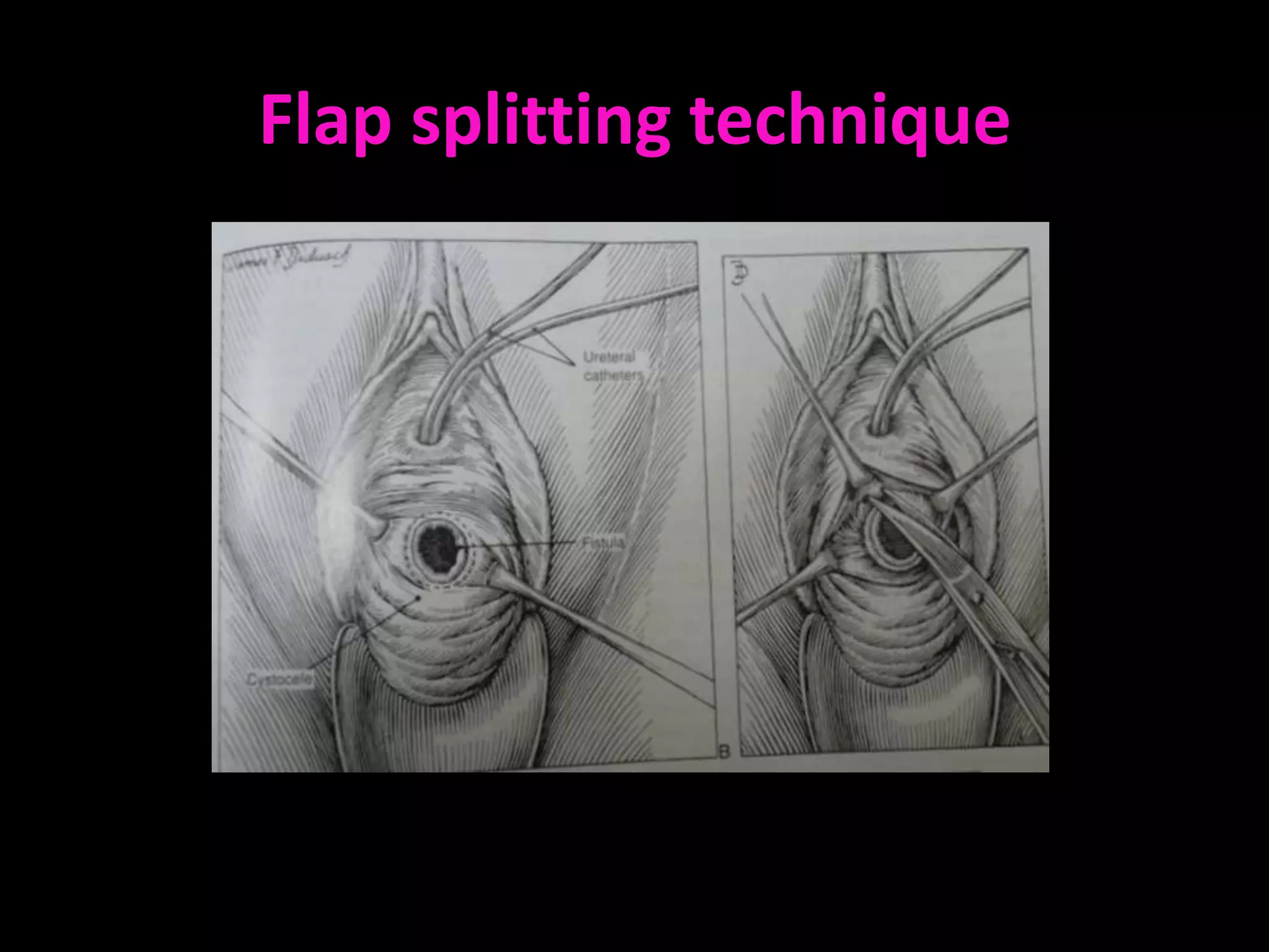
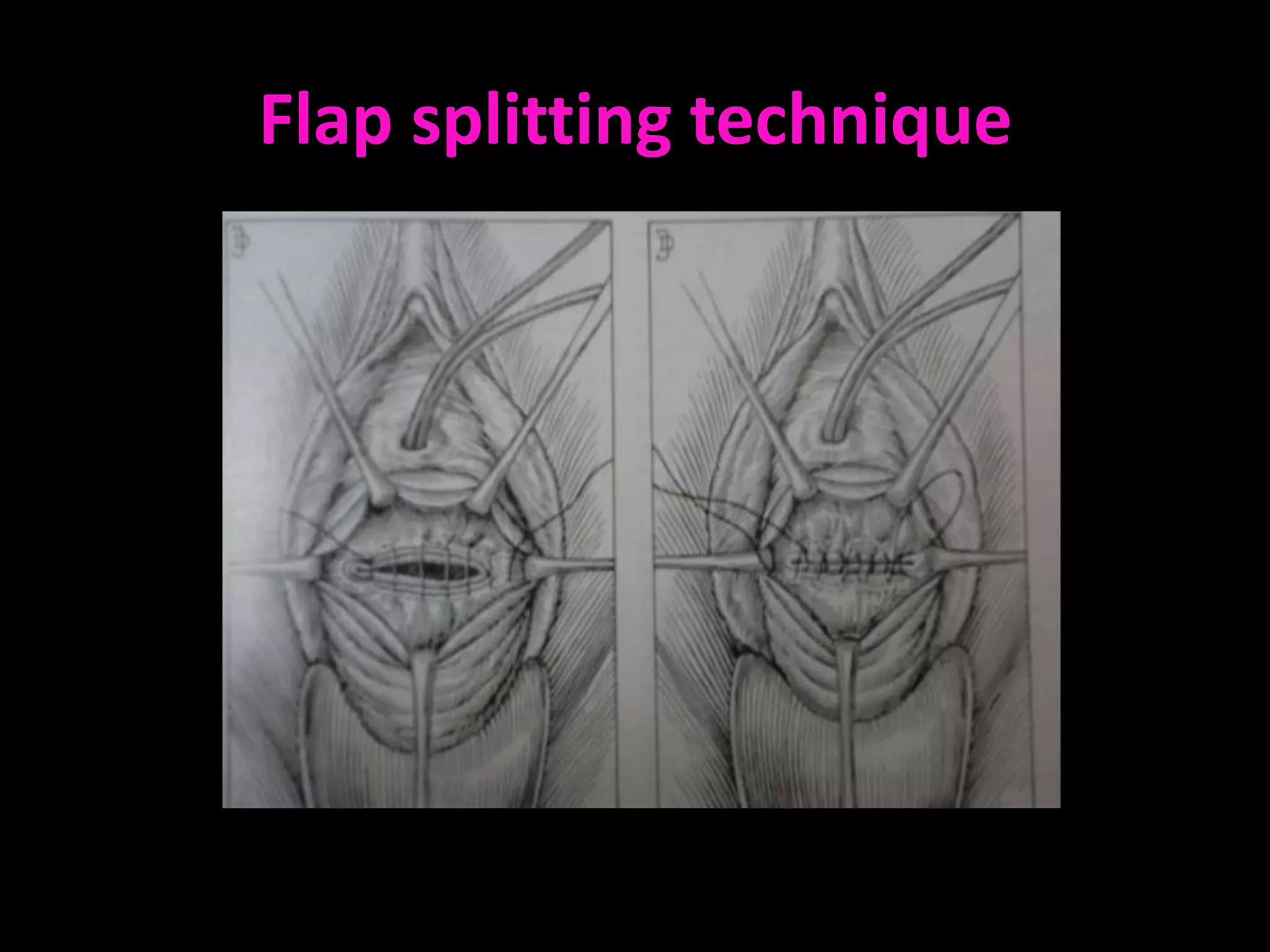
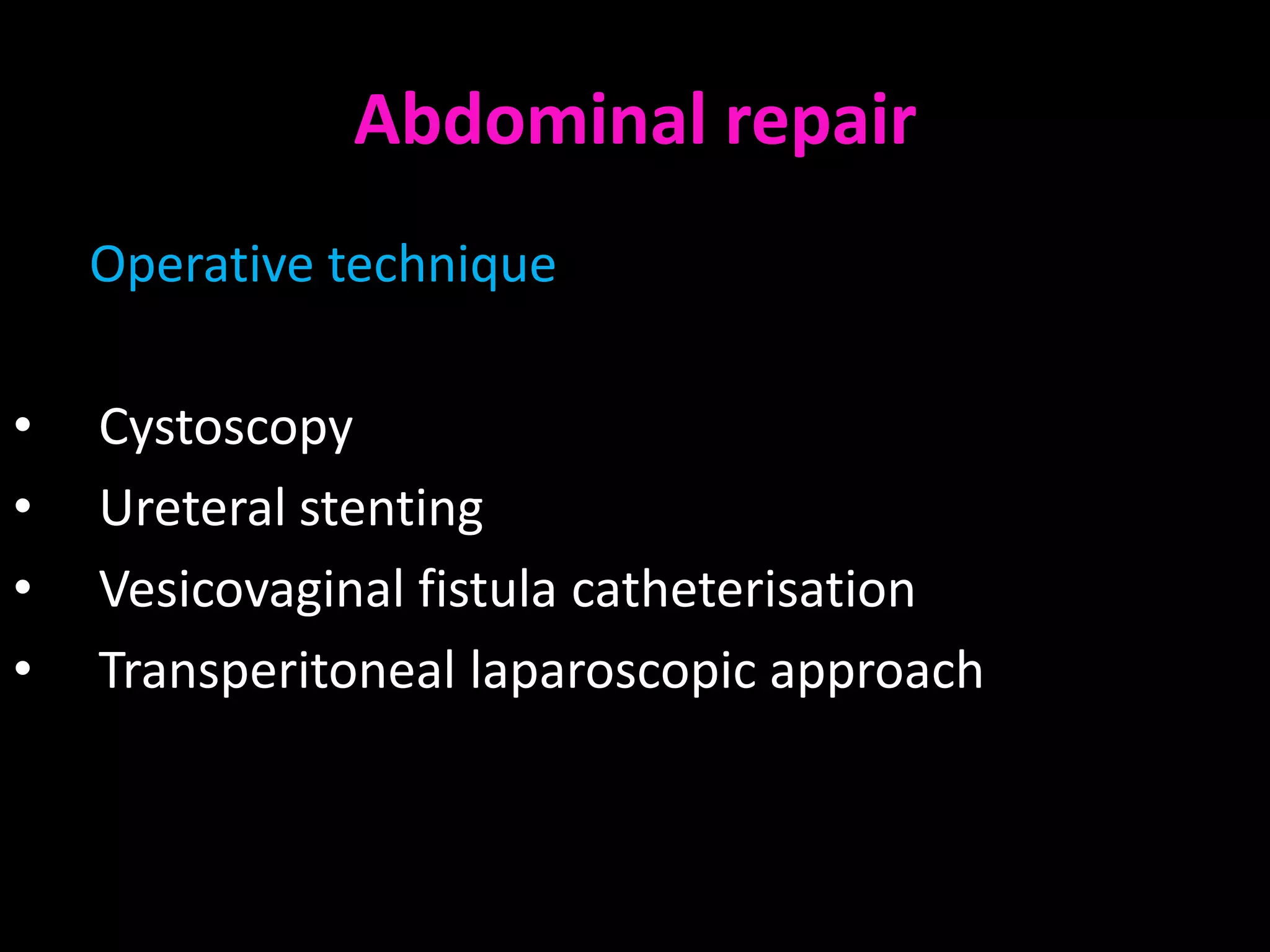
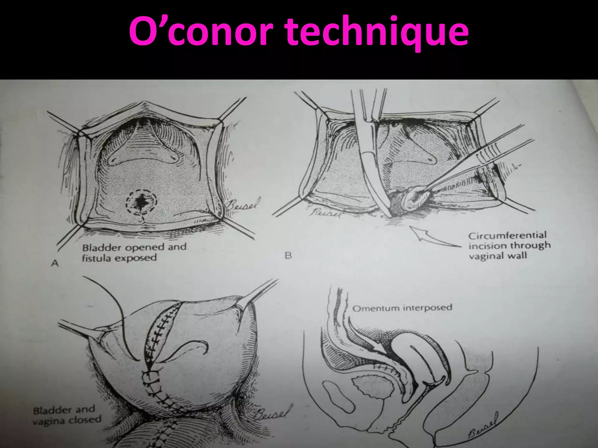
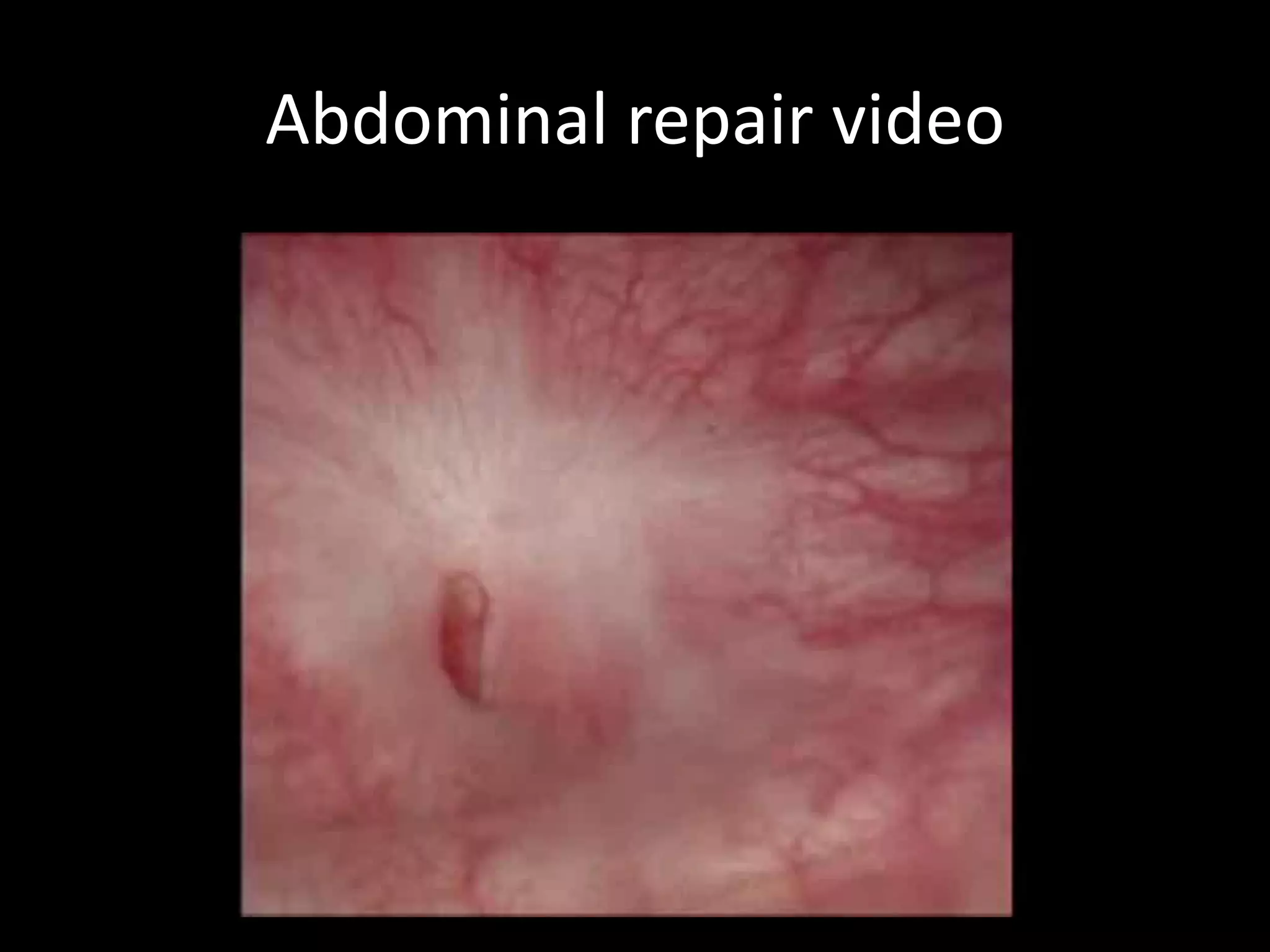
Urinary Bladder
Location & relations
• Located in the ant pelvis


• Rests on anterior part of
pelvic floor, behind the
symphysis pubis and below
the peritoneum
Bladder anatomy
Size & shape varies with amount of urine
Hollow muscular organ, urine reservoir
PARTS OF BLADDER

Body with a fundus or base
Bladder neck
Apex
A superior surface
Two inferolateral sufaces
Superior surface
Related to Peritoneum of utero-vesical pouch,
               uterus and bowel
Base of the bladder


Related with the
supravaginal cervix &
the anterior fornix.
Inferolateral surface


Related with the
space of Retzius.
Bladder neck


Rests on superior layer
of the urogenital
diaphragm
Bladder bed
Angles of the Bladder
• Apex - continuous with    • 2 Lateral angles where
  the obliterated urachus     the ureters enter the
                              bladder


• Neck - most inferior
  part, related to the
  superior pelvic fascia
Trigone of the Bladder

Triangular area marked by
three openings


         Two ureteral orifices
         Urethral opening
Bladder trigone


                  cervix


                  uterus
Female Urethra
     • 3 to 4 cm long
     • External urethral orifice
       – between vaginal orifice and
         clitoris
     • Internal urethral sphincter
       – detrussor muscle, thickened
         smooth muscle, involuntary
         control

     • External urethral sphincter
       – skeletal muscle, voluntary
         control
Histology of bladder
Blood Supply

        Vesical arteries

Superior VA
              Arises from the proximal part of ant div of Int I A
              Divides into numerous br & supply dome of bladder

Middle VA
              Br of SVA
              Supplies the base of bladder

Inferior VA
              Arises from middle rectal or vaginal artery
              Base & the Trigone
Venous drainage of bladder
 Vesical venous plexus


   Internal Iliac veins


Internal vertebral venous
          plexus
Lymphatic supply
• Superior part - external
  iliac lymph nodes

• Inferior part - internal iliac
  lymph nodes

• Bladder neck - sacral or
  common iliac lymph nodes
Micturition
Results from a complex interplay of sympathetic , parasympathetis &
                               higher centre
Micturition reflex

Filling of urinary bladder → stretch receptors → sensory
impulse via pelvic nerve to S2 – S4 → Parasympathetic
impulse via pelvic nerve → Contraction of detrusor muscle &
relaxation of internal sphincter → urine in urethra stimulates
stretch receptors → sensory impulse via pelvic nerve to S2 –
S4 → inhibition of somatic fibers in pudendal nerve →
relaxation of external sphincter → results in urination
Micturition reflex

Sympathetic (through hypogastric nerve)
stimulation of beta receptors on detrusor muscle
causes relaxation & of alpha receptors on internal
sphincter causes constriction of sphincter, hence
sympathetic stimulation causes filling & referred to
as nerve of filling.
Higher brain centers of Micturition
• Facilitatory & inhibitory centers in brain stem
  especially pons
• Centers located in cerebral cortex is normally
  inhibitory but can become excitatory
• For voluntary urination, cortical centers can
  facilitate the sacral micturition centers to help
  initiate a micturition reflex & at the same time
  inhibit the external urinary sphincter.
Voluntary Control of Micturition
• Micturition center in pons receives stretch signals and
  integrates cortical input (voluntary control)

• Sends signal for stimulation of detrussor and relaxes internal
  urethral sphincter

• To delay urination impulses sent through pudendal nerve to
  external urethral sphincter keep it contracted until you wish
  to urinate

• Valsalva maneuver
   – aids in expulsion of urine by pressure on bladder
   – can also activate micturition reflex voluntarily
BLADDER INJURIES

       Risk factors
Distorted pelvic anatomy
Previous Cesarean sections
Previous gynecologic surgeries
Extensive pelvic adhesion
( Severe endometriosis, PID etc)
Large myomas
Pelvic malignancies
Extensive surgical dissection
(e.g, RH, Retropubic procedure)
BLADDER INJURIESINJURY
              BLADDER


More frequent than
Ureteral Injuries


Rate -   1-1.8%
Mechanism of
  Bladder injury

Perforation of bladder dome during
Veress needle/trocar insertion

Incidental cystotomy during
development of bladder flap & VVS
 in routine/radical Hysterectomy

Adhesiolysis or dissection with
endoscopic scissors with or
without electrosurgery
Bladder injury in a case with previous
              C-section
Bladder injury during TLH for Big
      fibroid (20 weeks)
Diagnosis of bladder injuries


Unlike ureteral injuries,
almost all the bladder
injuries are diagnosed
intra-operatively
Signs of intra-operative
                  bladder injuries
•   Visualization of the Foley catheter bulb
•   Distention of urine collection bag with CO2 (Pneumaturia)
•   Urine drainage from accessory trocar site
•   Intraperitoneal leakage of Methylene Blue
•   Haematuria
•   Suprapubic bruising
•   Abdominal wall or pelvic mass
•   Cystoscopy – size & location
Intraoperative bladder injury
identification by Methylene blue test
Post-operative identification of Bladder injury


Bladder injury is suspected in the presence of:

•       Haematuria

•       Leakage of urine per vagina ( fistula)

•       Fever, flank pain, ileus, abdominal distension

•       Sepsis
Post-operative
     Diagnosis

Cystoscopy
     POST-OPERATIVE
                                VVF
Cystogram
Pad test
IVP
Diagnostic laparoscopy


                         Cystogram showing VVF
Sequelae of Undiagnosed Injuries
• Voiding dysfunction

• Detrusor instability

• Bladder stone formation with recurrent UTI

• Uro-genital fistula formation

• Renal damage
Management

    Intra-operative bladder injury
Depends on :
Size & location


• Small cystotomy (<10 mm)           -     Closure followed by
                                           drainage for 5-7 days

• Larger injuries                    -   Laparoscopic or open repair
Laparoscopic Bladder injury repair

Cystoscopy
         - Exclude injury to trigone
         - Check proximity of the defect to the ureter


Remove necrotic tissue, adhesions or areas of
endometriosis before actual repair
Laparoscopic repair of small
intraoperative bladder injury
Laparoscopic Bladder suturing
• Interrupted or continuous absorbable sutures through full
  thickness of bladder wall

• Polyglactin or Polydioxanone , no 3-0

• Single layer closure is sufficient

• Repair should include mucosa, muscularis & serosa

• Peritoneal imbrication or omental graft placement between
  suture lines may decrease risk of fistula formation
Post-operative Period
Bladder drainage with large caliber urethral or
suprapubic catheter

     5-7 days - simple fundal laceration

     14 days - closer to trigone or vaginal vault
             - significant thermal damage

Retrograde cystogram to confirm healing
Vesico-vaginal fistula
• Delayed bladder injury presents as a VVF
• Abnormal connection b/w bladder and vagina
• Seen in first 7-10 days post operatively
Incidence 0.3-2%

    Abdominal
hysterectomy- 83%

    Vaginal-8%

Urological surgeries-
       6.9%

   Radiation-4%

  Obstetric- 6.5%
Demographic variation

Obstetric injuries are most common cause of
VVF in developing countries whereas in developed
countries, gynecological surgical injuries are the
commonest cause of VVF.
What causes fistula ?

•   Direct trauma
•   Tissue devacularisation during dissection
•   Inadvertent suture placement
•   Infection- > tissue necrosis
•   Overdistention of bladder post operatively
Risk factors
•   Previous surgery
•   h/o sepsis
•   Endometriosis
•   Malignancy
•   Adhesions with bladder and uterus or cervix
•   Anatomical distortion within pelvis
•   Radiation
Clinical features
Depend on site and size of fistula

•   Vaginal leakage
•   Recurrent cystitis
•   Pyelonephritis
•   Unexplained pyrexia
•   Hematuria
•   Pain: flank, vaginal or supra pubic
•   Abnormal urinary stream
•   Irritation of vagina and perineum
•   Foul odour
Type of fistula
Simple -   Tissue healthy, good vaginal access

Complicated – large (> 5cms)
              scarring
              Impaired access
              Involvement of ureteric orifices
classification of urogenital fistulas

•   Urethral
•   Bladder neck
•   Sub symphysial
•   Midvaginal
•   Juxtacervical/vault
•   Vesicouterine
•   Vesicocervical
Presentation

•   Continuous urinary incontinence
•   Limited sensation of bladder fullness
•   Infrequent voiding
Timings of presentation


   5-14 days post-operatively
Investigations

• Dye test
• Cysto urethroscopy
• IVP
• Retrograde pyelogram
• Vaginal fluid collection
      to see conc. of urea
• Urine analysis and culture
Basic principles for fistulae repair
• Ensure that there is no cellulitis, edema, or
  infection at the fistula site prior to closing the
  fistula
• Excision of avascular scar tissue
• Wide mobilisation of bladder
• Tension free layer closure of bladder and
  vagina
• Good hemostasis with bladder drainage
• Using transplanted blood supply
Techniques of repair
•   Conservative
•   Abdominal approach
•   Vaginal approach
•   Laparoscopic
•   Combined
•   Electrocautery
•   Fibrin glue
•   Using interposition flaps or grafts
Various approaches

Vaginal
             Flap splitting
             Latzko’s procedure

Abdominal
             O’conor technique
             Modified O’Conor
             Laparoscopic transperitoneal repair
Vaginal vs abdominal approach
        Vaginal                     Abdominal

• In simple fistula        •   Inadequate vaginal exposure
• When easy access to      •   For complicated fistula
  anterior vaginal wall    •   Recurrent fistula
  e.g, trigonal fistula    •   Failure of vaginal repair
• Less morbiditiy          •   Multiple fistula
• Shorter operative time   •   Larger fistula
• Minimal blood loss       •   Associated pelvic pathology
• Quicker recovery         •   In close proximity to ureter
Timings of repair

• If diagnosed within 48 hrs post operatively –
                                immediate repair

Early repair    1-3 months
Late repair     2-4 months
Pre operative care

•   Urinary or vaginal infection- treated
•   Early attempts to divert urinary stream
•   Catheter drainage( spontaneous healing in 7 %)
•   Care for perineal skin
Flap splitting technique
• Adequate exposure made.
• Fistula tract excised with a scalpel
• The entire tract is dissected
• The layers of the bladder wall and vagina
  adequately delineated and mobilized
• The bladder mucosa closed with interrupted 4-0
  synthetic absorbable suture
• A second layer, the bladder muscle, is closed with
  2-0 synthetic absorbable suture.
Flap splitting technique
• Vaginal incision closed separately

• The bulbocavernosus muscle transplant ±

• The bladder filled with 200 mL of methylene
  blue to ascertain fistula closure.

• Catheter for 3 wks
Latzko’s repair
Prerequisites
      - Adequate preoperative vaginal vault length
      - Fistula located at vaginal apex

Success rate -   89% at first attempt
Latzko’s repair
• Obliterates upper vagina for 2-3 cm around the
  fistula ( partial colpocleisis)
• An elliptical portion of vaginal epithelium is
  stripped in all directions around fistula tract
• Pubovesical fascia closed in two layers
• Vaginal epithelium closed in interrupted sutures
• Posterior vaginal wall becomes the posterior
  bladder wall
Latzko’s repair
Abdominal repair
    Operative technique

•   Cystoscopy
•   Ureteral stenting
•   Vesicovaginal fistula catheterisation
•   Transperitoneal laparoscopic approach
O’conor technique
Abdominal repair video
Post operative care
• Supra pubic drain for distal fistula
• Urethral catheterization
• Adequate hydration
Interposition grafting

•   Brings in new blood supply to the area
•   Supports fistula repair site
•   Creates additional layer
•   Fill the dead space
Tissues used..
•   Martius graft- ( bulbocavernous muscle used)
•   Gracilis muscle
•   Omental pedicle graft
•   Peritoneal flap graft (paravesical area)
Complications of Fistula Repair

•   Post Operative Failure
•   Recurrent Fistula Formation
•   Injury to Ureter, Bowel, or Intestines
•   Vaginal Shortening
Prevention of bladder injuries
• Routine drainage of bladder prior to trocar insertion

• Identify the boundaries of the bladder (fill with 200-300 ml NS)

• Meticulous & careful sharp dissection in the presence of
• adhesion, endometriosis or previous LSCS

• Be careful with the use of cautery & while suturing the vault

• Be intrafascial in approach

                           CYSTOSCOPY at the end
Thank You

Bladder and injuries

  • 1.
  • 2.
    Location & relations •Located in the ant pelvis • Rests on anterior part of pelvic floor, behind the symphysis pubis and below the peritoneum
  • 3.
    Bladder anatomy Size &shape varies with amount of urine Hollow muscular organ, urine reservoir
  • 4.
    PARTS OF BLADDER Bodywith a fundus or base Bladder neck Apex A superior surface Two inferolateral sufaces
  • 5.
    Superior surface Related toPeritoneum of utero-vesical pouch, uterus and bowel
  • 6.
    Base of thebladder Related with the supravaginal cervix & the anterior fornix.
  • 7.
  • 8.
    Bladder neck Rests onsuperior layer of the urogenital diaphragm
  • 9.
  • 10.
    Angles of theBladder • Apex - continuous with • 2 Lateral angles where the obliterated urachus the ureters enter the bladder • Neck - most inferior part, related to the superior pelvic fascia
  • 11.
    Trigone of theBladder Triangular area marked by three openings Two ureteral orifices Urethral opening
  • 12.
    Bladder trigone cervix uterus
  • 13.
    Female Urethra • 3 to 4 cm long • External urethral orifice – between vaginal orifice and clitoris • Internal urethral sphincter – detrussor muscle, thickened smooth muscle, involuntary control • External urethral sphincter – skeletal muscle, voluntary control
  • 14.
  • 15.
    Blood Supply Vesical arteries Superior VA Arises from the proximal part of ant div of Int I A Divides into numerous br & supply dome of bladder Middle VA Br of SVA Supplies the base of bladder Inferior VA Arises from middle rectal or vaginal artery Base & the Trigone
  • 16.
    Venous drainage ofbladder Vesical venous plexus Internal Iliac veins Internal vertebral venous plexus
  • 17.
    Lymphatic supply • Superiorpart - external iliac lymph nodes • Inferior part - internal iliac lymph nodes • Bladder neck - sacral or common iliac lymph nodes
  • 18.
    Micturition Results from acomplex interplay of sympathetic , parasympathetis & higher centre
  • 19.
    Micturition reflex Filling ofurinary bladder → stretch receptors → sensory impulse via pelvic nerve to S2 – S4 → Parasympathetic impulse via pelvic nerve → Contraction of detrusor muscle & relaxation of internal sphincter → urine in urethra stimulates stretch receptors → sensory impulse via pelvic nerve to S2 – S4 → inhibition of somatic fibers in pudendal nerve → relaxation of external sphincter → results in urination
  • 20.
    Micturition reflex Sympathetic (throughhypogastric nerve) stimulation of beta receptors on detrusor muscle causes relaxation & of alpha receptors on internal sphincter causes constriction of sphincter, hence sympathetic stimulation causes filling & referred to as nerve of filling.
  • 21.
    Higher brain centersof Micturition • Facilitatory & inhibitory centers in brain stem especially pons • Centers located in cerebral cortex is normally inhibitory but can become excitatory • For voluntary urination, cortical centers can facilitate the sacral micturition centers to help initiate a micturition reflex & at the same time inhibit the external urinary sphincter.
  • 22.
    Voluntary Control ofMicturition • Micturition center in pons receives stretch signals and integrates cortical input (voluntary control) • Sends signal for stimulation of detrussor and relaxes internal urethral sphincter • To delay urination impulses sent through pudendal nerve to external urethral sphincter keep it contracted until you wish to urinate • Valsalva maneuver – aids in expulsion of urine by pressure on bladder – can also activate micturition reflex voluntarily
  • 23.
    BLADDER INJURIES Risk factors Distorted pelvic anatomy Previous Cesarean sections Previous gynecologic surgeries Extensive pelvic adhesion ( Severe endometriosis, PID etc) Large myomas Pelvic malignancies Extensive surgical dissection (e.g, RH, Retropubic procedure)
  • 24.
    BLADDER INJURIESINJURY BLADDER More frequent than Ureteral Injuries Rate - 1-1.8%
  • 25.
    Mechanism of Bladder injury Perforation of bladder dome during Veress needle/trocar insertion Incidental cystotomy during development of bladder flap & VVS in routine/radical Hysterectomy Adhesiolysis or dissection with endoscopic scissors with or without electrosurgery
  • 26.
    Bladder injury ina case with previous C-section
  • 27.
    Bladder injury duringTLH for Big fibroid (20 weeks)
  • 28.
    Diagnosis of bladderinjuries Unlike ureteral injuries, almost all the bladder injuries are diagnosed intra-operatively
  • 29.
    Signs of intra-operative bladder injuries • Visualization of the Foley catheter bulb • Distention of urine collection bag with CO2 (Pneumaturia) • Urine drainage from accessory trocar site • Intraperitoneal leakage of Methylene Blue • Haematuria • Suprapubic bruising • Abdominal wall or pelvic mass • Cystoscopy – size & location
  • 30.
  • 31.
    Post-operative identification ofBladder injury Bladder injury is suspected in the presence of: • Haematuria • Leakage of urine per vagina ( fistula) • Fever, flank pain, ileus, abdominal distension • Sepsis
  • 32.
    Post-operative Diagnosis Cystoscopy POST-OPERATIVE VVF Cystogram Pad test IVP Diagnostic laparoscopy Cystogram showing VVF
  • 33.
    Sequelae of UndiagnosedInjuries • Voiding dysfunction • Detrusor instability • Bladder stone formation with recurrent UTI • Uro-genital fistula formation • Renal damage
  • 34.
    Management Intra-operative bladder injury Depends on : Size & location • Small cystotomy (<10 mm) - Closure followed by drainage for 5-7 days • Larger injuries - Laparoscopic or open repair
  • 35.
    Laparoscopic Bladder injuryrepair Cystoscopy - Exclude injury to trigone - Check proximity of the defect to the ureter Remove necrotic tissue, adhesions or areas of endometriosis before actual repair
  • 36.
    Laparoscopic repair ofsmall intraoperative bladder injury
  • 37.
    Laparoscopic Bladder suturing •Interrupted or continuous absorbable sutures through full thickness of bladder wall • Polyglactin or Polydioxanone , no 3-0 • Single layer closure is sufficient • Repair should include mucosa, muscularis & serosa • Peritoneal imbrication or omental graft placement between suture lines may decrease risk of fistula formation
  • 38.
    Post-operative Period Bladder drainagewith large caliber urethral or suprapubic catheter 5-7 days - simple fundal laceration 14 days - closer to trigone or vaginal vault - significant thermal damage Retrograde cystogram to confirm healing
  • 39.
    Vesico-vaginal fistula • Delayedbladder injury presents as a VVF • Abnormal connection b/w bladder and vagina • Seen in first 7-10 days post operatively
  • 40.
    Incidence 0.3-2% Abdominal hysterectomy- 83% Vaginal-8% Urological surgeries- 6.9% Radiation-4% Obstetric- 6.5%
  • 41.
    Demographic variation Obstetric injuriesare most common cause of VVF in developing countries whereas in developed countries, gynecological surgical injuries are the commonest cause of VVF.
  • 42.
    What causes fistula? • Direct trauma • Tissue devacularisation during dissection • Inadvertent suture placement • Infection- > tissue necrosis • Overdistention of bladder post operatively
  • 43.
    Risk factors • Previous surgery • h/o sepsis • Endometriosis • Malignancy • Adhesions with bladder and uterus or cervix • Anatomical distortion within pelvis • Radiation
  • 44.
    Clinical features Depend onsite and size of fistula • Vaginal leakage • Recurrent cystitis • Pyelonephritis • Unexplained pyrexia • Hematuria • Pain: flank, vaginal or supra pubic • Abnormal urinary stream • Irritation of vagina and perineum • Foul odour
  • 45.
    Type of fistula Simple- Tissue healthy, good vaginal access Complicated – large (> 5cms) scarring Impaired access Involvement of ureteric orifices
  • 46.
    classification of urogenitalfistulas • Urethral • Bladder neck • Sub symphysial • Midvaginal • Juxtacervical/vault • Vesicouterine • Vesicocervical
  • 47.
    Presentation • Continuous urinary incontinence • Limited sensation of bladder fullness • Infrequent voiding
  • 48.
    Timings of presentation 5-14 days post-operatively
  • 49.
    Investigations • Dye test •Cysto urethroscopy • IVP • Retrograde pyelogram • Vaginal fluid collection to see conc. of urea • Urine analysis and culture
  • 50.
    Basic principles forfistulae repair • Ensure that there is no cellulitis, edema, or infection at the fistula site prior to closing the fistula • Excision of avascular scar tissue • Wide mobilisation of bladder • Tension free layer closure of bladder and vagina • Good hemostasis with bladder drainage • Using transplanted blood supply
  • 51.
    Techniques of repair • Conservative • Abdominal approach • Vaginal approach • Laparoscopic • Combined • Electrocautery • Fibrin glue • Using interposition flaps or grafts
  • 52.
    Various approaches Vaginal Flap splitting Latzko’s procedure Abdominal O’conor technique Modified O’Conor Laparoscopic transperitoneal repair
  • 53.
    Vaginal vs abdominalapproach Vaginal Abdominal • In simple fistula • Inadequate vaginal exposure • When easy access to • For complicated fistula anterior vaginal wall • Recurrent fistula e.g, trigonal fistula • Failure of vaginal repair • Less morbiditiy • Multiple fistula • Shorter operative time • Larger fistula • Minimal blood loss • Associated pelvic pathology • Quicker recovery • In close proximity to ureter
  • 54.
    Timings of repair •If diagnosed within 48 hrs post operatively – immediate repair Early repair 1-3 months Late repair 2-4 months
  • 55.
    Pre operative care • Urinary or vaginal infection- treated • Early attempts to divert urinary stream • Catheter drainage( spontaneous healing in 7 %) • Care for perineal skin
  • 56.
    Flap splitting technique •Adequate exposure made. • Fistula tract excised with a scalpel • The entire tract is dissected • The layers of the bladder wall and vagina adequately delineated and mobilized • The bladder mucosa closed with interrupted 4-0 synthetic absorbable suture • A second layer, the bladder muscle, is closed with 2-0 synthetic absorbable suture.
  • 57.
  • 58.
  • 59.
    Flap splitting technique •Vaginal incision closed separately • The bulbocavernosus muscle transplant ± • The bladder filled with 200 mL of methylene blue to ascertain fistula closure. • Catheter for 3 wks
  • 60.
    Latzko’s repair Prerequisites - Adequate preoperative vaginal vault length - Fistula located at vaginal apex Success rate - 89% at first attempt
  • 61.
    Latzko’s repair • Obliteratesupper vagina for 2-3 cm around the fistula ( partial colpocleisis) • An elliptical portion of vaginal epithelium is stripped in all directions around fistula tract • Pubovesical fascia closed in two layers • Vaginal epithelium closed in interrupted sutures • Posterior vaginal wall becomes the posterior bladder wall
  • 62.
  • 63.
    Abdominal repair Operative technique • Cystoscopy • Ureteral stenting • Vesicovaginal fistula catheterisation • Transperitoneal laparoscopic approach
  • 64.
  • 65.
  • 66.
    Post operative care •Supra pubic drain for distal fistula • Urethral catheterization • Adequate hydration
  • 67.
    Interposition grafting • Brings in new blood supply to the area • Supports fistula repair site • Creates additional layer • Fill the dead space
  • 68.
    Tissues used.. • Martius graft- ( bulbocavernous muscle used) • Gracilis muscle • Omental pedicle graft • Peritoneal flap graft (paravesical area)
  • 69.
    Complications of FistulaRepair • Post Operative Failure • Recurrent Fistula Formation • Injury to Ureter, Bowel, or Intestines • Vaginal Shortening
  • 70.
    Prevention of bladderinjuries • Routine drainage of bladder prior to trocar insertion • Identify the boundaries of the bladder (fill with 200-300 ml NS) • Meticulous & careful sharp dissection in the presence of • adhesion, endometriosis or previous LSCS • Be careful with the use of cautery & while suturing the vault • Be intrafascial in approach CYSTOSCOPY at the end
  • 71.

Editor's Notes

  • #53 In O’Connors technique, bladder is bi-valved but in modified approach, bi-valving is avoided and and repair is done by making a small incision on the posterior aspect to reachupto the fistula.