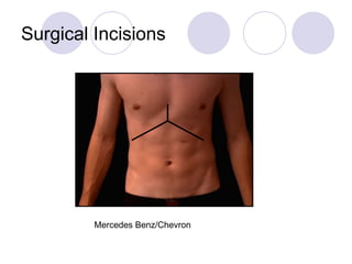
The Surgical Wound Cormac Joyce Final Med 2009
Surgery It is the branch of medicine concerned with diseases and conditions which require or are amenable to operative procedures It is derived from Greek “cheirourgia” “ cheir” = the hand “ ergon” = work
Surgery It is often said that it is “controlled trauma” Carried out in a sterile environment Under aseptic conditions
Surgery Many protocols are put in place to prevent infections in surgical wounds Hand washing Gowns and gloves Painting and draping Drains Antibiotics Laminar flow theatres Sterile instruments Sterile dressings
Surgery But wound infections can occur despite these measures causing: Death Morbidity Longer hospital stays Cosmetically displeasing wounds
Surgery Surgical wound infections are common Comprising 12% of nosocomial infections The rate of infection depends on the type of surgery undertaken
Operation Types The risk of a wound infection depends on the operation For that reason, operations are classified into distinct types Clean Clean-Contaminated Contaminated Dirty
Class I :Clean wounds Elective operations (non emergency) Non traumatic injury Good surgical technique Respiratory, gastrointestinal, biliary and genitourinary tracts not breached Risk of infection < 2% Eg: mastectomy, hernia repair
Class II: Clean - Contaminated Urgent or emergency case that is otherwise clean GI, GU or respiratory tracts entered electively, no spillage or unusual contamination Minor break in sterile technique occurred Endogenous flora involved Risk of infection: <10 % Eg: appendicectomy, bowel resection
Class III: Contaminated Non-purulent inflammation Gross spillage from GIT, entry into GU or biliary tract in the presence of infected bile/urine. Major break in technique Penetrating trauma < 4hrs old Chronic open wounds Risk of infection: 20% Eg: GSW, rectal surgery
Class IV : Dirty Purulent inflammation (abscess) Pre-operative perforation of GI, GU, biliary or respiratory tract Penetrating trauma > 4 hrs Existing acute bacterial infection or a perforated viscera is encountered (clean tissue is transected to gain access to pus). Risk of infection: 40%
Signs of Infection The cardinal points of acute inflammation Calor Rubor Dolor Tumour Functio Laesa
Signs of Infection Patient may be systemically unwell ↑  Temp Tachycardic Hypotension Wound breakdown Wound discharge Warm peripheries Septic shock
Prevention Aseptic technique Good technique Prophylactic antibiotics where appropriate Microbiology input Clean operating theatre Elective surgery Good post op care
Wound Healing Wound healing is a complex and dynamic process of restoring cellular structures and tissue layers There are 3 distinct phases There are various categories of wound healing the ultimate outcome of any healing process is repair of a tissue defect
Wound Healing The types of wound healing: 1° healing Delayed 1° healing 2° healing (Epithelialisation) Even though different categories exist, the interactions of cellular and extracellular constituents are similar.
Primary wound healing Also known as “healing by primary intention” Think of a typical surgical wound: the wound eges are approximated Minimal number of cellular constituents die Results in a small line of scar tissue Minimizes the need for granulation tissue so scarring is minimized
The importance of good… Technique Choice of suture Choice of needle Training Instruments Antibiotics Aftercare
Delayed Primary healing Occurs if wound egdes are not approximated immediately May be desired in contaminated wounds By day 4: phagocytosis of contaminated tissues has occurred Usually wound is closed surgically at this stage If contamination is present still : chronic inflammation ensues leading to prominent scar eventually
Secondary Healing Also called healing by secondary intention A full thickness wound is allowed to heal by itself: there is no approximation of wound edges Large amounts of granulation tissue formed Wound eventually very contracted Takes much longer to heal
Epithelialization Epithelization is the process by which epithelial cells migrate and replicate via mitosis and traverse the wound  Occurs by one of 2 mechanisms Common in the healing of ulcers and erosions
Epithelialization: Mechanisms Mechanism 1 If basement membrane is intact ie some dermis or dermal appendages remain Epithelialization occurs by epithelial cells migrating upwards
Epithelialization: Mechanisms Mechanism 2 Occurs in a deeper wound A single layer of epithelial cells advance from the wound edges to cover the wound They then stratify so wound cover is complete
Normal Wound Healing There are 3 phases Inflammatory phase: Days 0-4 Proliferative phase : Days 5-21 Remodelling phase: Days 22-60
Wound Healing It can also be classified in 4 stages: Haemostasis Inflammation Granulation Remodelling
Haemostasis Injury causes local bleeding Vasoconstriction is mediated by : Adrenaline Thrombaxane A2 Prostaglandin 2 α
Haemostasis Platelets then adhere to damaged endothelium and discharge ADP Which promotes thrombocyte clumping and “dams” the wound Inflammation is initiated by cytokine release from platelets
Haemostasis α -granules from platelets release: Platelet Derived Growth Factor (PDGF) Platelet factor IV Transforming Growth Factor  β Thrombocyte dense bodies release: Histamine Serotonin
Haemostasis PDGF attracts fibroblasts chemotactically Leading to collagen deposition in later stages of wound healing Fibrinogen  -> Fibrin Thus providing the structural support for the cellular components of inflammation
Inflammatory Phase Capillary dilatation occurs due to: Histamine Bradykinin Prostaglandins NO This dilatation allows inflammatory cells to reach the wound site
Inflammatory Phase These PMNs or leukocytes have several functions: Scavenge for debris Debride the wound Help to kill bacteria by: -oxidative burst mechanisms -opsonization
Inflammatory Phase Opsonin “ factor which enhances the efficiency of phagocytosis because it is recognized by receptors on leucocytes 2 major opsonins are: Fc fragment of IgG A product of complement, C3b
Inflammatory Phase Monocytes now enter the wound and become macrophages They have numerous functions
Macrophage functions in healing Secretion of numerous enzymes and cytokines  Collagenases and elastases To break down injured tissues PDGF, TGF β , IL, TNF To stimulate proliferation of fibroblasts, endothelial and smooth muscle cells
Proliferative Phase Angiogenesis The formation of new blood vessels Formed by endothelial cells becoming new capillaries within the wound bed Angiogenesis stimulated by TNF α
Proliferative Phase Collagen deposition Type III collagen is laid down by fibroblasts Fibroblasts are attracted by TGF β  and PDGF Total collagen content increases until day 21
Proliferative Phase Granulation Tissue Is the combination of collagen deposition and angiogenesis
Granulation Tissue Definition: Newly formed connective tissue, often found at the edge or base of ulcers and wounds made up of : capillaries, fibroblasts, myofibroblasts, and inflammatory cells embedded in a mucin rich ground substance during healing
Proliferative Phase Re-epithelialization occurs next: By upward migration of epithelial cells if BM is intact Or from wound edges
Remodelling Phase Fibroblasts become myofibroblasts And wound begins to contract Can contract 0.75mm per day Can over contract however Contraction allows wound to become smaller A large wound can contract by up to 40-80%
Remodelling Phase Type III collagen is degraded And replaced with Type I Water is removed from the scar, allowing collagen to cross-link Wound vascularity decreases Collagen cross linkage allows: Increased scar strength Scar contracture Decreased scar thickness
Wound Strength During phase 1 and 2 (inflammatory and proliferative phases) Wounds have very little strength During remodelling: Wounds rapidly gain strength @ 6 weeks: wound is 50% of final strength @12 months: wound is maximal strength: but this is only 75% of pre-injury tissue strength
Abnormal Scars Hypertrophic Scars Keloid Scars
Hypertrophic Scars Raised, red and thickened Limited to boundaries of scar Occurs shortly after injury Common on anterior chest and deltoids Regresses over time Related to wound tension and prolonged inflammatory phase of healing
Hypertrophic Scars Treatment: Surgical excision Intralesional Triamcenelone acetate injection
Hypertrophic Scars No racial or familial preponderance Electron microscopy: flattened collagen bundles parallel in orientation
Keloid Scars Raised, red and thickened scar Extends beyond original scar boundary Occurs months after injury Does not regress Commoner in darker skinned people Familial tendency ? Autoimmune phenomenon Worsened by surgery and in pregnancy Regresses post menopause
Keloid scar Treatment: Surgical excision : caveat- recurrence = 65% Compression treatment CO2 lasers Cryotherapy
Factors influencing scarring These can be broken down into: Patient factors Surgical factors
Patient Factors Age Elderly scar well -? 2° wrinkles Skin type Celtics : hypertrophic scar tendency Dark skinned: keloid scars
Patient Factors Anatomic region Midline Deltoid region Sternotomy post CABG
Patient Factors Patient morbidity Nutritional state Diabetes Wound infections
Patient Factors Local tissue Oedema Previous radiotherapy Vascular insufficiency
Surgical factors Atraumatic skin handling Eversion of wound edges Inversion places keratinised epidermis between the healing surfaces = delayed healing Tension free closure Clean and healthy wound edges
Everted Edges
Inverted Edges
Surgical factors Scar orientation Parallel to lines of relaxed skin tension Langers lines Suture tension “ Thou shall not commit tension” Over-tight: pressure necrosis Under-tight: wound gaping and widened scar
Langers Lines
Acute Inflammation Definition The cellular and vascular response to injury Short in duration Has cellular and chemical components
Acute Inflammation: Causes Injury by: Pathogens Bacteria, viruses, parasites Chemical agents Acids, alkalis Physical agents Heat, trauma (surgery), radiation Tissue death Infarction
Stages of Acute Inflammation Dilatation of local capillaries   endothelial permeability Leakage of protein-rich fluid into interstitial space – including fibrinogen Fibrinogen -> fibrin Margination of leukocytes to peripheries of capillaries Mostly neutrophils
Stages of Acute Inflammation Acute Inflammation is mediated by: Chemicals: interleukins and histamine Proteins: complement cascade
Complement Cascade Component of innate immune system Cascade of proteins Resulting in formation of Membrane-Attack-Complex (MAC) which can Destroy invading bacteria Recruit other cells ie neutrophils Can also act as opsonins: enhancing phagocytosis
Complement Cascade 2 main activating arms of CC: Classic pathway: consists of antigen-antibody complexes Alternative pathway: activated directly by contact with micro-organisms
Acute Inflammation: Neutrophils The role of the Neutrophil First cellular component to appear Attracted by inflammatory mediators By chemotaxis They can move: margination in blood vessels – by adhering to vascular endothelium: roll between endothelial cells: emigrate to interstitium
Acute Inflammation: Neutrophils Function: Phagocytosis of micro-organisms With lysosomal free radical degradation of pathogens
Chemical messengers in AI These allow cells to communicate with each other and mediate the immune response
Chemical messengers in AI Chemokines Cause direct migration of target cells to site of release Cytokines Soluble, biologically active molecules secreted by cells which have a variety of effects on the target cells
Cytokines: Examples IL-1: neutrophil adhesion and vascular adhesion molecules IL-2: proliferation of B cells and NK cells TNF: causes fever and promotes inflammation IFN: activates macrophages Histamine: vasodilation and   permeability
Effects of AI: beneficial Dilution of bacterial toxin Defence mechanisms are brought to the pathogen neutrophils;: phagocytosis Complement: cell lysis Antibodies Drug delivery
Effects of AI: beneficial Drainage to LNs: immune response stimulated Fibrin traps the pathogen in place so it can be attacked
Effects of AI: non beneficial Destruction of normal tissue: RA Lethal swelling in certain parts of the body ie epiglottitis Hypersensitivity reactions Asthma Anaphylaxis
Outcomes of Acute Inflammation Resolution Tissues restored to normal Pus/abscess Organization Tissues replaced by granulation tissues Chronic Inflammation If causative agent not removed
Chronic Inflammation Definition Tissue response to persistent injury Long in duration Cellular components differ from acute inflammation
Chronic Inflammation Causes Foreign bodies: ie sutures Bacteria: ie TB Chronic abscess: ie osteomyelitis Transplant: ie chronic rejection IBD Progression from AI
Chronic Inflammation Key points Histological pattern not as predictable as acute inflammation There may be areas of acute inflammation occurring simultaneously Granulation tissue and fibrosis may both be present: indicating the tissues attempts at repair
Chronic Inflammation Lymphocytes predominate Macrophages present too In granulomatous inflammation they fuse forming multinucleate Langhans giant cells Plasma cells are also present
Chronic Inflammation Macrophages Derived from monocytes Phagocytosis and killing of pathogens by lysosomes Antigen presentation Langhans giant cell formation
Chronic Inflammation: Effects Secondary infection ie chronic epithelial injury Scarring Resolution: restoration of normality Local lymphadenopathy
Surgical Incisions Gridiron: appendix
Surgical Incisions Lanz: appendix
Surgical Incisions Nephrectomy/ loin
Surgical Incisions Kochers
Surgical Incisions Inguinal : hernia, orchidectomy (for Ca) not for torsion
Surgical Incisions Midline sternotomy
Surgical Incisions Mercedes Benz/Chevron
Surgical Incisions Midline laparotomy
Surgical Incisions Rutherford Morrison : renal transplant
Surgical Incisions Lap Chole
Surgical Incisions Lap appendix
 

Fwd: lecture

  • 1.
    The Surgical WoundCormac Joyce Final Med 2009
  • 2.
    Surgery It isthe branch of medicine concerned with diseases and conditions which require or are amenable to operative procedures It is derived from Greek “cheirourgia” “ cheir” = the hand “ ergon” = work
  • 3.
    Surgery It isoften said that it is “controlled trauma” Carried out in a sterile environment Under aseptic conditions
  • 4.
    Surgery Many protocolsare put in place to prevent infections in surgical wounds Hand washing Gowns and gloves Painting and draping Drains Antibiotics Laminar flow theatres Sterile instruments Sterile dressings
  • 5.
    Surgery But woundinfections can occur despite these measures causing: Death Morbidity Longer hospital stays Cosmetically displeasing wounds
  • 6.
    Surgery Surgical woundinfections are common Comprising 12% of nosocomial infections The rate of infection depends on the type of surgery undertaken
  • 7.
    Operation Types Therisk of a wound infection depends on the operation For that reason, operations are classified into distinct types Clean Clean-Contaminated Contaminated Dirty
  • 8.
    Class I :Cleanwounds Elective operations (non emergency) Non traumatic injury Good surgical technique Respiratory, gastrointestinal, biliary and genitourinary tracts not breached Risk of infection < 2% Eg: mastectomy, hernia repair
  • 9.
    Class II: Clean- Contaminated Urgent or emergency case that is otherwise clean GI, GU or respiratory tracts entered electively, no spillage or unusual contamination Minor break in sterile technique occurred Endogenous flora involved Risk of infection: <10 % Eg: appendicectomy, bowel resection
  • 10.
    Class III: ContaminatedNon-purulent inflammation Gross spillage from GIT, entry into GU or biliary tract in the presence of infected bile/urine. Major break in technique Penetrating trauma < 4hrs old Chronic open wounds Risk of infection: 20% Eg: GSW, rectal surgery
  • 11.
    Class IV :Dirty Purulent inflammation (abscess) Pre-operative perforation of GI, GU, biliary or respiratory tract Penetrating trauma > 4 hrs Existing acute bacterial infection or a perforated viscera is encountered (clean tissue is transected to gain access to pus). Risk of infection: 40%
  • 12.
    Signs of InfectionThe cardinal points of acute inflammation Calor Rubor Dolor Tumour Functio Laesa
  • 13.
    Signs of InfectionPatient may be systemically unwell ↑ Temp Tachycardic Hypotension Wound breakdown Wound discharge Warm peripheries Septic shock
  • 14.
    Prevention Aseptic techniqueGood technique Prophylactic antibiotics where appropriate Microbiology input Clean operating theatre Elective surgery Good post op care
  • 15.
    Wound Healing Woundhealing is a complex and dynamic process of restoring cellular structures and tissue layers There are 3 distinct phases There are various categories of wound healing the ultimate outcome of any healing process is repair of a tissue defect
  • 16.
    Wound Healing Thetypes of wound healing: 1° healing Delayed 1° healing 2° healing (Epithelialisation) Even though different categories exist, the interactions of cellular and extracellular constituents are similar.
  • 17.
    Primary wound healingAlso known as “healing by primary intention” Think of a typical surgical wound: the wound eges are approximated Minimal number of cellular constituents die Results in a small line of scar tissue Minimizes the need for granulation tissue so scarring is minimized
  • 18.
    The importance ofgood… Technique Choice of suture Choice of needle Training Instruments Antibiotics Aftercare
  • 19.
    Delayed Primary healingOccurs if wound egdes are not approximated immediately May be desired in contaminated wounds By day 4: phagocytosis of contaminated tissues has occurred Usually wound is closed surgically at this stage If contamination is present still : chronic inflammation ensues leading to prominent scar eventually
  • 20.
    Secondary Healing Alsocalled healing by secondary intention A full thickness wound is allowed to heal by itself: there is no approximation of wound edges Large amounts of granulation tissue formed Wound eventually very contracted Takes much longer to heal
  • 21.
    Epithelialization Epithelization isthe process by which epithelial cells migrate and replicate via mitosis and traverse the wound Occurs by one of 2 mechanisms Common in the healing of ulcers and erosions
  • 22.
    Epithelialization: Mechanisms Mechanism1 If basement membrane is intact ie some dermis or dermal appendages remain Epithelialization occurs by epithelial cells migrating upwards
  • 23.
    Epithelialization: Mechanisms Mechanism2 Occurs in a deeper wound A single layer of epithelial cells advance from the wound edges to cover the wound They then stratify so wound cover is complete
  • 24.
    Normal Wound HealingThere are 3 phases Inflammatory phase: Days 0-4 Proliferative phase : Days 5-21 Remodelling phase: Days 22-60
  • 25.
    Wound Healing Itcan also be classified in 4 stages: Haemostasis Inflammation Granulation Remodelling
  • 26.
    Haemostasis Injury causeslocal bleeding Vasoconstriction is mediated by : Adrenaline Thrombaxane A2 Prostaglandin 2 α
  • 27.
    Haemostasis Platelets thenadhere to damaged endothelium and discharge ADP Which promotes thrombocyte clumping and “dams” the wound Inflammation is initiated by cytokine release from platelets
  • 28.
    Haemostasis α -granulesfrom platelets release: Platelet Derived Growth Factor (PDGF) Platelet factor IV Transforming Growth Factor β Thrombocyte dense bodies release: Histamine Serotonin
  • 29.
    Haemostasis PDGF attractsfibroblasts chemotactically Leading to collagen deposition in later stages of wound healing Fibrinogen -> Fibrin Thus providing the structural support for the cellular components of inflammation
  • 30.
    Inflammatory Phase Capillarydilatation occurs due to: Histamine Bradykinin Prostaglandins NO This dilatation allows inflammatory cells to reach the wound site
  • 31.
    Inflammatory Phase ThesePMNs or leukocytes have several functions: Scavenge for debris Debride the wound Help to kill bacteria by: -oxidative burst mechanisms -opsonization
  • 32.
    Inflammatory Phase Opsonin“ factor which enhances the efficiency of phagocytosis because it is recognized by receptors on leucocytes 2 major opsonins are: Fc fragment of IgG A product of complement, C3b
  • 33.
    Inflammatory Phase Monocytesnow enter the wound and become macrophages They have numerous functions
  • 34.
    Macrophage functions inhealing Secretion of numerous enzymes and cytokines Collagenases and elastases To break down injured tissues PDGF, TGF β , IL, TNF To stimulate proliferation of fibroblasts, endothelial and smooth muscle cells
  • 35.
    Proliferative Phase AngiogenesisThe formation of new blood vessels Formed by endothelial cells becoming new capillaries within the wound bed Angiogenesis stimulated by TNF α
  • 36.
    Proliferative Phase Collagendeposition Type III collagen is laid down by fibroblasts Fibroblasts are attracted by TGF β and PDGF Total collagen content increases until day 21
  • 37.
    Proliferative Phase GranulationTissue Is the combination of collagen deposition and angiogenesis
  • 38.
    Granulation Tissue Definition:Newly formed connective tissue, often found at the edge or base of ulcers and wounds made up of : capillaries, fibroblasts, myofibroblasts, and inflammatory cells embedded in a mucin rich ground substance during healing
  • 39.
    Proliferative Phase Re-epithelializationoccurs next: By upward migration of epithelial cells if BM is intact Or from wound edges
  • 40.
    Remodelling Phase Fibroblastsbecome myofibroblasts And wound begins to contract Can contract 0.75mm per day Can over contract however Contraction allows wound to become smaller A large wound can contract by up to 40-80%
  • 41.
    Remodelling Phase TypeIII collagen is degraded And replaced with Type I Water is removed from the scar, allowing collagen to cross-link Wound vascularity decreases Collagen cross linkage allows: Increased scar strength Scar contracture Decreased scar thickness
  • 42.
    Wound Strength Duringphase 1 and 2 (inflammatory and proliferative phases) Wounds have very little strength During remodelling: Wounds rapidly gain strength @ 6 weeks: wound is 50% of final strength @12 months: wound is maximal strength: but this is only 75% of pre-injury tissue strength
  • 43.
    Abnormal Scars HypertrophicScars Keloid Scars
  • 44.
    Hypertrophic Scars Raised,red and thickened Limited to boundaries of scar Occurs shortly after injury Common on anterior chest and deltoids Regresses over time Related to wound tension and prolonged inflammatory phase of healing
  • 45.
    Hypertrophic Scars Treatment:Surgical excision Intralesional Triamcenelone acetate injection
  • 46.
    Hypertrophic Scars Noracial or familial preponderance Electron microscopy: flattened collagen bundles parallel in orientation
  • 47.
    Keloid Scars Raised,red and thickened scar Extends beyond original scar boundary Occurs months after injury Does not regress Commoner in darker skinned people Familial tendency ? Autoimmune phenomenon Worsened by surgery and in pregnancy Regresses post menopause
  • 48.
    Keloid scar Treatment:Surgical excision : caveat- recurrence = 65% Compression treatment CO2 lasers Cryotherapy
  • 49.
    Factors influencing scarringThese can be broken down into: Patient factors Surgical factors
  • 50.
    Patient Factors AgeElderly scar well -? 2° wrinkles Skin type Celtics : hypertrophic scar tendency Dark skinned: keloid scars
  • 51.
    Patient Factors Anatomicregion Midline Deltoid region Sternotomy post CABG
  • 52.
    Patient Factors Patientmorbidity Nutritional state Diabetes Wound infections
  • 53.
    Patient Factors Localtissue Oedema Previous radiotherapy Vascular insufficiency
  • 54.
    Surgical factors Atraumaticskin handling Eversion of wound edges Inversion places keratinised epidermis between the healing surfaces = delayed healing Tension free closure Clean and healthy wound edges
  • 55.
  • 56.
  • 57.
    Surgical factors Scarorientation Parallel to lines of relaxed skin tension Langers lines Suture tension “ Thou shall not commit tension” Over-tight: pressure necrosis Under-tight: wound gaping and widened scar
  • 58.
  • 59.
    Acute Inflammation DefinitionThe cellular and vascular response to injury Short in duration Has cellular and chemical components
  • 60.
    Acute Inflammation: CausesInjury by: Pathogens Bacteria, viruses, parasites Chemical agents Acids, alkalis Physical agents Heat, trauma (surgery), radiation Tissue death Infarction
  • 61.
    Stages of AcuteInflammation Dilatation of local capillaries  endothelial permeability Leakage of protein-rich fluid into interstitial space – including fibrinogen Fibrinogen -> fibrin Margination of leukocytes to peripheries of capillaries Mostly neutrophils
  • 62.
    Stages of AcuteInflammation Acute Inflammation is mediated by: Chemicals: interleukins and histamine Proteins: complement cascade
  • 63.
    Complement Cascade Componentof innate immune system Cascade of proteins Resulting in formation of Membrane-Attack-Complex (MAC) which can Destroy invading bacteria Recruit other cells ie neutrophils Can also act as opsonins: enhancing phagocytosis
  • 64.
    Complement Cascade 2main activating arms of CC: Classic pathway: consists of antigen-antibody complexes Alternative pathway: activated directly by contact with micro-organisms
  • 65.
    Acute Inflammation: NeutrophilsThe role of the Neutrophil First cellular component to appear Attracted by inflammatory mediators By chemotaxis They can move: margination in blood vessels – by adhering to vascular endothelium: roll between endothelial cells: emigrate to interstitium
  • 66.
    Acute Inflammation: NeutrophilsFunction: Phagocytosis of micro-organisms With lysosomal free radical degradation of pathogens
  • 67.
    Chemical messengers inAI These allow cells to communicate with each other and mediate the immune response
  • 68.
    Chemical messengers inAI Chemokines Cause direct migration of target cells to site of release Cytokines Soluble, biologically active molecules secreted by cells which have a variety of effects on the target cells
  • 69.
    Cytokines: Examples IL-1:neutrophil adhesion and vascular adhesion molecules IL-2: proliferation of B cells and NK cells TNF: causes fever and promotes inflammation IFN: activates macrophages Histamine: vasodilation and  permeability
  • 70.
    Effects of AI:beneficial Dilution of bacterial toxin Defence mechanisms are brought to the pathogen neutrophils;: phagocytosis Complement: cell lysis Antibodies Drug delivery
  • 71.
    Effects of AI:beneficial Drainage to LNs: immune response stimulated Fibrin traps the pathogen in place so it can be attacked
  • 72.
    Effects of AI:non beneficial Destruction of normal tissue: RA Lethal swelling in certain parts of the body ie epiglottitis Hypersensitivity reactions Asthma Anaphylaxis
  • 73.
    Outcomes of AcuteInflammation Resolution Tissues restored to normal Pus/abscess Organization Tissues replaced by granulation tissues Chronic Inflammation If causative agent not removed
  • 74.
    Chronic Inflammation DefinitionTissue response to persistent injury Long in duration Cellular components differ from acute inflammation
  • 75.
    Chronic Inflammation CausesForeign bodies: ie sutures Bacteria: ie TB Chronic abscess: ie osteomyelitis Transplant: ie chronic rejection IBD Progression from AI
  • 76.
    Chronic Inflammation Keypoints Histological pattern not as predictable as acute inflammation There may be areas of acute inflammation occurring simultaneously Granulation tissue and fibrosis may both be present: indicating the tissues attempts at repair
  • 77.
    Chronic Inflammation Lymphocytespredominate Macrophages present too In granulomatous inflammation they fuse forming multinucleate Langhans giant cells Plasma cells are also present
  • 78.
    Chronic Inflammation MacrophagesDerived from monocytes Phagocytosis and killing of pathogens by lysosomes Antigen presentation Langhans giant cell formation
  • 79.
    Chronic Inflammation: EffectsSecondary infection ie chronic epithelial injury Scarring Resolution: restoration of normality Local lymphadenopathy
  • 80.
  • 81.
  • 82.
  • 83.
  • 84.
    Surgical Incisions Inguinal: hernia, orchidectomy (for Ca) not for torsion
  • 85.
  • 86.
  • 87.
  • 88.
    Surgical Incisions RutherfordMorrison : renal transplant
  • 89.
  • 90.
  • 91.