1.2 ครูเริ่มเปิดอภิปรำยโดยให้นักเรียนร่วมกันแสดงควำมคิดเห็นว่ำเซลล์ควรจะมี
กิจกรรมใดบ้ำงเพื่อให้สิ่งมีชีวิตนั้นสำมำรถดำรงชีวิตอยู่ได้ในสภำพแวดล้อมธรรมชำติ
1.3 นักเรียนสำมำรถตั้งคำถำมที่อยำกรู้เพิ่มเติมหลังจำกได้ร่วมกันอภิปรำยใน
ห้องเรียนแล้ว เช่น ภำยในเซลล์นั้นสำมำรถแบ่งออกได้เป็นกี่ส่วน อะไรบ้ำง
ขั้นสอน : ครูอธิบำยเนื้อหำ “โครงสร้ำงพื้นฐำนภำยในเซลล์” ว่ำ
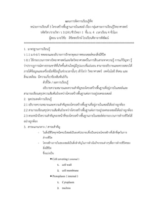
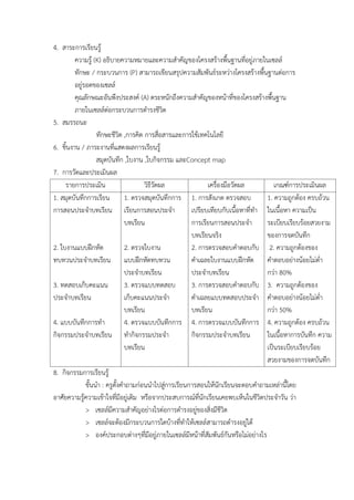
> cell covering ( external ) protoplasm ( internal )
> The covering of cell
Cell membrane / plasma membrane / cytoplasmic
membrane
o 60% protein = glycoprotein , mucoprotein
Integral protein
Peripheral protein
o 40% lipid = phospholipid , cholesterol
o Unitmenbrane / lipid bilayer
o Semipermeable membrane
o Function = covering of cell and organelle , fluid mosaic
model
Cell wall
o Function = protection and strength increasing of cell
o Ex. = plant : cellulose ( plasmodesmata )
= fungi : chitin
= diatom : silica
= bacteria : peptidoglycan
@@@ animal cell = glycoprotein ( tissue formimg ) abnormal is form cancer cell
> The protoplasm of cell
Cytoplasm = the liquid that between cell membrane and
nucleus
o Organelle = the functional unit of cell
double unit membrane
mitochondria ( energy production )
o oval shape
o matrix ( cellular respiration )
22.
o DNA ,RNA , protein
Choloplast ( photosynthesis )
o Cholophyll ( light reaction )
o Stroma ( dark reaction )
o DNA , RNA , protein
Golgi body ( modified and contain )
o Vertebrate > invertebrane
o Vesicle formation
Another : acrosome of sperm : nematocyst of hydra
one unit membrane
Endoplasmic reticulum ( ER )
o Tube structure that connecting of cell
o Hyaloplasm
o 2 types : rough ER ( RER ) = ER + ribosome : protein synthesis
: smooth ER ( SER ) = ER : lipid and steroid synthesis
Lysosome
o Vesicle with contained hydrolytic enzyme
o Usually found in phagocytic cell ex. Leucocyte , amoebocyte
o Function : substrate / nutrient
: microbe / antigen
: cell damage / exprire
: mmetamorphosis
Vacuole
o Vesicle with covered tonoplast
o Conaining chemical substrate
o Uaually found in plant and lower animal
o 3 types : sap vacuole = plant , liquid containing ( water , solution )
: food vacuole = protozoa / phagocyte , food containing
: contractile vacuole = fresh water protozoa , excretion /
water balance
Non unit membrane
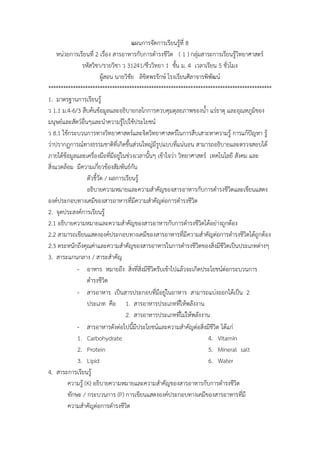
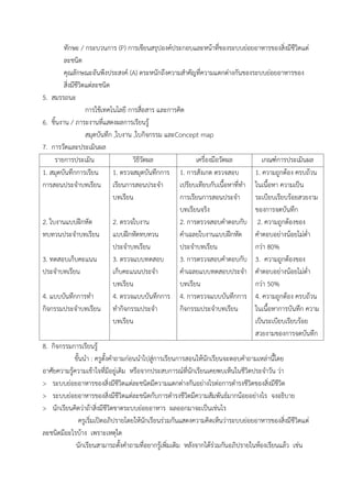
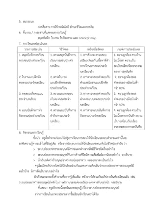
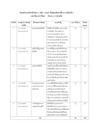
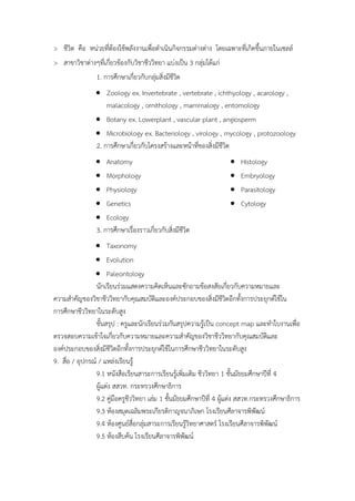
Ribosome
o Can found in all organism ( except : virus , viroid )
23.
o Ribonucleoprotein (rRNA + protein )
o Small subunit + large subunit
o 2 types : prokaryote = 70s ( 30s + 50s )
: eukaryote = 80s ( 40s + 60s )
o 3 location : free = protein synthesis for cell
: RER = protein synthesis for export
: nuclear membrane = protein synthesis for nucleus
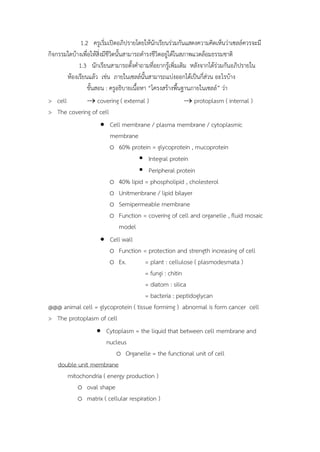
Centriole
o 2 tube ที่ตั้งฉำกกัน (can found in animal and some protozoa only)
o Cell division = chromosome movement ( spidle fiber )
o 9 + 0
o DNA , RAN , protein
o Basal body / kinetosome = cilia and flagella movement ( 9 + 2 )
Cytoskeleton
o Structure and movement of cell
o Protein fiber
o 3 types : microtubule = large size , tubulin , motion of cell /
chromosome / organelle
: intermedia filament = middle size , supercoil มีส่วนห่อหุ้ม , คง
ตำแหน่ง organelle / nucleus.
: microfilament = small size , actin , cytoplasmic streaming /
pseudopodium / muscle contraction / cell conformation
change / cytokinesis of animal cell.
o Cytoplasmic inclusion = chemical substrate in cytoplasm
= non-living part of cytoplasm
= ex. Starch grain , protein , waste product of
metabolism
Nucleus = the central of cell
Robert Brown ( 1831 )
ก้อนทึบแสงมักอยู่กลำงเซล์
Usually found single nucleus of cell ( except : RBC [non] ,
paramecium[2 nucleus] , skeleton muscle[many] )
Contain genetic material : cell activities controlled
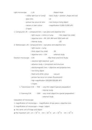
Light microscope -L.M. - Robert Hook
– visible light (sun or lump) - basic study = position ,shape and size
- glass lens - air
- picture has occur on lens - non-living or living object
- nature or stain colour - magnification (1,000-15,000 เท่ำ)
- 2 types
1. Compound L.M. – compound lens = eye piece and objective lens
- light source = mirror or lump - thin object (on slide)
- objective lens = 4X ,10X ,40X and 100X (with oil)
- internal study - 2D
2. Stereoscopic L.M.– compound lens = eye piece and objective lens
- light source = lump
- thick object (no slide) - 3D
- objective lens < 10X - external study
Electron microscope - E.M. - Max Knoll and Ernst Ruska
– electron light (electron gun)
- advance study = component and structure
- electromagnetic lens = objective and projector lens
- non-living object
- black and white colour - vacuum
- picture has occur on screen (fluorescent)
- high magnification (200,000-500,000 เท่ำ)
- 2 types
1. Transmission E.M. – TEM – very thin object (special preparation)
- internal study
2. Scanning E.M. - SEM - very small object (no special preparation)
- external study
Calaulation of microscope
1. magnification of microscope = magnification of eye piece x objective lens
2. magnification of microscope = image / object
@ the same unit of image and object
@ the important unit : cm = 10-2
m mm = 10-3
m um = 10-6
m nm = 10-9
m


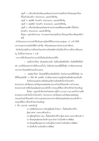
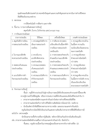
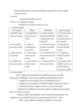
![o Ribonucleoprotein ( rRNA + protein )
o Small subunit + large subunit
o 2 types : prokaryote = 70s ( 30s + 50s )
: eukaryote = 80s ( 40s + 60s )
o 3 location : free = protein synthesis for cell
: RER = protein synthesis for export
: nuclear membrane = protein synthesis for nucleus
Centriole
o 2 tube ที่ตั้งฉำกกัน (can found in animal and some protozoa only)
o Cell division = chromosome movement ( spidle fiber )
o 9 + 0
o DNA , RAN , protein
o Basal body / kinetosome = cilia and flagella movement ( 9 + 2 )
Cytoskeleton
o Structure and movement of cell
o Protein fiber
o 3 types : microtubule = large size , tubulin , motion of cell /
chromosome / organelle
: intermedia filament = middle size , supercoil มีส่วนห่อหุ้ม , คง
ตำแหน่ง organelle / nucleus.
: microfilament = small size , actin , cytoplasmic streaming /
pseudopodium / muscle contraction / cell conformation
change / cytokinesis of animal cell.
o Cytoplasmic inclusion = chemical substrate in cytoplasm
= non-living part of cytoplasm
= ex. Starch grain , protein , waste product of
metabolism
Nucleus = the central of cell
Robert Brown ( 1831 )
ก้อนทึบแสงมักอยู่กลำงเซล์
Usually found single nucleus of cell ( except : RBC [non] ,
paramecium[2 nucleus] , skeleton muscle[many] )
Contain genetic material : cell activities controlled](https://image.slidesharecdn.com/bio-140505103842-phpapp01/85/Bio-4-2-23-320.jpg)