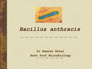
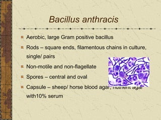
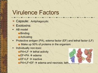
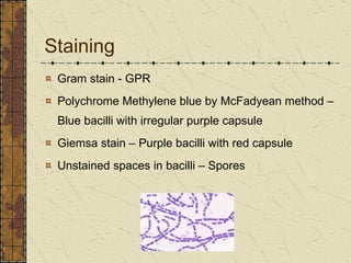
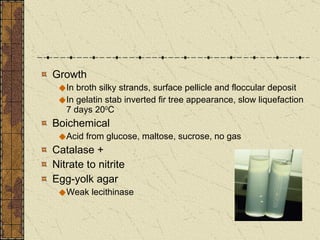
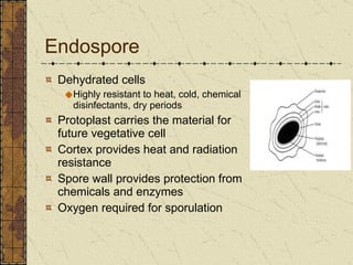
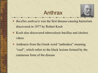
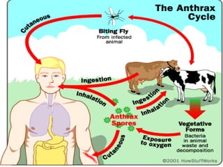
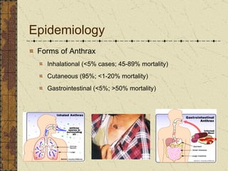
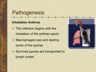
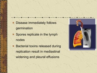
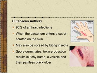
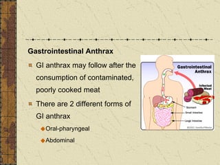
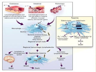
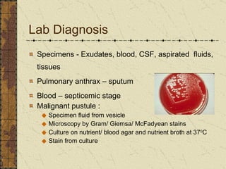
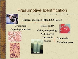
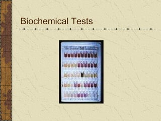
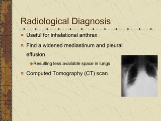
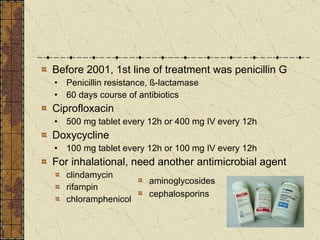
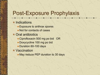
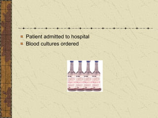
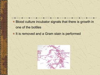
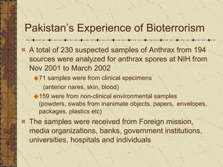
Bacillus anthracis Dr Kamran Afzal Asst Prof Microbiology
Clinically Important GPRs Spore forming Bacillus Clostridium Non-spore forming Corynebacterium Listeria Lactobacillus Bacilli with branching filaments Actinomyces Nocardia
Identity of Genus Bacillus Growth – large, rough/ irregular colonies GPR with spores (sporangium) Spores –resistant to heat, drying, chemical disinfectants, UV radiation, staining, may lie freely Inclusions – lipid, volutin or crystalline proteins
Species 34 species of Bacillus B. anthracis  – only true infectious pathogen B. cereus  – saprophyte with pathogenic potential Most species – saprophytes, resident in soil, some opportunistic infections
Bacillus anthracis Aerobic, large Gram positive bacillus Rods – square ends, filamentous chains in culture, single/ pairs Non-motile and non-flagellate Spores – central and oval Capsule – sheep/ horse blood agar, nutrient agar with10% serum
Virulence Factors Capsule:  Antiphagocytic Exotoxins: AB model Binding Activating Protective antigen (PA), edema factor (EF) and lethal factor (LF) Make up 50% of proteins in the organism Individually non-toxic PA+LF    lethal activity EF+PA    edema  EF+LF    inactive PA+LF+EF    edema and necrosis; lethal activity
Staining Gram stain - GPR Polychrome Methylene blue by McFadyean method –Blue bacilli with irregular purple capsule  Giemsa stain – Purple bacilli with red capsule Unstained spaces in bacilli – Spores
Culture Aerobe/ facultative anaerobe Temp range – 12 to 45 0 C Nutrient agar, blood agar, selective media Colonies – grey granular discs 2-3 mm after 24 hrs on nutrient agar (Medusa head), sticky membranous consistency Non-hemolytic – BA
Growth In broth silky strands, surface pellicle and floccular deposit In gelatin stab inverted fir tree appearance, slow liquefaction 7 days 20 0 C  Boichemical Acid from glucose, maltose, sucrose, no gas  Catalase + Nitrate to nitrite Egg-yolk agar Weak lecithinase
Endospore Dehydrated cells Highly resistant to heat, cold, chemical disinfectants, dry periods Protoplast carries the material for future vegetative cell Cortex provides heat and radiation resistance Spore wall provides protection from chemicals and enzymes Oxygen required for sporulation
Anthrax Bacillus anthracis  was the first disease-causing bacterium discovered in 1877 by Robert Koch Koch also discovered tuberculosis bacillus and cholera vibrio Anthracis from the Greek word “anthrakos” meaning “coal”, which refers to the black lesions formed by the cutaneous form of the disease
Caused by spores Primarily a disease of domesticated and wild animals Herbivores such as sheep, cows, horses, goats Natural reservoir is soil Anthrax zones Soil rich in organic matter (pH < 6.0)
 
Who gets it? People who work with imported animal hides or furs in areas where standards are insufficient to prevent exposure to anthrax spores People who handle potentially infected animal products in high-incidence areas People who work directly with it in the lab Military personnel deployed to sensitive areas are at high risk for exposure to the organism
Epidemiology Forms of Anthrax Inhalational (<5% cases; 45-89% mortality) Cutaneous (95%; <1-20% mortality) Gastrointestinal (<5%; >50% mortality)
Pathogenesis Inhalation Anthrax The infection begins with the inhalation of the anthrax spore Macrophages lyse and destroy some of the spores Survived spores are transported to lymph nodes
Disease immediately follows germination Spores replicate in the lymph nodes Bacterial toxins released during replication result in mediastinal  widening and pleural effusions
Inhalation Anthrax is the most lethal type of Anthrax Incubation period 1–7 days Possibly ranging up to 42 days (depending on how many spores were inhaled) Case fatality  Untreated (97%) With antimicrobial therapy (75%)
Cutaneous Anthrax  95% of anthrax infections When the bacterium enters a cut or scratch on the skin May also be spread by biting insects  Spore germinates, toxin production results in itchy bump, a vesicle and then painless black ulcer
Gastrointestinal Anthrax  GI anthrax may follow after the consumption of contaminated, poorly cooked meat There are 2 different forms of GI anthrax Oral-pharyngeal Abdominal
 
Lab Diagnosis Specimens -  Exudates, blood, CSF, aspirated  fluids, tissues Pulmonary anthrax – sputum Blood – septicemic stage Malignant pustule : Specimen fluid from vesicle Microscopy by Gram/ Giemsa/ McFadyean stains Culture on nutrient/ blood agar and nutrient broth at 37 0 C Stain from culture
Presumptive Identification Clinical   specimen (blood, CSF, etc.) Isolate on BA Colony morphology No hemolysis Non motile Spores Gram stain Malachite green Gram stain Capsule production
Confirmatory Identification Isolate Phage lysis Capsule Bicarbonate  media Horse  blood (McFadyean  Stain) (McFadyean stain  India ink stain) DFA Capsule antigen Cell wall
Biochemical Tests
Rapid Detection Methods - ELISA assay for antigen / antibody detection - Immunofluorescence examinations - PCR for detection of nucleic acid
Radiological Diagnosis Useful for inhalational anthrax  Find  a widened mediastinum and pleural effusion Resulting less available space in lungs  Computed Tomography (CT) scan
Treatment Hospitalization IV antibiotics Empirical until sensitivities known Intensive supportive care Electrolyte and acid-base imbalances Mechanical ventilation Hemodynamic support
Before 2001, 1st line of treatment was penicillin G Penicillin resistance, ß-lactamase 60 days course of antibiotics Ciprofloxacin 500 mg tablet every 12h or 400 mg IV every 12h  Doxycycline 100 mg tablet every 12h or 100 mg IV every 12h  For inhalational, need another antimicrobial agent clindamycin  rifampin  chloramphenicol aminoglycosides cephalosporins
Post-Exposure Prophylaxis Indications Exposure to anthrax spores Not for contacts of cases Oral antibiotics Ciprofloxacin 500 mg po bid  OR Doxycycline 100 mg po bid Duration 60-100 days Vaccination May reduce PEP duration to 30 days
Case Study 63 year-old man was admitted to the emergency department  because of fever, vomiting and confusion On physical examination, the patient was lethargic and disoriented. His temperature was 39 o C, blood pressure 150/80 mm Hg, pulse 110/min and respiration 18/min Rhonchi on auscultation of the chest No neck rigidity
Patient admitted to hospital Blood cultures ordered
Blood culture incubator signals that there is growth in one of the bottles It is removed and a Gram stain is performed
Lab Diagnosis In vitro tests – to differentiate from  B. cereus ,  capsule demonstration, McFadyean stain, motility, pencillin sensitivity, egg-yolk lecithinase activity, virulence in mice In vivo test – intra-peritoneal inoculation in mice
As Biological Warfare Agent Why is this agent considered to be the number-one biological threat?
Benefits of Using Anthrax as a BW Highly lethal (Inhalational Anthrax) Long shelf-life Relatively easy availability Short incubation period (Relative to most other BW) Anthrax is stable in various types of weapons systems Dangerous as both powder or aerosolized forms Only need a small amount for a ‘Mass Effect’
Pakistan’s Experience of Bioterrorism  A total of 230 suspected samples of Anthrax from 194 sources were analyzed for anthrax spores at NIH from Nov 2001 to March 2002 71 samples were from clinical specimens  (anterior nares, skin, blood) 159 were from non-clinical environmental samples  (powders, swabs from inanimate objects, papers,  envelopes, packages, plastics etc) The samples were received from Foreign mission, media organizations, banks, government institutions, universities, hospitals and individuals
Out of these, 141 samples yielded growth suggestive of Bacillus species On the basis of colony morphology, Gram’s stain and other preliminary laboratory tests 62 isolates were found suspicious for  B. anthracis , however all the samples were negative by animal inoculation The suspected anthrax parcel/letter bombs in Pakistan during the investigation period were hoaxes

Bacillus anthracis

  • 1.
    Bacillus anthracis DrKamran Afzal Asst Prof Microbiology
  • 2.
    Clinically Important GPRsSpore forming Bacillus Clostridium Non-spore forming Corynebacterium Listeria Lactobacillus Bacilli with branching filaments Actinomyces Nocardia
  • 3.
    Identity of GenusBacillus Growth – large, rough/ irregular colonies GPR with spores (sporangium) Spores –resistant to heat, drying, chemical disinfectants, UV radiation, staining, may lie freely Inclusions – lipid, volutin or crystalline proteins
  • 4.
    Species 34 speciesof Bacillus B. anthracis – only true infectious pathogen B. cereus – saprophyte with pathogenic potential Most species – saprophytes, resident in soil, some opportunistic infections
  • 5.
    Bacillus anthracis Aerobic,large Gram positive bacillus Rods – square ends, filamentous chains in culture, single/ pairs Non-motile and non-flagellate Spores – central and oval Capsule – sheep/ horse blood agar, nutrient agar with10% serum
  • 6.
    Virulence Factors Capsule: Antiphagocytic Exotoxins: AB model Binding Activating Protective antigen (PA), edema factor (EF) and lethal factor (LF) Make up 50% of proteins in the organism Individually non-toxic PA+LF  lethal activity EF+PA  edema EF+LF  inactive PA+LF+EF  edema and necrosis; lethal activity
  • 7.
    Staining Gram stain- GPR Polychrome Methylene blue by McFadyean method –Blue bacilli with irregular purple capsule Giemsa stain – Purple bacilli with red capsule Unstained spaces in bacilli – Spores
  • 8.
    Culture Aerobe/ facultativeanaerobe Temp range – 12 to 45 0 C Nutrient agar, blood agar, selective media Colonies – grey granular discs 2-3 mm after 24 hrs on nutrient agar (Medusa head), sticky membranous consistency Non-hemolytic – BA
  • 9.
    Growth In brothsilky strands, surface pellicle and floccular deposit In gelatin stab inverted fir tree appearance, slow liquefaction 7 days 20 0 C Boichemical Acid from glucose, maltose, sucrose, no gas Catalase + Nitrate to nitrite Egg-yolk agar Weak lecithinase
  • 10.
    Endospore Dehydrated cellsHighly resistant to heat, cold, chemical disinfectants, dry periods Protoplast carries the material for future vegetative cell Cortex provides heat and radiation resistance Spore wall provides protection from chemicals and enzymes Oxygen required for sporulation
  • 11.
    Anthrax Bacillus anthracis was the first disease-causing bacterium discovered in 1877 by Robert Koch Koch also discovered tuberculosis bacillus and cholera vibrio Anthracis from the Greek word “anthrakos” meaning “coal”, which refers to the black lesions formed by the cutaneous form of the disease
  • 12.
    Caused by sporesPrimarily a disease of domesticated and wild animals Herbivores such as sheep, cows, horses, goats Natural reservoir is soil Anthrax zones Soil rich in organic matter (pH < 6.0)
  • 13.
  • 14.
    Who gets it?People who work with imported animal hides or furs in areas where standards are insufficient to prevent exposure to anthrax spores People who handle potentially infected animal products in high-incidence areas People who work directly with it in the lab Military personnel deployed to sensitive areas are at high risk for exposure to the organism
  • 15.
    Epidemiology Forms ofAnthrax Inhalational (<5% cases; 45-89% mortality) Cutaneous (95%; <1-20% mortality) Gastrointestinal (<5%; >50% mortality)
  • 16.
    Pathogenesis Inhalation AnthraxThe infection begins with the inhalation of the anthrax spore Macrophages lyse and destroy some of the spores Survived spores are transported to lymph nodes
  • 17.
    Disease immediately followsgermination Spores replicate in the lymph nodes Bacterial toxins released during replication result in mediastinal widening and pleural effusions
  • 18.
    Inhalation Anthrax isthe most lethal type of Anthrax Incubation period 1–7 days Possibly ranging up to 42 days (depending on how many spores were inhaled) Case fatality Untreated (97%) With antimicrobial therapy (75%)
  • 19.
    Cutaneous Anthrax 95% of anthrax infections When the bacterium enters a cut or scratch on the skin May also be spread by biting insects Spore germinates, toxin production results in itchy bump, a vesicle and then painless black ulcer
  • 20.
    Gastrointestinal Anthrax GI anthrax may follow after the consumption of contaminated, poorly cooked meat There are 2 different forms of GI anthrax Oral-pharyngeal Abdominal
  • 21.
  • 22.
    Lab Diagnosis Specimens- Exudates, blood, CSF, aspirated fluids, tissues Pulmonary anthrax – sputum Blood – septicemic stage Malignant pustule : Specimen fluid from vesicle Microscopy by Gram/ Giemsa/ McFadyean stains Culture on nutrient/ blood agar and nutrient broth at 37 0 C Stain from culture
  • 23.
    Presumptive Identification Clinical specimen (blood, CSF, etc.) Isolate on BA Colony morphology No hemolysis Non motile Spores Gram stain Malachite green Gram stain Capsule production
  • 24.
    Confirmatory Identification IsolatePhage lysis Capsule Bicarbonate media Horse blood (McFadyean Stain) (McFadyean stain India ink stain) DFA Capsule antigen Cell wall
  • 25.
  • 26.
    Rapid Detection Methods- ELISA assay for antigen / antibody detection - Immunofluorescence examinations - PCR for detection of nucleic acid
  • 27.
    Radiological Diagnosis Usefulfor inhalational anthrax Find a widened mediastinum and pleural effusion Resulting less available space in lungs Computed Tomography (CT) scan
  • 28.
    Treatment Hospitalization IVantibiotics Empirical until sensitivities known Intensive supportive care Electrolyte and acid-base imbalances Mechanical ventilation Hemodynamic support
  • 29.
    Before 2001, 1stline of treatment was penicillin G Penicillin resistance, ß-lactamase 60 days course of antibiotics Ciprofloxacin 500 mg tablet every 12h or 400 mg IV every 12h Doxycycline 100 mg tablet every 12h or 100 mg IV every 12h For inhalational, need another antimicrobial agent clindamycin rifampin chloramphenicol aminoglycosides cephalosporins
  • 30.
    Post-Exposure Prophylaxis IndicationsExposure to anthrax spores Not for contacts of cases Oral antibiotics Ciprofloxacin 500 mg po bid OR Doxycycline 100 mg po bid Duration 60-100 days Vaccination May reduce PEP duration to 30 days
  • 31.
    Case Study 63year-old man was admitted to the emergency department because of fever, vomiting and confusion On physical examination, the patient was lethargic and disoriented. His temperature was 39 o C, blood pressure 150/80 mm Hg, pulse 110/min and respiration 18/min Rhonchi on auscultation of the chest No neck rigidity
  • 32.
    Patient admitted tohospital Blood cultures ordered
  • 33.
    Blood culture incubatorsignals that there is growth in one of the bottles It is removed and a Gram stain is performed
  • 34.
    Lab Diagnosis Invitro tests – to differentiate from B. cereus , capsule demonstration, McFadyean stain, motility, pencillin sensitivity, egg-yolk lecithinase activity, virulence in mice In vivo test – intra-peritoneal inoculation in mice
  • 35.
    As Biological WarfareAgent Why is this agent considered to be the number-one biological threat?
  • 36.
    Benefits of UsingAnthrax as a BW Highly lethal (Inhalational Anthrax) Long shelf-life Relatively easy availability Short incubation period (Relative to most other BW) Anthrax is stable in various types of weapons systems Dangerous as both powder or aerosolized forms Only need a small amount for a ‘Mass Effect’
  • 37.
    Pakistan’s Experience ofBioterrorism A total of 230 suspected samples of Anthrax from 194 sources were analyzed for anthrax spores at NIH from Nov 2001 to March 2002 71 samples were from clinical specimens (anterior nares, skin, blood) 159 were from non-clinical environmental samples (powders, swabs from inanimate objects, papers, envelopes, packages, plastics etc) The samples were received from Foreign mission, media organizations, banks, government institutions, universities, hospitals and individuals
  • 38.
    Out of these,141 samples yielded growth suggestive of Bacillus species On the basis of colony morphology, Gram’s stain and other preliminary laboratory tests 62 isolates were found suspicious for B. anthracis , however all the samples were negative by animal inoculation The suspected anthrax parcel/letter bombs in Pakistan during the investigation period were hoaxes