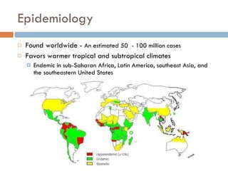
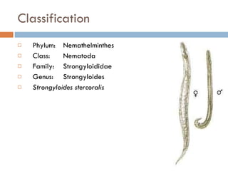
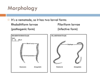
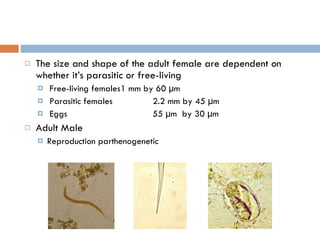
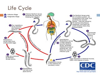
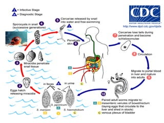
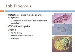
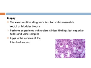
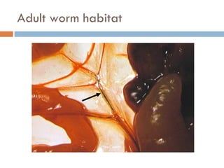
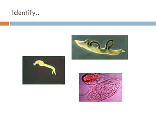
STRONGYLOIDES ‘STRONGYLOIDOSIS’ SCHISTOSOMA ‘SCHISTOSOMIASIS’ Dr Kamran Afzal,  Asst Prof Microbiology
Strongyloides stercoralis   (Threadworm)
Epidemiology Found worldwide -  An estimated 50  - 100 million cases Favors warmer tropical and subtropical climates Endemic in sub-Saharan Africa, Latin America, southeast Asia, and the southeastern United States
Worms can be free-living in the soil or live in a host Only females are parasitic The definitive host is human, but may also affect other primates and dogs Habitat Parasitic adult in duodenum and jejunum of man  Larvae not eggs are passed in human faeces Infective larvae found in soil, intestine and perianal skin Mode of Transmission Skin penetration by larva
Classification Phylum:  Nemathelminthes Class: Nematoda Family:   Strongyloididae Genus:  Strongyloides Strongyloides stercoralis
Morphology It’s a nematode, so it has two larval forms Rhabditiform larvae Filariform larvae (pathogenic form) (infective form)
The size and shape of the adult female are dependent on whether it’s parasitic or free-living Free-living females 1 mm by 60 µm Parasitic females 2.2 mm by 45 µm Eggs 55 µm  by 30 µm Adult Male Reproduction parthenogenetic
Life Cycle
Strongyloidiasis Strongyloides stercoralis has a direct parasitic life cycle, meaning it can complete its entire life cycle in the definitive host This causes an  autoinfection  in the human, because the worm keeps infecting them without ever leaving Disseminated strongyloidiasis, or  hyperinfection Immunocomprised, when the worms spread throughout the body Massive larval invasion in tissues e.g. lungs and serous cavities Leading to sepsis and secondary bacterial infections Hyperinfection has an 85% fatality rate
Symptoms and Signs of Hyperinfection Diarrhea Pleuritic pain Peripheral eosinophilia Severe generalized abdominal pain, diffuse pulmonary infiltrates, ileus, shock, sepsis may occur
Symptoms Dependent on the stage in the worm’s life cycle Stage Site Symptoms egg intestine --- rhabditiform intestine, soil --- (“free living”) filariform soil ground itch larva currens lungs wheezing, cough hemoptysis! eosinophilia adult intestines abdominal pain ulcers diarrhea/constipation weight loss bowel obstruction malabsorption
1/3 of patients are asymptomatic Ground itch Pruritic papulovesicular cutaneous eruption Larva Currens (“racing larvae”) Pathognomonic rash Pruritic wheal or linear urticaria Creeps 5-15 cm/hr!
Lab Diagnosis Direct microscopy Rhabditiform larvae in stool Larvae are seen in stool approximately 1 month after skin penetration >90% sensitivity for stool samples if 7 or more samples are examined Eggs Larvae in sputum sample Serology   ovo
With hyperinfection Filariform larvae may also be recovered in stool if fixed rapidly Sputum samples Bronchioalveolar lavage Urinalysis Semen analysis CSF analysis NO eosinophilia!
Features of Rhabditiform larvae Actively motile 200-250µm x 16 µm in size Short buccal cavity Rhabditiform large bulbed esophagus
Microscopic (biopsy) findings Intestinal biopsy shows adult worms, eggs and larvae Lungs and other organs show only larvae
Treatment Ivermectin (drug of choice) 200 mcg/kg/d PO for 2 days Binds to chloride ion channels Causes hyperpolarization  ->  paralysis Affects adults only… Cure rate of up to 97% Albendazole (2nd line drug) 400 mg BID x 7 days Thiabendazole (2nd line drug before) Hyperinfection - treatment for 2-3 weeks may be life saving but the mortality is very high
Prevention Properly dispose of human wastes Wear Shoes … Don’t eat dirt
Schistosoma  species ‘Bilharziasis’
Epidemiology Second most prevalent tropical parasitic disease in the world (behind Malaria only) 200 M people in 74 countries 120 M have symptoms 20 M have severe illness Between 200,000 and 800,000 deaths/year Described by Theodore Bilharz in Cairo in 1851
The disease is often associated with water resource development projects, such as dams and irrigation schemes, fresh water reservoirs, artificial lakes, cultivation of rice, where the snail intermediate hosts of the parasite breed
Etiology The Schistosomes are blood trematodes (blood flukes) belonging to the Phylum Platyhelmintha Human disease caused mainly by 3 species of flat worms S. haematobium : affects 54 countries in Africa and the Middle East, urinary disease S. mansoni : most common (Africa), intestinal disease S. japonicum : Asia-Pacific, intestinal disease
Transmission: direct penetration of skin by fork-tailed cercaria in water Pathogenic potential: high, based on worm populations and location in veins, capability of eggs to erode tissue
 
Schistosomiasis S. haematobium  (urogenital) In urinary schistosomiasis damage to the urinary tract is done by schistosome eggs in the urine Bladder cancer is common in advanced cases Macrohematuria
S. mansoni, S. japonicum In intestinal schistosomiasis disease is slower to develop Progressive enlargement of the liver and spleen
They differ from other trematodes in that they have separate sexes The schistosomes remain in copula throughout their life span, the male surrounding and holding the female within its gynephoric canal
Clinical presentation of severe schistosomiasis Portal hypertension Hepatosplenic shunt Esophageal varices Hemorrhages
Acute Schistosomiasis Katayama Fever Fever, headache, generalized myalgias Right upper quadrant pain, tender hepatomegaly Bloody diarrhea Respiratory symptoms: 70%  S. mansoni Interstitial pneumonia (radiologic) Splenomegaly: 30% Aseptic meningitis
Lab Diagnosis Detection of eggs in stools or urine: Diagnostic 3 specimens may be needed: Intermittent shedding CBC with eosinophilia Anemia Fe-deficiency Anemia of chronic disease Marocytic
Biopsy The most sensitive diagnostic test for schistosomiasis is  rectal or bladder biopsy Perform on patients with typical clinical findings but negative feces and urine samples Eggs in the venules of the intestinal mucosa
Serology Useful in specific circumstances Useful in pts with no eggs: Katayama fever Commercially available assays: Less sensitive and specific than multiple stool sample exams Immunoblot assays to detect circulating egg or worm Ag: highly sensitive and specific
Treatment The only way to reduce disease symptoms Praziquantel: effective in a single dose against all species Oxamniquine: effective in a single dose, but only against  S. mansoni
Prevention and control Educate people to not urinate or defecate in fresh water supplies  Eliminate snail vectors by making the water habitat unsuitable (increase water flow, remove vegetation) Provision of safe, adequate water supply and sanitation Provide piped water to avoid direct contact with cercariae Snail control through focal mollusciciding Mass drug treatment of communities to reduce reservoir of infection
Adult worm habitat
Identify..

Strongyloides schistosoma

  • 1.
    STRONGYLOIDES ‘STRONGYLOIDOSIS’ SCHISTOSOMA‘SCHISTOSOMIASIS’ Dr Kamran Afzal, Asst Prof Microbiology
  • 2.
  • 3.
    Epidemiology Found worldwide- An estimated 50 - 100 million cases Favors warmer tropical and subtropical climates Endemic in sub-Saharan Africa, Latin America, southeast Asia, and the southeastern United States
  • 4.
    Worms can befree-living in the soil or live in a host Only females are parasitic The definitive host is human, but may also affect other primates and dogs Habitat Parasitic adult in duodenum and jejunum of man Larvae not eggs are passed in human faeces Infective larvae found in soil, intestine and perianal skin Mode of Transmission Skin penetration by larva
  • 5.
    Classification Phylum: Nemathelminthes Class: Nematoda Family: Strongyloididae Genus: Strongyloides Strongyloides stercoralis
  • 6.
    Morphology It’s anematode, so it has two larval forms Rhabditiform larvae Filariform larvae (pathogenic form) (infective form)
  • 7.
    The size andshape of the adult female are dependent on whether it’s parasitic or free-living Free-living females 1 mm by 60 µm Parasitic females 2.2 mm by 45 µm Eggs 55 µm by 30 µm Adult Male Reproduction parthenogenetic
  • 8.
  • 9.
    Strongyloidiasis Strongyloides stercoralishas a direct parasitic life cycle, meaning it can complete its entire life cycle in the definitive host This causes an autoinfection in the human, because the worm keeps infecting them without ever leaving Disseminated strongyloidiasis, or hyperinfection Immunocomprised, when the worms spread throughout the body Massive larval invasion in tissues e.g. lungs and serous cavities Leading to sepsis and secondary bacterial infections Hyperinfection has an 85% fatality rate
  • 10.
    Symptoms and Signsof Hyperinfection Diarrhea Pleuritic pain Peripheral eosinophilia Severe generalized abdominal pain, diffuse pulmonary infiltrates, ileus, shock, sepsis may occur
  • 11.
    Symptoms Dependent onthe stage in the worm’s life cycle Stage Site Symptoms egg intestine --- rhabditiform intestine, soil --- (“free living”) filariform soil ground itch larva currens lungs wheezing, cough hemoptysis! eosinophilia adult intestines abdominal pain ulcers diarrhea/constipation weight loss bowel obstruction malabsorption
  • 12.
    1/3 of patientsare asymptomatic Ground itch Pruritic papulovesicular cutaneous eruption Larva Currens (“racing larvae”) Pathognomonic rash Pruritic wheal or linear urticaria Creeps 5-15 cm/hr!
  • 13.
    Lab Diagnosis Directmicroscopy Rhabditiform larvae in stool Larvae are seen in stool approximately 1 month after skin penetration >90% sensitivity for stool samples if 7 or more samples are examined Eggs Larvae in sputum sample Serology ovo
  • 14.
    With hyperinfection Filariformlarvae may also be recovered in stool if fixed rapidly Sputum samples Bronchioalveolar lavage Urinalysis Semen analysis CSF analysis NO eosinophilia!
  • 15.
    Features of Rhabditiformlarvae Actively motile 200-250µm x 16 µm in size Short buccal cavity Rhabditiform large bulbed esophagus
  • 16.
    Microscopic (biopsy) findingsIntestinal biopsy shows adult worms, eggs and larvae Lungs and other organs show only larvae
  • 17.
    Treatment Ivermectin (drugof choice) 200 mcg/kg/d PO for 2 days Binds to chloride ion channels Causes hyperpolarization -> paralysis Affects adults only… Cure rate of up to 97% Albendazole (2nd line drug) 400 mg BID x 7 days Thiabendazole (2nd line drug before) Hyperinfection - treatment for 2-3 weeks may be life saving but the mortality is very high
  • 18.
    Prevention Properly disposeof human wastes Wear Shoes … Don’t eat dirt
  • 19.
    Schistosoma species‘Bilharziasis’
  • 20.
    Epidemiology Second mostprevalent tropical parasitic disease in the world (behind Malaria only) 200 M people in 74 countries 120 M have symptoms 20 M have severe illness Between 200,000 and 800,000 deaths/year Described by Theodore Bilharz in Cairo in 1851
  • 21.
    The disease isoften associated with water resource development projects, such as dams and irrigation schemes, fresh water reservoirs, artificial lakes, cultivation of rice, where the snail intermediate hosts of the parasite breed
  • 22.
    Etiology The Schistosomesare blood trematodes (blood flukes) belonging to the Phylum Platyhelmintha Human disease caused mainly by 3 species of flat worms S. haematobium : affects 54 countries in Africa and the Middle East, urinary disease S. mansoni : most common (Africa), intestinal disease S. japonicum : Asia-Pacific, intestinal disease
  • 23.
    Transmission: direct penetrationof skin by fork-tailed cercaria in water Pathogenic potential: high, based on worm populations and location in veins, capability of eggs to erode tissue
  • 24.
  • 25.
    Schistosomiasis S. haematobium (urogenital) In urinary schistosomiasis damage to the urinary tract is done by schistosome eggs in the urine Bladder cancer is common in advanced cases Macrohematuria
  • 26.
    S. mansoni, S.japonicum In intestinal schistosomiasis disease is slower to develop Progressive enlargement of the liver and spleen
  • 27.
    They differ fromother trematodes in that they have separate sexes The schistosomes remain in copula throughout their life span, the male surrounding and holding the female within its gynephoric canal
  • 28.
    Clinical presentation ofsevere schistosomiasis Portal hypertension Hepatosplenic shunt Esophageal varices Hemorrhages
  • 29.
    Acute Schistosomiasis KatayamaFever Fever, headache, generalized myalgias Right upper quadrant pain, tender hepatomegaly Bloody diarrhea Respiratory symptoms: 70% S. mansoni Interstitial pneumonia (radiologic) Splenomegaly: 30% Aseptic meningitis
  • 30.
    Lab Diagnosis Detectionof eggs in stools or urine: Diagnostic 3 specimens may be needed: Intermittent shedding CBC with eosinophilia Anemia Fe-deficiency Anemia of chronic disease Marocytic
  • 31.
    Biopsy The mostsensitive diagnostic test for schistosomiasis is rectal or bladder biopsy Perform on patients with typical clinical findings but negative feces and urine samples Eggs in the venules of the intestinal mucosa
  • 32.
    Serology Useful inspecific circumstances Useful in pts with no eggs: Katayama fever Commercially available assays: Less sensitive and specific than multiple stool sample exams Immunoblot assays to detect circulating egg or worm Ag: highly sensitive and specific
  • 33.
    Treatment The onlyway to reduce disease symptoms Praziquantel: effective in a single dose against all species Oxamniquine: effective in a single dose, but only against S. mansoni
  • 34.
    Prevention and controlEducate people to not urinate or defecate in fresh water supplies Eliminate snail vectors by making the water habitat unsuitable (increase water flow, remove vegetation) Provision of safe, adequate water supply and sanitation Provide piped water to avoid direct contact with cercariae Snail control through focal mollusciciding Mass drug treatment of communities to reduce reservoir of infection
  • 35.
  • 36.