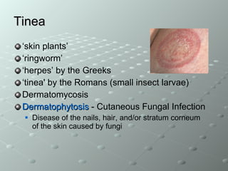
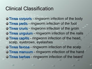
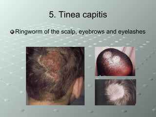
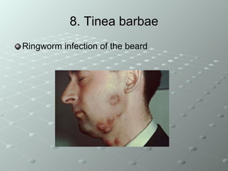
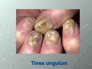
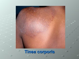
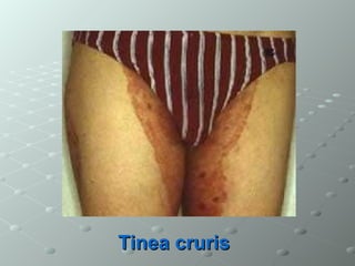
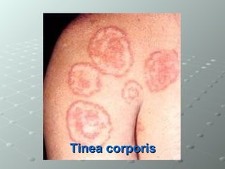
Tinea – The Dermatophytes Dr Kamran Afzal Asst Prof Microbiology
Tinea ‘ skin plants’ ‘ ringworm’ ‘ herpes’ by the Greeks ‘ tinea' by the Romans (small insect larvae) Dermatomycosis Dermatophytosis  -  Cutaneous Fungal Infection Disease of the nails, hair, and/or stratum corneum of the skin caused by fungi
Three important genera  Trichophyton  - Skin, hair, nail Epidermophyton  - Skin, nail Microsporum  - Skin, hair
Clinical Classification Tinea corporis   - ringworm infection of the body   Tinea pedis   - ringworm infection of the foot Tinea cruris   - ringworm infection of the groin   Tinea unguium   - ringworm infection of the nails   Tinea capitis   - ringworm infection of the head, scalp, eyebrows, eyelashes   Tinea favosa   - ringworm infection of the scalp   Tinea manuum   - ringworm infection of the hand   Tinea barbae   - ringworm infection of the beard
1. Tinea corporis Body ringworm Circular rash Does not fade Can appear on all surfaces Spreads by direct contact
CLINICAL MANIFESTATIONS Concentric or ring-like lesions   on skin Generally restricted to stratum corneum of the smooth skin In severe cases these are raised and may become inflamed
Tinea corporis resolves itself in several months Symptoms result from fungal metabolites such as toxin/allergens
 
2. Tinea pedis Athlete’s Foot Three causal agents T. rubrum source are people with chronic infections, because fungus not long-lived in squames T. mentagrophytes Epidermophyton floccosum source are long-lived arthrospores, that reside in squames deposited in rugs and carpets (fomites)
CLINICAL MANIFESTATIONS Between toes or toe webs (releasing of clear fluid) 4th and 5th toes are most common In one study - 85 % of college students carried a ringworm fungus Soreness and itching of any part of the foot
3. Tinea cruris Jock Itch Ringworm infection of the groin, scrotum and inner thigh Causes of Tinea cruris include  T. rubrum T. mentagrophytes E. flocossum
CLINICAL MANIFESTATIONS More common in men than women Predisposing factors include persistent perspiration, high humidity, irritation of skin from clothes, such as tight fitting underwear or athletic supporters, pre-existing disease such as diabetes and obesity
4. Tinea unguium Nail Infection Most commonly caused by  T. rubrum E. floccosum Trichophyton  species Can follow Tinea pedis
Nail changes Begins distal    proximal Toes > fingers Thickening Yellowing Onycholysis, crumbling Associated Tinea pedis
5. Tinea capitis Ringworm of the scalp, eyebrows and eyelashes
CLINICAL MANIFESTATIONS Caused by species of Microsporum  and  Trichophyton Fungus grows into hair follicle Tinea capitis is very contagious and can be transmitted by people, animals or objects carrying the fungus It always requires systemic medication - griseofulvin Fungistatic agents are somewhat effective (miconazole, clotrimazole)
Alopecia in affected areas Endothrix invasion of hair shaft   Using a  Wood's lamp  on hair Microsporum  species tend to fluoresce green Trichophyton  species generally do not fluoresce
The clinical hallmark is single or multiple patches of hair loss, sometimes with a “black dot” pattern, which may be accompanied by signs of inflammation such as scaling, pustules and itching
6. Tinea favosa Ringworm infection of the scalp (crusty hair)
7. Tinea manuum Ringworm infection of the hand
8. Tinea barbae Ringworm   infection of the beard
Transmission Close human contact (Direct) Sharing clothes, combs, brushes, towels, bedsheets... (Indirect) Animal-to-human contact (Zoophilic)
Pathology Dermatophytes use keratin as a source of nutrition Therefore they infect skin, hair, nails All 3 organisms infect /attack skin Microsporum  does not infect nails Epidermophyton  does not infect hair, they do not invade underlying non-keratinized tissues
Clinical Significance DERMATOPHYTOSES Characterized by  Itching, scaling of skin patches that can become inflamed and weeping Infection in different sites may be due to different organisms but is given one name
Clinical manifestations Skin:   Circular, dry, erythematous, scaly, itchy lesions  Hair:   Typical lesions, ”kerion”, scarring, “alopecia” Nail:  Thickened, deformed, friable, discolored nails, sub-ungual debris accumulation  Favus   (Tinea favosa)
Lab Diagnosis Nail clippings, skin scrapings, hair /follicle Placed in sterile container preferably, or between 2 slides No role for swabs KOH   ( 10-25% )   will be added in the lab to dissolve tissue material Lactophenol blue stain   to see if fungal hyphae seen
For full identification culture   on  selective media  required  Sabouraud dextrose agar (SDA) SDA with cycloheximide or chloramphenicol Low pH 5.0 May require  10-14  days for growth Macroscopic and microscopic identification of colonies
T. mentagrophytes
Treatment Topical  Miconazole, clotrimazole, econazole, terbinafine... Oral Griseofulvin Ketaconazole Itraconazole Terbinafine
Superficial Mycoses Reddish spots if fair skinned  Caused by  Malazzesia furfur   “ Spaghetti Meatballs”  appearance in Lactophenol Cotton Blue stain Tinea versicolor Treatment Ketoconazole, fluconazole, itraconazole
QUIZ..
Tinea pedis
Tinea unguium
Tinea corporis
Tinea cruris
Tinea barbae
Tinea corporis

Tinea dermatophytes

  • 1.
    Tinea – TheDermatophytes Dr Kamran Afzal Asst Prof Microbiology
  • 2.
    Tinea ‘ skinplants’ ‘ ringworm’ ‘ herpes’ by the Greeks ‘ tinea' by the Romans (small insect larvae) Dermatomycosis Dermatophytosis - Cutaneous Fungal Infection Disease of the nails, hair, and/or stratum corneum of the skin caused by fungi
  • 3.
    Three important genera Trichophyton - Skin, hair, nail Epidermophyton - Skin, nail Microsporum - Skin, hair
  • 4.
    Clinical Classification Tineacorporis - ringworm infection of the body Tinea pedis - ringworm infection of the foot Tinea cruris - ringworm infection of the groin Tinea unguium - ringworm infection of the nails Tinea capitis - ringworm infection of the head, scalp, eyebrows, eyelashes Tinea favosa - ringworm infection of the scalp Tinea manuum - ringworm infection of the hand Tinea barbae - ringworm infection of the beard
  • 5.
    1. Tinea corporisBody ringworm Circular rash Does not fade Can appear on all surfaces Spreads by direct contact
  • 6.
    CLINICAL MANIFESTATIONS Concentricor ring-like lesions on skin Generally restricted to stratum corneum of the smooth skin In severe cases these are raised and may become inflamed
  • 7.
    Tinea corporis resolvesitself in several months Symptoms result from fungal metabolites such as toxin/allergens
  • 8.
  • 9.
    2. Tinea pedisAthlete’s Foot Three causal agents T. rubrum source are people with chronic infections, because fungus not long-lived in squames T. mentagrophytes Epidermophyton floccosum source are long-lived arthrospores, that reside in squames deposited in rugs and carpets (fomites)
  • 10.
    CLINICAL MANIFESTATIONS Betweentoes or toe webs (releasing of clear fluid) 4th and 5th toes are most common In one study - 85 % of college students carried a ringworm fungus Soreness and itching of any part of the foot
  • 11.
    3. Tinea crurisJock Itch Ringworm infection of the groin, scrotum and inner thigh Causes of Tinea cruris include T. rubrum T. mentagrophytes E. flocossum
  • 12.
    CLINICAL MANIFESTATIONS Morecommon in men than women Predisposing factors include persistent perspiration, high humidity, irritation of skin from clothes, such as tight fitting underwear or athletic supporters, pre-existing disease such as diabetes and obesity
  • 13.
    4. Tinea unguiumNail Infection Most commonly caused by T. rubrum E. floccosum Trichophyton species Can follow Tinea pedis
  • 14.
    Nail changes Beginsdistal  proximal Toes > fingers Thickening Yellowing Onycholysis, crumbling Associated Tinea pedis
  • 15.
    5. Tinea capitisRingworm of the scalp, eyebrows and eyelashes
  • 16.
    CLINICAL MANIFESTATIONS Causedby species of Microsporum and Trichophyton Fungus grows into hair follicle Tinea capitis is very contagious and can be transmitted by people, animals or objects carrying the fungus It always requires systemic medication - griseofulvin Fungistatic agents are somewhat effective (miconazole, clotrimazole)
  • 17.
    Alopecia in affectedareas Endothrix invasion of hair shaft Using a Wood's lamp on hair Microsporum species tend to fluoresce green Trichophyton species generally do not fluoresce
  • 18.
    The clinical hallmarkis single or multiple patches of hair loss, sometimes with a “black dot” pattern, which may be accompanied by signs of inflammation such as scaling, pustules and itching
  • 19.
    6. Tinea favosaRingworm infection of the scalp (crusty hair)
  • 20.
    7. Tinea manuumRingworm infection of the hand
  • 21.
    8. Tinea barbaeRingworm infection of the beard
  • 22.
    Transmission Close humancontact (Direct) Sharing clothes, combs, brushes, towels, bedsheets... (Indirect) Animal-to-human contact (Zoophilic)
  • 23.
    Pathology Dermatophytes usekeratin as a source of nutrition Therefore they infect skin, hair, nails All 3 organisms infect /attack skin Microsporum does not infect nails Epidermophyton does not infect hair, they do not invade underlying non-keratinized tissues
  • 24.
    Clinical Significance DERMATOPHYTOSESCharacterized by Itching, scaling of skin patches that can become inflamed and weeping Infection in different sites may be due to different organisms but is given one name
  • 25.
    Clinical manifestations Skin: Circular, dry, erythematous, scaly, itchy lesions Hair: Typical lesions, ”kerion”, scarring, “alopecia” Nail: Thickened, deformed, friable, discolored nails, sub-ungual debris accumulation Favus (Tinea favosa)
  • 26.
    Lab Diagnosis Nailclippings, skin scrapings, hair /follicle Placed in sterile container preferably, or between 2 slides No role for swabs KOH ( 10-25% ) will be added in the lab to dissolve tissue material Lactophenol blue stain to see if fungal hyphae seen
  • 27.
    For full identificationculture on selective media required Sabouraud dextrose agar (SDA) SDA with cycloheximide or chloramphenicol Low pH 5.0 May require 10-14 days for growth Macroscopic and microscopic identification of colonies
  • 28.
  • 29.
    Treatment Topical Miconazole, clotrimazole, econazole, terbinafine... Oral Griseofulvin Ketaconazole Itraconazole Terbinafine
  • 30.
    Superficial Mycoses Reddishspots if fair skinned Caused by Malazzesia furfur “ Spaghetti Meatballs” appearance in Lactophenol Cotton Blue stain Tinea versicolor Treatment Ketoconazole, fluconazole, itraconazole
  • 31.
  • 32.
  • 33.
  • 34.
  • 35.
  • 36.
  • 37.