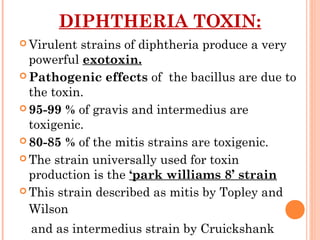
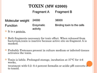
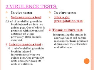
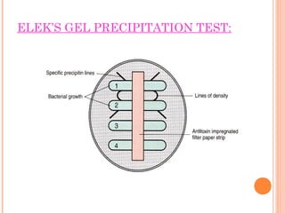
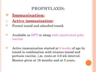
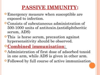
CORYNEBACTERI
UM
DIPHTHERIAE
Dr V S Vatkar
Asso Prof
Microbiology Department
D Y Patil Medical College, Kolhapur
CORYNEBACTERIUM
Kingdom: Bacteria
Phylum: Actinobacteria
Order: Actinomycetales
Family: Corynebacteriaceae
Genus: Corynebacterium
Species: C. diphtheriae
HISTORY:
BRETONNEAU(1826):
RECOGNIZED AS CLINICAL ENTITY, WHO CALLED IT AS
DIPHTHERITE.
 Greek word koryne means club & Diphtheros meaning
leather i e leathery pseudo memb over tonsils.
 Diphthria bacillus first observed & described by
Klebs(1883) & First cultivated by Loeffler(1884): Hence known as
Klebs-Loeffler bacillus.
 Roux & Yersin (1888): show pathogenic effect of diphtheria toxin
 Its antitoxin : described by Von Behring in 1890
CORYNEBACTERIUM
DIPHTHERIAE
 Morphology:
 Slender rod with tendency of clubbing at one or both
ends
 Measuring 3-6µm ×0.6-0.8µm.
 Pleomorphic
 Nonsporing, nonmotile, noncapsulated.
 Cells often show septa, and branching is infrequently
observed.
 Bacilli being at various angles to each other, resembling
the letter V or L Called Chinese letter or
cuneiform arrangement due to bacilli divides
horizontally & daughter cell lie at acute angle to parent
cell (snapping type of division)
 Granules: composed of polymetaphosphotase seen in
the cells & present at the ends or poles of the bacilli,
metachromatic granules also called volutin or Babes
Ernst granules.(polar bodies)
Granules are more strongly Gram positive than rest of
the cell.
Loeffler’s methylene blue: granules take bluish
purple colour
MORPHOLOGY
 Albert’s, Neisser’s and Ponder’s
special stains for demonstrating granules
clearly.
 Well developed in enriched media like
blood agar or Loeffler’s serum slope
GRAM STAINING
ARRANGEMENT OF
BACILLI
STAINING WITH METHYLNE BLUE :
ALBERT STAIN
•CULTURAL CHARACTERISTICS:
 Media:
growth scanty on ordinary media.
Enrichment with blood, serum or egg is necessary for good growth.
Loeffler’s serum slope & tellurite blood agar used for cultivation.
Loeffler’s serum slope Tellurite blood agar
colonies within 6-8 hrs -small, colonies takes 2 days to appear
circular white opaque discs, enlarge gray or black colonies
on incubation acquire yellow tint reduce tellurite to metallic
tellurium
 Optimum temperature:
for growth is 37° (15-40°C)
 Optimum pH: 7.2
 Aerobe and facultative anaerobe
 Modifications such as McLeod’s and Hoyle’s media have been utilised
•A medium containing tellurite should be used to select for
Corynebacterium and other Gm+ organisms. It inhibits
Gm­ organisms.
•Two kinds are used:
•Cystine tellurite ­ has a longer shelf life
•Tinsdale ­ helps to differentiate amongst the
Corynebacterium.
Colonies on either appear black or gray due to
tellurite reduction.
S. aureus and Listeria also grow as black colonies.
Black colonies on potassium tellurite medium
LOEFFLER’S SERUM SLOPE
 Based on colony morphology on tellurite
medium & other properties, McLeod classified
diphtheria bacilli into three types:
 Gravis: causing most serious disease
 Mitis: the mildest variety
 Intermedius: disease of intermediate severity.
 Gravis and intermedius types are associated with high
case fatality rates.
 Paralytic complications common with gravis,
hemorragic complication are common with gravis and
intermedius.
 Obstructive lesions in air passage in mitis infections.
 Diphtheria bacilli ferment glucose, galactose, maltose
and dextrin producing acid but no gas
 Some strains ferment sucrose.
 Hiss serum water used for testing sugar fermentation.
GravisGravis IntermediusIntermedius MitisMitis
MorphologyMorphology ShortShort
rodsrods,uniform,uniform
staining,pleomo-staining,pleomo-
rphism.rphism.
Long barredLong barred
forms,clubbedforms,clubbed
ends,veryends,very
pleomorphic.pleomorphic.
Long,curved,Long,curved, pleple
omorphicomorphic
rods,prominentrods,prominent
granules.granules.
Colony onColony on
tellurite agartellurite agar
In 2-3 days,flatIn 2-3 days,flat
colonies withcolonies with
raised darkraised dark
centrecentre andand
crenated edgecrenated edge
with radialwith radial
striation- ‘striation- ‘daisydaisy
headhead’’ colonycolony
Dull granularDull granular
centre withcentre with
smoother, moresmoother, more
glisteningglistening
peripheryperiphery andand
lighter ring nearlighter ring near
the edge-the edge- ‘‘frog’sfrog’s
egg’egg’ colonycolony
In 2-3In 2-3
days,coloniesdays,colonies
becomebecome flat,flat,withwith
aa centralcentral
elevationelevation
‘‘poached egg’poached egg’
colonycolony
Consistency ofConsistency of
coloniescolonies
Like ‘Like ‘coldcold
margarine’,margarine’,
brittle, move as abrittle, move as a
whole onwhole on
plate,not easilyplate,not easily
picked out orpicked out or
IntermediateIntermediate
between gravisbetween gravis
and mitisand mitis
Soft, buttery andSoft, buttery and
easilyeasily
emulsifiableemulsifiable
TABLE CONTINUED………..
HomolysiHomolysi
ss
variablevariable Non hemolyticNon hemolytic UsuallyUsually
hemolytichemolytic
Growth inGrowth in
brothbroth
Surface pellicleSurface pellicle,,
granulargranular
deposit, little ordeposit, little or
no turbidityno turbidity
Turbidity in 24Turbidity in 24
hourshours, clearing, clearing
in 48 hours, within 48 hours, with
fine granularfine granular
sedimentsediment
Diffuse turbidityDiffuse turbidity
with soft pelliclewith soft pellicle
laterlater
GlycogenGlycogen
andand
starchstarch
fermentatifermentati
onon
PositivePositive NegativeNegative NegativeNegative
DIPHTHERIA TOXIN:
 Virulent strains of diphtheria produce a very
powerful exotoxin.
 Pathogenic effects of the bacillus are due to
the toxin.
 95-99 % of gravis and intermedius are
toxigenic.
 80-85 % of the mitis strains are toxigenic.
 The strain universally used for toxin
production is the ‘park williams 8’ strain
 This strain described as mitis by Topley and
Wilson
and as intermedius strain by Cruickshank
TOXIN (MW 62000)
Fragment AFragment A Fragment BFragment B
Molecular weightMolecular weight 2400024000 3800038000
FunctionFunction EnzymaticEnzymatic
activityactivity
Binding toxin to the cellsBinding toxin to the cells
 It is a protein.
 Both fragments necessary for toxic effect. When released from
bacterium,toxin is inactive because active site on fragment A is
masked.
 Probably Proteases present in culture medium or infected tissues
activates the toxin.
 Toxin is labile. Prolonged storage, incubation at 37°C for 4-6
weeks,
treatment with 0.2- 0.4 percent formalin or acidic pH converts it
to toxoid.
•TOXIN PRODUCTION DEPENDS
UPON:
 Corynephages (tox +):
acts as genetic determinant controlling toxin production.
Non toxigenic strains may be rendered toxigenic by
infecting them with beta phage or some other toxlarger
phage- known as lysogenic conversion.
when bacillus cured of its phage,it loses toxigenic capacity.
 Concentration of iron:
optimum level of iron for toxin production is 0.1 mg per
litre.
Conc. Of 0.5 mg per litre inhibits formation of toxin
ACTION:  Toxin acts by ihibiting
protein synthesis.
 Fragment A inhibits
polypeptide chain
elongation in presence
of nicotinamide
adenine dinucleotide
(NAD) by inactivating
elongation factor 2
(EF2)
RESISTANCE :
 Colonies remain viable for two or more weeks
at 25-30°C.
 Readily destroyed by heat in 10 minutes at
58°C. and in a minute at 100°C.
 Resistant to action of light, dessication and
freezing.
 It has been cultured from dried bits of
pseudomembrane after 14 weeks.
 Remains virulent in blankets, floor dust for 5
weeks.
 Easily destroyed by antiseptics.
 Susceptible to penicillin, erythromycin and
broad spectrum antibiotics.
ANTIGENIC STRUCTURE:
 Antigenically heterogenous.
 Gravis strains classified into 13 types.
 Intermedius into 4 types.
 And mitis into 40 types.
Gravis strains of:
 Types I and III common in Great Britain,
 Type II worldwide,
 Type IV mainly in Egypt,
 Type V in USA.
TYPING
1.Bacteriophage typing: There are 15 bacteriophage types have
been described.
 Types I and III are mitis,
 IV and VI are intermedius,
 VII is avirulent gravis,
 Remainder are virulent gravis.
2.bacteriocin(diphthericin) typing:
3.Bacterial polypeptide analysis:
4.DNA restriction patterns and hybridisation
with DNA probes
PATHOGENESIS:
 Incubation period 3-4
days.
 Carriers have longer
incubation period
 Route of entry: upper
respiratory tract.
 Adhere by pillus
shaft(spa A) and two
small pili(spaB & spaC)
 Grow locally & produce
toxin
SITE OF INFECTION:
1. Faucial: commonest type
2. Laryngeal
3. Nasal
4. Otitic
5. Conjunctival
6. Genital- vulval, vaginal or prepucial.
7. cutaneous
BULL NECK PSEUDOMEMBRANE
CLINICAL TYPES:
 Malignant or hypertoxic:
Severe toxemia with marked adenitis (bull neck)
Death is due to circulatory failure.
 Septic:
leads to ulceration,cellulitis and even gangrene
around pseudomembrane.
 Hemorrhagic:
Bleeding from the edge of the membrane,
epistaxis, conjunctival hemorrhage, purpura and
generalised bleeding tendency.
COMPLICATIONS:
 Asphyxia due to mechanical obstruction
 Acute circulatory failure
 Post diphtheritic paralysis:
seen in 3rd
or 4th
week of disease
palatine or ciliary but not pupillary
 Septic : pneumonia and otitis media
 Relapse in 1% cases.
LABORATORY DIAGNOSIS:
1. Isolation of diphtheria bacillus:
 One or two swabs from lesion collected under vision
 Gram staining, Leishman staining, done.
 Inoculation done on Loeffler’s serum slope, tellurite
blood agar and on ordinary blood agar.
 Colony morphology seen after incubation.
 Colony smear done & methylene blue staining,
neisser or Albert staining done to see metachromatic
granules.
2.VIRULENCE TESTS:
 In vivo tests:
1. Subcutaneous test:
0.8 ml of emulsified growth in
broth injected s.c. into two
guinea pigs. One of which is
protected with 500 units of
antitoxin 18-24 hrs
previously. Both animals
observed.
2. Intracutaneous test :
0 .1 ml of emlsified growth in
broth in injected
intracutaneously in two
guinea pigs. One given 500
units and other given 50
units of antitoxin.
 In vitro tests:
1. Elek’s gel
precipitation test:
2. Tissue culture test:
incorporating the strains in
agar overlay of cell culture
monolayers. Toxin produced
diffuses into the cells below
and kills them.
ELEK’S GEL PRECIPITATION TEST:
PROPHYLAXIS:
 Immunisation:
1. Active immunisation:
 Formol toxoid and adsorbed toxoid.
 Available as DPT or along with inactivated polio
vaccine
 Active immunisation started at 6 weeks of age by
toxoid in combination with tetanus toxoid and
pertusis vaccine. i.m. route.at 4-6 wk interval.
Booster given at 18 months and at 5 years.
PASSIVE IMMUNITY:
 Emergency measure when susceptible are
exposed to infection.
 Consists of subcutaneous administration of
500-1000 units of antitoxin.(antidiphtheritic
serum, ADS)
 This is horse serum, precaution against
hypersensitivity should be observed.
Combined immunisation:
 Administration of first dose of adsorbed toxoid
in one arm, while ADS is given in other arm.
 Followed by full course of active immunisation.
TREATMENT :
 Antitoxic and antibiotic therapy.
 Antitoxin given immediately in suspected cases.
Recommended dose is 20,000 to 1,00,000 units for
serious cases.
 Half dose if given i.v.
 C. diphtheriae sensitive to penicillin and can be
cleared from throat within few days of treatment.
 Erythromycin effective in carriers.
OTHER PATHOGENIC
CORYNEBACTERIA:
 C. ulcerance:
can cause diphtheria like lesion.
it liquefies gelatin, ferments trehalos, does not reduce
nitrate to nitrite.
 Arcanobacterium hemolyticum cause pharyngitis, skin
ulcers.
 C. Jakeium cause cutaneous and blood infections
 Corynebacteria causing superficial skin
infections:
 C. minutissimum
 C.tenuis
DIPHTHEROIDS:
 Normal commensals in throat, skin,
conjunctiva and other areas.
 e.g. c.pseudodiphtheriticum.
C. xerosis.
Corynebacterium

Corynebacterium

  • 1.
    CORYNEBACTERI UM DIPHTHERIAE Dr V SVatkar Asso Prof Microbiology Department D Y Patil Medical College, Kolhapur
  • 2.
    CORYNEBACTERIUM Kingdom: Bacteria Phylum: Actinobacteria Order:Actinomycetales Family: Corynebacteriaceae Genus: Corynebacterium Species: C. diphtheriae
  • 3.
    HISTORY: BRETONNEAU(1826): RECOGNIZED AS CLINICALENTITY, WHO CALLED IT AS DIPHTHERITE.  Greek word koryne means club & Diphtheros meaning leather i e leathery pseudo memb over tonsils.  Diphthria bacillus first observed & described by Klebs(1883) & First cultivated by Loeffler(1884): Hence known as Klebs-Loeffler bacillus.  Roux & Yersin (1888): show pathogenic effect of diphtheria toxin  Its antitoxin : described by Von Behring in 1890
  • 4.
    CORYNEBACTERIUM DIPHTHERIAE  Morphology:  Slenderrod with tendency of clubbing at one or both ends  Measuring 3-6µm ×0.6-0.8µm.  Pleomorphic  Nonsporing, nonmotile, noncapsulated.  Cells often show septa, and branching is infrequently observed.
  • 5.
     Bacilli beingat various angles to each other, resembling the letter V or L Called Chinese letter or cuneiform arrangement due to bacilli divides horizontally & daughter cell lie at acute angle to parent cell (snapping type of division)  Granules: composed of polymetaphosphotase seen in the cells & present at the ends or poles of the bacilli, metachromatic granules also called volutin or Babes Ernst granules.(polar bodies) Granules are more strongly Gram positive than rest of the cell. Loeffler’s methylene blue: granules take bluish purple colour MORPHOLOGY
  • 6.
     Albert’s, Neisser’sand Ponder’s special stains for demonstrating granules clearly.  Well developed in enriched media like blood agar or Loeffler’s serum slope
  • 7.
  • 8.
  • 9.
  • 10.
    •CULTURAL CHARACTERISTICS:  Media: growthscanty on ordinary media. Enrichment with blood, serum or egg is necessary for good growth. Loeffler’s serum slope & tellurite blood agar used for cultivation. Loeffler’s serum slope Tellurite blood agar colonies within 6-8 hrs -small, colonies takes 2 days to appear circular white opaque discs, enlarge gray or black colonies on incubation acquire yellow tint reduce tellurite to metallic tellurium  Optimum temperature: for growth is 37° (15-40°C)  Optimum pH: 7.2  Aerobe and facultative anaerobe  Modifications such as McLeod’s and Hoyle’s media have been utilised
  • 11.
    •A medium containingtellurite should be used to select for Corynebacterium and other Gm+ organisms. It inhibits Gm­ organisms. •Two kinds are used: •Cystine tellurite ­ has a longer shelf life •Tinsdale ­ helps to differentiate amongst the Corynebacterium. Colonies on either appear black or gray due to tellurite reduction. S. aureus and Listeria also grow as black colonies.
  • 12.
    Black colonies onpotassium tellurite medium
  • 13.
  • 15.
     Based oncolony morphology on tellurite medium & other properties, McLeod classified diphtheria bacilli into three types:  Gravis: causing most serious disease  Mitis: the mildest variety  Intermedius: disease of intermediate severity.
  • 16.
     Gravis andintermedius types are associated with high case fatality rates.  Paralytic complications common with gravis, hemorragic complication are common with gravis and intermedius.  Obstructive lesions in air passage in mitis infections.  Diphtheria bacilli ferment glucose, galactose, maltose and dextrin producing acid but no gas  Some strains ferment sucrose.  Hiss serum water used for testing sugar fermentation.
  • 17.
    GravisGravis IntermediusIntermedius MitisMitis MorphologyMorphologyShortShort rodsrods,uniform,uniform staining,pleomo-staining,pleomo- rphism.rphism. Long barredLong barred forms,clubbedforms,clubbed ends,veryends,very pleomorphic.pleomorphic. Long,curved,Long,curved, pleple omorphicomorphic rods,prominentrods,prominent granules.granules. Colony onColony on tellurite agartellurite agar In 2-3 days,flatIn 2-3 days,flat colonies withcolonies with raised darkraised dark centrecentre andand crenated edgecrenated edge with radialwith radial striation- ‘striation- ‘daisydaisy headhead’’ colonycolony Dull granularDull granular centre withcentre with smoother, moresmoother, more glisteningglistening peripheryperiphery andand lighter ring nearlighter ring near the edge-the edge- ‘‘frog’sfrog’s egg’egg’ colonycolony In 2-3In 2-3 days,coloniesdays,colonies becomebecome flat,flat,withwith aa centralcentral elevationelevation ‘‘poached egg’poached egg’ colonycolony Consistency ofConsistency of coloniescolonies Like ‘Like ‘coldcold margarine’,margarine’, brittle, move as abrittle, move as a whole onwhole on plate,not easilyplate,not easily picked out orpicked out or IntermediateIntermediate between gravisbetween gravis and mitisand mitis Soft, buttery andSoft, buttery and easilyeasily emulsifiableemulsifiable
  • 18.
    TABLE CONTINUED……….. HomolysiHomolysi ss variablevariable NonhemolyticNon hemolytic UsuallyUsually hemolytichemolytic Growth inGrowth in brothbroth Surface pellicleSurface pellicle,, granulargranular deposit, little ordeposit, little or no turbidityno turbidity Turbidity in 24Turbidity in 24 hourshours, clearing, clearing in 48 hours, within 48 hours, with fine granularfine granular sedimentsediment Diffuse turbidityDiffuse turbidity with soft pelliclewith soft pellicle laterlater GlycogenGlycogen andand starchstarch fermentatifermentati onon PositivePositive NegativeNegative NegativeNegative
  • 19.
    DIPHTHERIA TOXIN:  Virulentstrains of diphtheria produce a very powerful exotoxin.  Pathogenic effects of the bacillus are due to the toxin.  95-99 % of gravis and intermedius are toxigenic.  80-85 % of the mitis strains are toxigenic.  The strain universally used for toxin production is the ‘park williams 8’ strain  This strain described as mitis by Topley and Wilson and as intermedius strain by Cruickshank
  • 20.
    TOXIN (MW 62000) FragmentAFragment A Fragment BFragment B Molecular weightMolecular weight 2400024000 3800038000 FunctionFunction EnzymaticEnzymatic activityactivity Binding toxin to the cellsBinding toxin to the cells  It is a protein.  Both fragments necessary for toxic effect. When released from bacterium,toxin is inactive because active site on fragment A is masked.  Probably Proteases present in culture medium or infected tissues activates the toxin.  Toxin is labile. Prolonged storage, incubation at 37°C for 4-6 weeks, treatment with 0.2- 0.4 percent formalin or acidic pH converts it to toxoid.
  • 21.
    •TOXIN PRODUCTION DEPENDS UPON: Corynephages (tox +): acts as genetic determinant controlling toxin production. Non toxigenic strains may be rendered toxigenic by infecting them with beta phage or some other toxlarger phage- known as lysogenic conversion. when bacillus cured of its phage,it loses toxigenic capacity.  Concentration of iron: optimum level of iron for toxin production is 0.1 mg per litre. Conc. Of 0.5 mg per litre inhibits formation of toxin
  • 22.
    ACTION:  Toxinacts by ihibiting protein synthesis.  Fragment A inhibits polypeptide chain elongation in presence of nicotinamide adenine dinucleotide (NAD) by inactivating elongation factor 2 (EF2)
  • 24.
    RESISTANCE :  Coloniesremain viable for two or more weeks at 25-30°C.  Readily destroyed by heat in 10 minutes at 58°C. and in a minute at 100°C.  Resistant to action of light, dessication and freezing.  It has been cultured from dried bits of pseudomembrane after 14 weeks.  Remains virulent in blankets, floor dust for 5 weeks.  Easily destroyed by antiseptics.  Susceptible to penicillin, erythromycin and broad spectrum antibiotics.
  • 25.
    ANTIGENIC STRUCTURE:  Antigenicallyheterogenous.  Gravis strains classified into 13 types.  Intermedius into 4 types.  And mitis into 40 types. Gravis strains of:  Types I and III common in Great Britain,  Type II worldwide,  Type IV mainly in Egypt,  Type V in USA.
  • 26.
    TYPING 1.Bacteriophage typing: Thereare 15 bacteriophage types have been described.  Types I and III are mitis,  IV and VI are intermedius,  VII is avirulent gravis,  Remainder are virulent gravis. 2.bacteriocin(diphthericin) typing: 3.Bacterial polypeptide analysis: 4.DNA restriction patterns and hybridisation with DNA probes
  • 27.
    PATHOGENESIS:  Incubation period3-4 days.  Carriers have longer incubation period  Route of entry: upper respiratory tract.  Adhere by pillus shaft(spa A) and two small pili(spaB & spaC)  Grow locally & produce toxin
  • 28.
    SITE OF INFECTION: 1.Faucial: commonest type 2. Laryngeal 3. Nasal 4. Otitic 5. Conjunctival 6. Genital- vulval, vaginal or prepucial. 7. cutaneous
  • 29.
  • 30.
    CLINICAL TYPES:  Malignantor hypertoxic: Severe toxemia with marked adenitis (bull neck) Death is due to circulatory failure.  Septic: leads to ulceration,cellulitis and even gangrene around pseudomembrane.  Hemorrhagic: Bleeding from the edge of the membrane, epistaxis, conjunctival hemorrhage, purpura and generalised bleeding tendency.
  • 31.
    COMPLICATIONS:  Asphyxia dueto mechanical obstruction  Acute circulatory failure  Post diphtheritic paralysis: seen in 3rd or 4th week of disease palatine or ciliary but not pupillary  Septic : pneumonia and otitis media  Relapse in 1% cases.
  • 32.
    LABORATORY DIAGNOSIS: 1. Isolationof diphtheria bacillus:  One or two swabs from lesion collected under vision  Gram staining, Leishman staining, done.  Inoculation done on Loeffler’s serum slope, tellurite blood agar and on ordinary blood agar.  Colony morphology seen after incubation.  Colony smear done & methylene blue staining, neisser or Albert staining done to see metachromatic granules.
  • 33.
    2.VIRULENCE TESTS:  Invivo tests: 1. Subcutaneous test: 0.8 ml of emulsified growth in broth injected s.c. into two guinea pigs. One of which is protected with 500 units of antitoxin 18-24 hrs previously. Both animals observed. 2. Intracutaneous test : 0 .1 ml of emlsified growth in broth in injected intracutaneously in two guinea pigs. One given 500 units and other given 50 units of antitoxin.  In vitro tests: 1. Elek’s gel precipitation test: 2. Tissue culture test: incorporating the strains in agar overlay of cell culture monolayers. Toxin produced diffuses into the cells below and kills them.
  • 34.
  • 35.
    PROPHYLAXIS:  Immunisation: 1. Activeimmunisation:  Formol toxoid and adsorbed toxoid.  Available as DPT or along with inactivated polio vaccine  Active immunisation started at 6 weeks of age by toxoid in combination with tetanus toxoid and pertusis vaccine. i.m. route.at 4-6 wk interval. Booster given at 18 months and at 5 years.
  • 36.
    PASSIVE IMMUNITY:  Emergencymeasure when susceptible are exposed to infection.  Consists of subcutaneous administration of 500-1000 units of antitoxin.(antidiphtheritic serum, ADS)  This is horse serum, precaution against hypersensitivity should be observed. Combined immunisation:  Administration of first dose of adsorbed toxoid in one arm, while ADS is given in other arm.  Followed by full course of active immunisation.
  • 37.
    TREATMENT :  Antitoxicand antibiotic therapy.  Antitoxin given immediately in suspected cases. Recommended dose is 20,000 to 1,00,000 units for serious cases.  Half dose if given i.v.  C. diphtheriae sensitive to penicillin and can be cleared from throat within few days of treatment.  Erythromycin effective in carriers.
  • 38.
    OTHER PATHOGENIC CORYNEBACTERIA:  C.ulcerance: can cause diphtheria like lesion. it liquefies gelatin, ferments trehalos, does not reduce nitrate to nitrite.  Arcanobacterium hemolyticum cause pharyngitis, skin ulcers.  C. Jakeium cause cutaneous and blood infections  Corynebacteria causing superficial skin infections:  C. minutissimum  C.tenuis
  • 39.
    DIPHTHEROIDS:  Normal commensalsin throat, skin, conjunctiva and other areas.  e.g. c.pseudodiphtheriticum. C. xerosis.