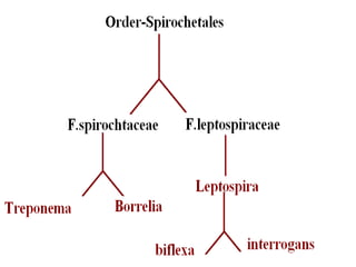
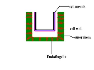
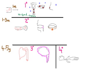
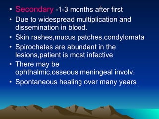
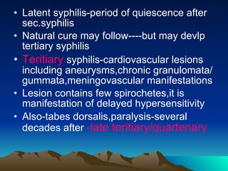
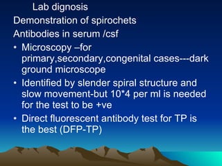
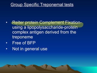
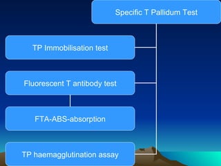
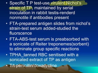
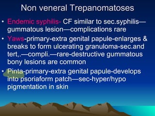
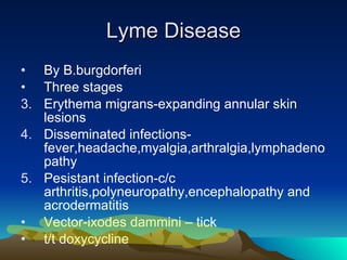
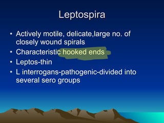
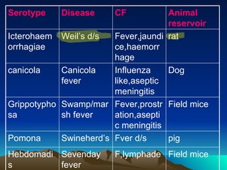
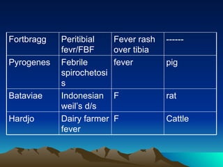
Spirochetes Gram -ve
 
Elongated ,motile,flexible,twisted spirally along the long axisendoflagella-polar,wound around helical potoplastic cylinder—between outer membrane and cell wall.
 
Treponema Short,slender,fine spirals,pointed/round ends Both commensals and pathogens Venereal syphilis-T.pallidum Endemic syphilis-T.pallidum/T.endemicum Yaws-T.pertenue Pinta-T.carateum
Treponma pallidum Pallidum-pale staining Morpho-thin ,delicate,ten regular spirals-sharp and angular(indian ink),motile(dark ground/phase contr)-sec. spirals appear & dis appear-but primary persists Silver impregnation method,fontana’s method,Leviditi’s method(tissue sections) Culture-do not grow in artificial media
 
Virulent strains-maintained by serial testicular passage in rabbits for decades In culture-isolates are mainly non-pathogenic-but similar morph. And antigenic property Best known is  REITER STRAIN  –Tr.phagedenis Imp for specific tr. Test for identification of syphilis
Resistance-delicate,fomites hav no influence in transfer Fever therapy  -killed in 1 to 3 days at 0-4C,so transfusion syphilis can be prevented by storing blood for 4 days Natural infection occurs only in humans
 
Antigenic structure-treponemal infection—three types Reagin-STS/Nonspecific tests-antigen is hapten/cardiolipin(diphosphatidyl glycerol) from beef heart Group antigen-in pathogenic and non pathogenic Species specific-PS  antigen-only positive in patients infected with pathogenic treponems
 
Primary- chancre-at the site of entry Painless, realtively avascular,superficial ,indurated,ulcerated lesion. Hard chancre-/Hunterian chancre—covered by exudate rich in spirochetes Regional lymph nodes are swollen,discrete,rubbery and non tender Chancre heals by 10-40 days even without t/t
Secondary  -1-3 months after first Due to widespread multiplication and dissemination in blood. Skin rashes,mucus patches,condylomata Spirochetes are abundent in the lesions,patient is most infective There may be ophthalmic,osseous,meningeal involv. Spontaneous healing over many years
Latent syphilis-period of quiescence after sec.syphilis Natural cure may follow----but may devlp tertiary syphilis Teritiary  syphilis-cardiovascular lesions including aneurysms,chronic granulomata/gummata,meningovascular manifestations Lesion contains few spirochetes,it is manifestation of delayed hypersensitivity Also-tabes dorsalis,paralysis-several decades after  -late teritiary/quartenary
Complications Neurosyphilis Cardiovascular syphilis Gummatous syphilis
Occupationally acquired-primary chancre in fingers Blood transfusion-absent Congenital- lesions develop only fourth month of gestation-when fetal immune competence starts appearing-pathogenesis requires immune response from fetus Can b prevented if treated before 4 th  month If untreated the obstetric history will b-one abortion,still birth,live birth with syphilis,and finally healthy infants
Lab dignosis Demonstration of spirochets Antibodies in serum /csf Microscopy –for primary,secondary,congenital cases---dark ground microscope Identified by slender spiral structure and slow movement-but 10*4 per ml is needed for the test to be +ve Direct fluorescent antibody test for TP is the best (DFP-TP)
Serological tests Reagin Antibody test Group specific treponemal test Specific Trponema pallidum test
STS/ Reagin antibody test Wassermann complement fixation test Kahn flocculation test VDRL Rapid Plasma Reagin
VDRL-inactivated serum(serum heated at 56C for 30 min)-is mixed with cardiolipin on a special slide—rotated for 4min---positive-visible clumps-by serial dilution titre can be known VDRL Can be tested for CSF, but not plasma Rapid plasma Reagin-VDRL antigen with fine carbon particles-more clear result-can be done with unheated serum /plasma—but not with CSF Reagin antibody becomes detectable  7-10  days after appearance of primary chancre /  3-5 weeks  after acquiring infection
Biological false positive in STS Acute infections, injuries, inflammations-recheck after it SLE and other collagen d/s Leprosy Malaria Relapsing fever IMN Hepatitis Tropical eosinophilia
Group Specific Treponemal tests Reiter protein Complement Fixation-using a lipopolysaccharide-protein complex antigen derived from the treponeme Free of BFP Not in general use
Specific T Pallidum Test TP Immobilisation test Fluorescent T antibody test TP haemagglutination assay FTA-ABS-absorption
Specific T P test-use virulent Nichol’s strain of TP, maintained by serial inoculation in rabbit testis-renderd nonmotile if antibodies present FTA-prepared antigen slides from nichol’s strain-test serum added-studied the fluorscence FTA-ABS-test serum is preabsorbed with a sonicate of Reiter treponemes(sorbent) to eliminate group specific reactions TPHA_tanned RBC senitised with a sonicated extract of TP as antigen T/t-penicillin/doxacycline
Non veneral Trepanomatoses Endemic syphilis-  CF similar to sec.syphilis—gummatous lesion—complications rare Yaws -primary-extra genital papule-enlarges & breaks to form ulcerating granuloma-sec.and tert,.—compli.—rare-destructive gummatous bony lesions are common Pinta-primary-extra genital papule-develops into psoriaform patch—sec-hyper/hypo pigmentation in skin
Non veneral treponema— T.denticole
Borrelia Large ,motile,refractile spirochetes with irregular ,wide open coils Those causing Relapsing fever B vincenti -fusospirochetosis B burgdorferi –Lyme disease
Relapsing fever Louse borne-B.recurrentis-vector-pediculus humanus corporis Tick borne-accidental human infection Antigenic prop-readily undergoes antigenic variations in vivo-that’s y relapses occurs-by DNA rearrangements in linear plasmids Ultimate recovery by developiong immunty against all Detected by lashing movemenmt RF- false positive for syphilis Agglutinins for proteus OXK are sometimes seen in high titres in louse born T/t-tetracycline,penicillin,chloram,erythro
Borrelia vincenti Normal mouth commensal Under malnutrion- Ulcerative gingivostomatitis/oropharyngitis/Vincent’s angina In these cases,it is associated with fusiform bacilli(Fusobacterium fusiforme)-this symbiotic infection is known as Fusospirochetosis T/t-penicillin,metronidazole
Lyme Disease By B.burgdorferi Three stages Erythema migrans-expanding annular skin lesions Disseminated infections-fever,headache,myalgia,arthralgia,lymphadenopathy Pesistant infection-c/c arthritis,polyneuropathy,encephalopathy and acrodermatitis Vector-ixodes dammini – tick t/t doxycycline
Leptospira Actively motile, delicate,large no. of closely wound spirals Characteristic hooked ends Leptos-thin L interrogans-pathogenic-divided into several sero groups
Field mice F,lymphade Sevenday fever Hebdomadis pig Fver d/s Swineherd’s Pomona Field mice Fever,prostration,aseptic meningitis Swamp/marsh fever Grippotyphosa Dog Influenza like,aseptic meningitis Canicola fever canicola rat Fever,jaundice,haemorrhage Weil’s d/s Icterohaemorrhagiae Animal reservoir CF Disease Serotype
Cattle F Dairy farmer fever Hardjo rat F Indonesian weil’s d/s Bataviae pig fever Febrile spirochetosis Pyrogenes ------ Fever rash over tibia Peritibial fevr/FBF Fortbragg
Morphology-leptospires are delicate flexible helical rods Num. coils-so close together so can be diff. only under dark ground illumination Ends are hooked resembles umbrella handles Actively motile Stain poorly with anilline dyes Stained with Geimsa stain Better results with silver impregnation methods
Culture-grown in media enriched with rabbit serum.use 5-fluro uracil to avoid contaminents Simple method to avoid contaminents-inoculate intraperitoneally in guinea pigs and culture the heart blood collected 10 min later----lepts are able to invade the blood stream more rapidly than other bacteria In natural host assymptomatic—accidentally reaches humans—d/s Liquid and semisolid media-Korthof’s media,Stuart’s media,Fletcher’s media Semisynthetic-EMJH Growth ch. Few mm below the surface
Antigenic properties-exhibits considerable antigenic cross reaction. A genus specific somatic antigen is present in all members of the genus Pathogenicity-in natural reservoir assymptomatic Infected urine—food –humans Entes body thru cuts/intact mucosa of mouth, nose or conjunctiva IP-10 days
Weil’s ds.-mild pyrexia ----fatal illness with hepatorenal damage In severe cases the onset is acute-Rigor,vomiting ,headache,intense irritation of eye Fever is irregular-subsides in about 10 days Jaundice occurs in 10-20 % by seond or third day. Purpuric he. May ccur in skin and mucosa Albuminuria is a constant feature
Clinically presentation –Icteric and nonicteric Many cases presents as aseptic meningitis ,sometimes abdominal symptoms predominate Clinical dg. Is impossible in majority of cases Leptosires are seen in blood in acute conditions, but not after 8-10 days Persists in  kidneys-demonstrated in urine in the later stages if the disease
Lab dig-demonstration micro.,culture ,inoculation,serology Examination of blood-only in early stages-and b4 antibiotics given— Dark field micro. 3-4 drops blood---EMJH----37C—2days—after tht room temp.—2weeks-------samples are examined on every third day under darkground for lept. Primary isolation may be dalayed-may take weeeks to months-
Blood—I/perit. –guinea pigs-dies of fever and jaundice in 8-12 days (if icterohaemorrhagiae) ,others(canicola,pomona)-no effect---from 3 rd  day-peritoneal fluid is examined daily Blood from cardiac puncture is inoculated into cultural media Examination of urine-appears in 2 nd  week of ds. And intermittently thr after for 4-6wks Examine immediately aftr voiding-as lysed in acidic urine/centrifuged deposits under dark field
Serological dg.-Antibodies appear in serum towards the end of the first week,and increase till the 4 th  week,declining thr aftr. Agglutinins are demostrable years after infection. Two types f tests Broadly reactive screening tests/genus specific tests Serotype specific tests
Broadly Specific-antigens from  L.biflexaPatoc 1  strain Tests- Sensitised erythrocyte lysis-(SEL) CF Agglutination IF Dip-stick assay ELISA-detect IgM &IgG-to indicate the stage of the disease
Type specific tests-to identify the infecting serovar Macrscopic Agglutination test-Formalinised Lpt.serovars * test serum---for agglutination –in serial dilns. Microscopic Agglutination test(MAT)-live culture*test serum—in lowpower dark field—more specific
Diag thru animals-by culturing pieces of kidney Ex. Of water-shaved and scarfified area of the skin of a guinea pig is immersed in water for an hour –infection take place thru abrasions Px-Doxycycline 200mg.p.o. per week T/t-if serious- Penicillin-1-2million units-iv-6 th  hourly -10 days

Spirochetes

  • 1.
  • 2.
  • 3.
    Elongated ,motile,flexible,twisted spirallyalong the long axisendoflagella-polar,wound around helical potoplastic cylinder—between outer membrane and cell wall.
  • 4.
  • 5.
    Treponema Short,slender,fine spirals,pointed/roundends Both commensals and pathogens Venereal syphilis-T.pallidum Endemic syphilis-T.pallidum/T.endemicum Yaws-T.pertenue Pinta-T.carateum
  • 6.
    Treponma pallidum Pallidum-palestaining Morpho-thin ,delicate,ten regular spirals-sharp and angular(indian ink),motile(dark ground/phase contr)-sec. spirals appear & dis appear-but primary persists Silver impregnation method,fontana’s method,Leviditi’s method(tissue sections) Culture-do not grow in artificial media
  • 7.
  • 8.
    Virulent strains-maintained byserial testicular passage in rabbits for decades In culture-isolates are mainly non-pathogenic-but similar morph. And antigenic property Best known is REITER STRAIN –Tr.phagedenis Imp for specific tr. Test for identification of syphilis
  • 9.
    Resistance-delicate,fomites hav noinfluence in transfer Fever therapy -killed in 1 to 3 days at 0-4C,so transfusion syphilis can be prevented by storing blood for 4 days Natural infection occurs only in humans
  • 10.
  • 11.
    Antigenic structure-treponemal infection—threetypes Reagin-STS/Nonspecific tests-antigen is hapten/cardiolipin(diphosphatidyl glycerol) from beef heart Group antigen-in pathogenic and non pathogenic Species specific-PS antigen-only positive in patients infected with pathogenic treponems
  • 12.
  • 13.
    Primary- chancre-at thesite of entry Painless, realtively avascular,superficial ,indurated,ulcerated lesion. Hard chancre-/Hunterian chancre—covered by exudate rich in spirochetes Regional lymph nodes are swollen,discrete,rubbery and non tender Chancre heals by 10-40 days even without t/t
  • 14.
    Secondary -1-3months after first Due to widespread multiplication and dissemination in blood. Skin rashes,mucus patches,condylomata Spirochetes are abundent in the lesions,patient is most infective There may be ophthalmic,osseous,meningeal involv. Spontaneous healing over many years
  • 15.
    Latent syphilis-period ofquiescence after sec.syphilis Natural cure may follow----but may devlp tertiary syphilis Teritiary syphilis-cardiovascular lesions including aneurysms,chronic granulomata/gummata,meningovascular manifestations Lesion contains few spirochetes,it is manifestation of delayed hypersensitivity Also-tabes dorsalis,paralysis-several decades after -late teritiary/quartenary
  • 16.
    Complications Neurosyphilis Cardiovascularsyphilis Gummatous syphilis
  • 17.
    Occupationally acquired-primary chancrein fingers Blood transfusion-absent Congenital- lesions develop only fourth month of gestation-when fetal immune competence starts appearing-pathogenesis requires immune response from fetus Can b prevented if treated before 4 th month If untreated the obstetric history will b-one abortion,still birth,live birth with syphilis,and finally healthy infants
  • 18.
    Lab dignosis Demonstrationof spirochets Antibodies in serum /csf Microscopy –for primary,secondary,congenital cases---dark ground microscope Identified by slender spiral structure and slow movement-but 10*4 per ml is needed for the test to be +ve Direct fluorescent antibody test for TP is the best (DFP-TP)
  • 19.
    Serological tests ReaginAntibody test Group specific treponemal test Specific Trponema pallidum test
  • 20.
    STS/ Reagin antibodytest Wassermann complement fixation test Kahn flocculation test VDRL Rapid Plasma Reagin
  • 21.
    VDRL-inactivated serum(serum heatedat 56C for 30 min)-is mixed with cardiolipin on a special slide—rotated for 4min---positive-visible clumps-by serial dilution titre can be known VDRL Can be tested for CSF, but not plasma Rapid plasma Reagin-VDRL antigen with fine carbon particles-more clear result-can be done with unheated serum /plasma—but not with CSF Reagin antibody becomes detectable 7-10 days after appearance of primary chancre / 3-5 weeks after acquiring infection
  • 22.
    Biological false positivein STS Acute infections, injuries, inflammations-recheck after it SLE and other collagen d/s Leprosy Malaria Relapsing fever IMN Hepatitis Tropical eosinophilia
  • 23.
    Group Specific Treponemaltests Reiter protein Complement Fixation-using a lipopolysaccharide-protein complex antigen derived from the treponeme Free of BFP Not in general use
  • 24.
    Specific T PallidumTest TP Immobilisation test Fluorescent T antibody test TP haemagglutination assay FTA-ABS-absorption
  • 25.
    Specific T Ptest-use virulent Nichol’s strain of TP, maintained by serial inoculation in rabbit testis-renderd nonmotile if antibodies present FTA-prepared antigen slides from nichol’s strain-test serum added-studied the fluorscence FTA-ABS-test serum is preabsorbed with a sonicate of Reiter treponemes(sorbent) to eliminate group specific reactions TPHA_tanned RBC senitised with a sonicated extract of TP as antigen T/t-penicillin/doxacycline
  • 26.
    Non veneral TrepanomatosesEndemic syphilis- CF similar to sec.syphilis—gummatous lesion—complications rare Yaws -primary-extra genital papule-enlarges & breaks to form ulcerating granuloma-sec.and tert,.—compli.—rare-destructive gummatous bony lesions are common Pinta-primary-extra genital papule-develops into psoriaform patch—sec-hyper/hypo pigmentation in skin
  • 27.
  • 28.
    Borrelia Large ,motile,refractilespirochetes with irregular ,wide open coils Those causing Relapsing fever B vincenti -fusospirochetosis B burgdorferi –Lyme disease
  • 29.
    Relapsing fever Louseborne-B.recurrentis-vector-pediculus humanus corporis Tick borne-accidental human infection Antigenic prop-readily undergoes antigenic variations in vivo-that’s y relapses occurs-by DNA rearrangements in linear plasmids Ultimate recovery by developiong immunty against all Detected by lashing movemenmt RF- false positive for syphilis Agglutinins for proteus OXK are sometimes seen in high titres in louse born T/t-tetracycline,penicillin,chloram,erythro
  • 30.
    Borrelia vincenti Normalmouth commensal Under malnutrion- Ulcerative gingivostomatitis/oropharyngitis/Vincent’s angina In these cases,it is associated with fusiform bacilli(Fusobacterium fusiforme)-this symbiotic infection is known as Fusospirochetosis T/t-penicillin,metronidazole
  • 31.
    Lyme Disease ByB.burgdorferi Three stages Erythema migrans-expanding annular skin lesions Disseminated infections-fever,headache,myalgia,arthralgia,lymphadenopathy Pesistant infection-c/c arthritis,polyneuropathy,encephalopathy and acrodermatitis Vector-ixodes dammini – tick t/t doxycycline
  • 32.
    Leptospira Actively motile,delicate,large no. of closely wound spirals Characteristic hooked ends Leptos-thin L interrogans-pathogenic-divided into several sero groups
  • 33.
    Field mice F,lymphadeSevenday fever Hebdomadis pig Fver d/s Swineherd’s Pomona Field mice Fever,prostration,aseptic meningitis Swamp/marsh fever Grippotyphosa Dog Influenza like,aseptic meningitis Canicola fever canicola rat Fever,jaundice,haemorrhage Weil’s d/s Icterohaemorrhagiae Animal reservoir CF Disease Serotype
  • 34.
    Cattle F Dairyfarmer fever Hardjo rat F Indonesian weil’s d/s Bataviae pig fever Febrile spirochetosis Pyrogenes ------ Fever rash over tibia Peritibial fevr/FBF Fortbragg
  • 35.
    Morphology-leptospires are delicateflexible helical rods Num. coils-so close together so can be diff. only under dark ground illumination Ends are hooked resembles umbrella handles Actively motile Stain poorly with anilline dyes Stained with Geimsa stain Better results with silver impregnation methods
  • 36.
    Culture-grown in mediaenriched with rabbit serum.use 5-fluro uracil to avoid contaminents Simple method to avoid contaminents-inoculate intraperitoneally in guinea pigs and culture the heart blood collected 10 min later----lepts are able to invade the blood stream more rapidly than other bacteria In natural host assymptomatic—accidentally reaches humans—d/s Liquid and semisolid media-Korthof’s media,Stuart’s media,Fletcher’s media Semisynthetic-EMJH Growth ch. Few mm below the surface
  • 37.
    Antigenic properties-exhibits considerableantigenic cross reaction. A genus specific somatic antigen is present in all members of the genus Pathogenicity-in natural reservoir assymptomatic Infected urine—food –humans Entes body thru cuts/intact mucosa of mouth, nose or conjunctiva IP-10 days
  • 38.
    Weil’s ds.-mild pyrexia----fatal illness with hepatorenal damage In severe cases the onset is acute-Rigor,vomiting ,headache,intense irritation of eye Fever is irregular-subsides in about 10 days Jaundice occurs in 10-20 % by seond or third day. Purpuric he. May ccur in skin and mucosa Albuminuria is a constant feature
  • 39.
    Clinically presentation –Ictericand nonicteric Many cases presents as aseptic meningitis ,sometimes abdominal symptoms predominate Clinical dg. Is impossible in majority of cases Leptosires are seen in blood in acute conditions, but not after 8-10 days Persists in kidneys-demonstrated in urine in the later stages if the disease
  • 40.
    Lab dig-demonstration micro.,culture,inoculation,serology Examination of blood-only in early stages-and b4 antibiotics given— Dark field micro. 3-4 drops blood---EMJH----37C—2days—after tht room temp.—2weeks-------samples are examined on every third day under darkground for lept. Primary isolation may be dalayed-may take weeeks to months-
  • 41.
    Blood—I/perit. –guinea pigs-diesof fever and jaundice in 8-12 days (if icterohaemorrhagiae) ,others(canicola,pomona)-no effect---from 3 rd day-peritoneal fluid is examined daily Blood from cardiac puncture is inoculated into cultural media Examination of urine-appears in 2 nd week of ds. And intermittently thr after for 4-6wks Examine immediately aftr voiding-as lysed in acidic urine/centrifuged deposits under dark field
  • 42.
    Serological dg.-Antibodies appearin serum towards the end of the first week,and increase till the 4 th week,declining thr aftr. Agglutinins are demostrable years after infection. Two types f tests Broadly reactive screening tests/genus specific tests Serotype specific tests
  • 43.
    Broadly Specific-antigens from L.biflexaPatoc 1 strain Tests- Sensitised erythrocyte lysis-(SEL) CF Agglutination IF Dip-stick assay ELISA-detect IgM &IgG-to indicate the stage of the disease
  • 44.
    Type specific tests-toidentify the infecting serovar Macrscopic Agglutination test-Formalinised Lpt.serovars * test serum---for agglutination –in serial dilns. Microscopic Agglutination test(MAT)-live culture*test serum—in lowpower dark field—more specific
  • 45.
    Diag thru animals-byculturing pieces of kidney Ex. Of water-shaved and scarfified area of the skin of a guinea pig is immersed in water for an hour –infection take place thru abrasions Px-Doxycycline 200mg.p.o. per week T/t-if serious- Penicillin-1-2million units-iv-6 th hourly -10 days