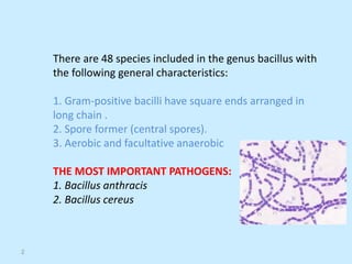
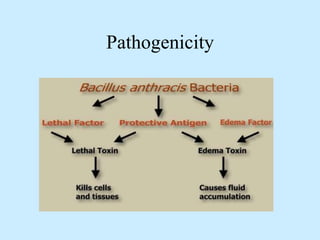
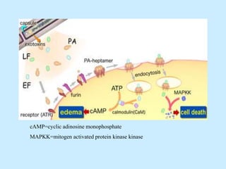
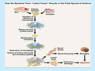
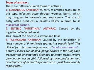
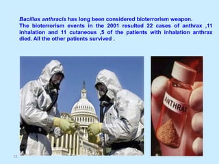
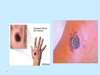
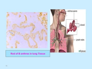
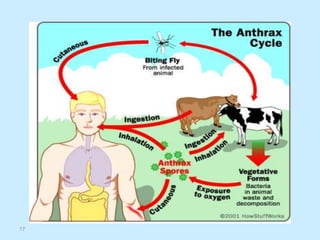
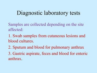
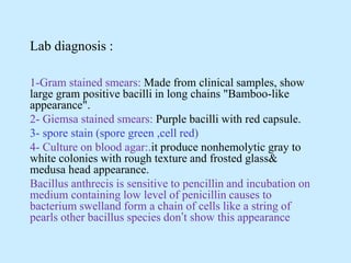
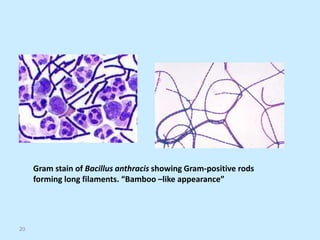
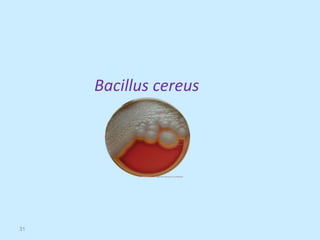
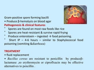
Spore forming Gram positive
bacilli
Bacillus species
There are 48 species included in the genus bacillus with
the following general characteristics:
1. Gram-positive bacilli have square ends arranged in
long chain .
2. Spore former (central spores).
3. Aerobic and facultative anaerobic
THE MOST IMPORTANT PATHOGENS:
1. Bacillus anthracis
2. Bacillus cereus
2
3
Bacillus anthracis
Morphology:
1. Large gram positive bacilli.
2. Non-motile.
3. Found singly, in pairs or in long chains.
4. Capsule could be demonstrated during growth in
infected animals.(poly peptide)
5. Spores are formed in culture, dead animal's tissue
,bone marrow and skin .
6. Spores are oval and centrally located.
Survival in Soil
• Spores remain viable in soil for years.
4
Pathogenesis
Anthrax is a zoonotic disease of –goats ,sheep ,cattle
,horses and human .
Human become infected incidentally by contact with
infected animals or their products.
In human the infection is usually acquired by the
entry of spores through injected skin ,or eating
contaminated food with spores ,or by inhalation
The spores germinate in the tissue at the site
of entry ,and growth of the vegetative
organisms results in formation of a
gelatinous edema and congestion .bacilli
spread by lymphatic to the bloodstream ,and
they multiply in the blood and tissues
shortly before and after the animal’s death.
ANTIGENIC STRUCTURE AND
PATHOGENIC DETERMINANTS:
1. The Capsular Polypeptide: Composed of poly
peptide of a high molecular weight consisting of
D-glutamic acid and it is antiphagocytic.
2. Polysaccharide Somatic Antigen: Composed of N
acetulglucos-eamine and Dgalactose.
3. Complex Protein Toxin: This toxin appear to be
responsible for signs and symptoms characteristic
of anthrax. Accumulation of the toxin in tissue and
its effect on the central nervous system results in
death by respiratory failure and anoxia.
Anthrax toxin is made up of three proteins
1-protective antigen(PA)
2-Edema factor (EF)
3-Lethal factor (LF)
(PA)binds to specific cell receptor ,and following
proteolytic activation it forms a membrane
channel that mediates entry of (EF) and (LF) into
the cell
(EF)is with (PA)it forms a toxin known as edema
toxin
(LF) plus (PA) form lethal toxin ,which is the major
virulence factor and cause death in infected animal
and human .
Pathogenicity
cAMP=cyclic adinosine monophosphate
MAPKK=mitogen activated protein kinase kinase
11
Types of anthrax :
There are different clinical forms of anthrax:
1. CUTANEOUS ANTHRAX: 95-98% of anthrax cases are of
this type. Infection occur through wounds, burns, which
may progress to toxaemia and septicemia. The site of
entry often produces a painless blister referred to as
Malignant pustule.
2. ENTERIC "INTESTINAL" ANTHRAX: Caused by the
ingestion of infected meat.
This form of the disease is severe and fatal.
3. PULMONARY ANTHRAX: Caused by the inhalation of
large number of B. anthracis spores. It is usually fetal. This
clinical form is commonly known as "wool sorter disease".
Anthrax spores are inhaled, phagocytosed in the lungs and
transported by lymphatic drainage to lymph nodes ,where
germination occurs ,this followed by toxin production and
development of hemorrhagic and sepsis ,which are usually
rapidly fatal.
12
13
Bacillus anthracis has long been considered bioterrorism weapon.
The bioterrorism events in the 2001 resulted 22 cases of anthrax ,11
inhalation and 11 cutaneous ,5 of the patients with inhalation anthrax
died. All the other patients survived .
14
15
Rod of B anthrax in lung Tissue
16
17
Diagnostic laboratory tests
Samples are collected depending on the site
affected:
1. Swab samples from cutaneous lesions and
blood cultures.
2. Sputum and blood for pulmonary anthrax
3. Gastric aspirate, feces and blood for enteric
anthrax.
Lab diagnosis :
1-Gram stained smears: Made from clinical samples, show
large gram positive bacilli in long chains "Bamboo-like
appearance".
2- Giemsa stained smears: Purple bacilli with red capsule.
3- spore stain (spore green ,cell red)
4- Culture on blood agar:.it produce nonhemolytic gray to
white colonies with rough texture and frosted glass&
medusa head appearance.
Bacillus anthrecis is sensitive to pencillin and incubation on
medium containing low level of penicillin causes to
bacterium swelland form a chain of cells like a string of
pearls other bacillus species don’t show this appearance
20
Gram stain of Bacillus anthracis showing Gram-positive rods
forming long filaments. “Bamboo –like appearance”
21
showing endospores as green and the vegetative cell as red
22
Colonial growth of Bacillus anthracis. When lifted by an inoculation
loop, colonies, in this case on sheep blood agar, show a tenacity
that allows them to be pulled up and stay upright with a texture
similar to egg whites.
5- Biochemical Identification:
1-catalase :positive.
2-Carbohydrate fermentation test positive for :
(glucose,maltose,ribose, sucrose ).
3- Gelatine liqefaction test: positive
4- Nitrate reduction test: Positive
5- Starch hydrolysis test: Positive
6- Voges-Proskauer test: Positive
7- Sensitivity to penicillin. Sensitive
8- Lysis by gamma phages: Positive. This test accurately
differentiate B.anthracis from other bacilllus species.
26
27
Lysis of Bacillus anthracis by the lytic phage gamma.
The plaque (clear area) in the region of confluent growth is where the
gamma phage was applied. The plaque results from the phage's .
6- Animal inoculation test: Experimental animals are
injected intraperitoneally by a suspension of the
test organism "Suspected B. anthracis culture".
The animal dies in 48-96 hours due to respiratory
failure.
Large number of typical bacilli can be found in the
blood and tissue of spleen of the infected animal.
7- ELISA measure antibodies against edema and
lethal toxins.
Vaccine
four countries produce vaccines for anthrax ,Russia
and china use attenuated spore –based vaccin by
scarification .
The united state and great britain use bacteria –free
filtrate of cultures adsorbed to aluminum
hydroxide .
The dose is (0,2 and 4 weeks ,then 6,12and 18
months followed by annual boosters .
The vaccine is available only to the USA department
of defense and persons at risk for repeated
exposure to Bacillus antheracis .
Treatment
Many antibiotics are effective against anthrax in humans ,but
treatment must be started early , ciprofloxacin is
recommended for treatment, pencillin G, along with
gentamycin or streptomycin .
In the setting of potential exposure to bacillus anthracis as an
agent of biologic war ,prophylaxis with ciprofloxacin
should be continued for 4 weeks while three doses of
vaccine are being given ,or for 8 weeks if no vaccine is
found .
Bacillus cereus
31
Gram-positive spore forming bacilli
• Produce β-hemolysis on blood agar
Pathogenesis & clinical features
° Spores are found on most raw foods like rice
° Spores are heat-resistant & survive rapid frying
° Produce enterotoxin – ingested → food poisoning.
° Short IP – 4-6 hours – similar to Staphylococcal food
poisoning (vomiting &diarrhoea)
TREATMENT
• fluid replacement
• Bacillus cereus are resistant to penicillin by produceβ-
lactamase ,so erythromycin or ciproflxacin may be effective
alternatives to pencillin .
32
33
B. cereus
B. anthracis
Characteristic
-
+
growth requirement for thiamin
+
-
hemolysis on sheep blood agar
-
+
polypeptide capsule
-
+
lysis by gamma phage
+
-
motility
-
+
string-of-pearls test

Bacillus spp..ppt....by..ali..rasool.badr

  • 1.
    Spore forming Grampositive bacilli Bacillus species
  • 2.
    There are 48species included in the genus bacillus with the following general characteristics: 1. Gram-positive bacilli have square ends arranged in long chain . 2. Spore former (central spores). 3. Aerobic and facultative anaerobic THE MOST IMPORTANT PATHOGENS: 1. Bacillus anthracis 2. Bacillus cereus 2
  • 3.
  • 4.
    Morphology: 1. Large grampositive bacilli. 2. Non-motile. 3. Found singly, in pairs or in long chains. 4. Capsule could be demonstrated during growth in infected animals.(poly peptide) 5. Spores are formed in culture, dead animal's tissue ,bone marrow and skin . 6. Spores are oval and centrally located. Survival in Soil • Spores remain viable in soil for years. 4
  • 5.
    Pathogenesis Anthrax is azoonotic disease of –goats ,sheep ,cattle ,horses and human . Human become infected incidentally by contact with infected animals or their products. In human the infection is usually acquired by the entry of spores through injected skin ,or eating contaminated food with spores ,or by inhalation
  • 6.
    The spores germinatein the tissue at the site of entry ,and growth of the vegetative organisms results in formation of a gelatinous edema and congestion .bacilli spread by lymphatic to the bloodstream ,and they multiply in the blood and tissues shortly before and after the animal’s death.
  • 7.
    ANTIGENIC STRUCTURE AND PATHOGENICDETERMINANTS: 1. The Capsular Polypeptide: Composed of poly peptide of a high molecular weight consisting of D-glutamic acid and it is antiphagocytic. 2. Polysaccharide Somatic Antigen: Composed of N acetulglucos-eamine and Dgalactose. 3. Complex Protein Toxin: This toxin appear to be responsible for signs and symptoms characteristic of anthrax. Accumulation of the toxin in tissue and its effect on the central nervous system results in death by respiratory failure and anoxia.
  • 8.
    Anthrax toxin ismade up of three proteins 1-protective antigen(PA) 2-Edema factor (EF) 3-Lethal factor (LF) (PA)binds to specific cell receptor ,and following proteolytic activation it forms a membrane channel that mediates entry of (EF) and (LF) into the cell (EF)is with (PA)it forms a toxin known as edema toxin (LF) plus (PA) form lethal toxin ,which is the major virulence factor and cause death in infected animal and human .
  • 9.
  • 10.
  • 11.
  • 12.
    Types of anthrax: There are different clinical forms of anthrax: 1. CUTANEOUS ANTHRAX: 95-98% of anthrax cases are of this type. Infection occur through wounds, burns, which may progress to toxaemia and septicemia. The site of entry often produces a painless blister referred to as Malignant pustule. 2. ENTERIC "INTESTINAL" ANTHRAX: Caused by the ingestion of infected meat. This form of the disease is severe and fatal. 3. PULMONARY ANTHRAX: Caused by the inhalation of large number of B. anthracis spores. It is usually fetal. This clinical form is commonly known as "wool sorter disease". Anthrax spores are inhaled, phagocytosed in the lungs and transported by lymphatic drainage to lymph nodes ,where germination occurs ,this followed by toxin production and development of hemorrhagic and sepsis ,which are usually rapidly fatal. 12
  • 13.
    13 Bacillus anthracis haslong been considered bioterrorism weapon. The bioterrorism events in the 2001 resulted 22 cases of anthrax ,11 inhalation and 11 cutaneous ,5 of the patients with inhalation anthrax died. All the other patients survived .
  • 14.
  • 15.
    15 Rod of Banthrax in lung Tissue
  • 16.
  • 17.
  • 18.
    Diagnostic laboratory tests Samplesare collected depending on the site affected: 1. Swab samples from cutaneous lesions and blood cultures. 2. Sputum and blood for pulmonary anthrax 3. Gastric aspirate, feces and blood for enteric anthrax.
  • 19.
    Lab diagnosis : 1-Gramstained smears: Made from clinical samples, show large gram positive bacilli in long chains "Bamboo-like appearance". 2- Giemsa stained smears: Purple bacilli with red capsule. 3- spore stain (spore green ,cell red) 4- Culture on blood agar:.it produce nonhemolytic gray to white colonies with rough texture and frosted glass& medusa head appearance. Bacillus anthrecis is sensitive to pencillin and incubation on medium containing low level of penicillin causes to bacterium swelland form a chain of cells like a string of pearls other bacillus species don’t show this appearance
  • 20.
    20 Gram stain ofBacillus anthracis showing Gram-positive rods forming long filaments. “Bamboo –like appearance”
  • 21.
    21 showing endospores asgreen and the vegetative cell as red
  • 22.
    22 Colonial growth ofBacillus anthracis. When lifted by an inoculation loop, colonies, in this case on sheep blood agar, show a tenacity that allows them to be pulled up and stay upright with a texture similar to egg whites.
  • 26.
    5- Biochemical Identification: 1-catalase:positive. 2-Carbohydrate fermentation test positive for : (glucose,maltose,ribose, sucrose ). 3- Gelatine liqefaction test: positive 4- Nitrate reduction test: Positive 5- Starch hydrolysis test: Positive 6- Voges-Proskauer test: Positive 7- Sensitivity to penicillin. Sensitive 8- Lysis by gamma phages: Positive. This test accurately differentiate B.anthracis from other bacilllus species. 26
  • 27.
    27 Lysis of Bacillusanthracis by the lytic phage gamma. The plaque (clear area) in the region of confluent growth is where the gamma phage was applied. The plaque results from the phage's .
  • 28.
    6- Animal inoculationtest: Experimental animals are injected intraperitoneally by a suspension of the test organism "Suspected B. anthracis culture". The animal dies in 48-96 hours due to respiratory failure. Large number of typical bacilli can be found in the blood and tissue of spleen of the infected animal. 7- ELISA measure antibodies against edema and lethal toxins.
  • 29.
    Vaccine four countries producevaccines for anthrax ,Russia and china use attenuated spore –based vaccin by scarification . The united state and great britain use bacteria –free filtrate of cultures adsorbed to aluminum hydroxide . The dose is (0,2 and 4 weeks ,then 6,12and 18 months followed by annual boosters . The vaccine is available only to the USA department of defense and persons at risk for repeated exposure to Bacillus antheracis .
  • 30.
    Treatment Many antibiotics areeffective against anthrax in humans ,but treatment must be started early , ciprofloxacin is recommended for treatment, pencillin G, along with gentamycin or streptomycin . In the setting of potential exposure to bacillus anthracis as an agent of biologic war ,prophylaxis with ciprofloxacin should be continued for 4 weeks while three doses of vaccine are being given ,or for 8 weeks if no vaccine is found .
  • 31.
  • 32.
    Gram-positive spore formingbacilli • Produce β-hemolysis on blood agar Pathogenesis & clinical features ° Spores are found on most raw foods like rice ° Spores are heat-resistant & survive rapid frying ° Produce enterotoxin – ingested → food poisoning. ° Short IP – 4-6 hours – similar to Staphylococcal food poisoning (vomiting &diarrhoea) TREATMENT • fluid replacement • Bacillus cereus are resistant to penicillin by produceβ- lactamase ,so erythromycin or ciproflxacin may be effective alternatives to pencillin . 32
  • 33.
    33 B. cereus B. anthracis Characteristic - + growthrequirement for thiamin + - hemolysis on sheep blood agar - + polypeptide capsule - + lysis by gamma phage + - motility - + string-of-pearls test