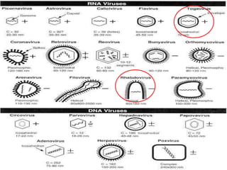
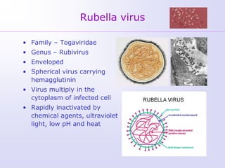
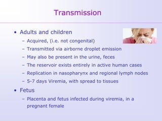
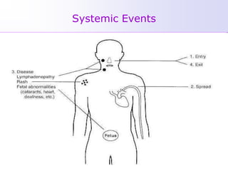
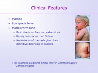
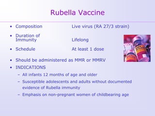
RUBELLA

RABIES

          Dr
          Kamran Afzal
          Asst Prof
          Microbiology
Rubella virus

• Family – Togaviridae
• Genus – Rubivirus
• Enveloped
• Spherical virus carrying
  hemagglutinin
• Virus multiply in the
  cytoplasm of infected cell
• Rapidly inactivated by
  chemical agents, ultraviolet
  light, low pH and heat
Transmission

• Adults and children
  – Acquired, (i.e. not congenital)
  – Transmitted via airborne droplet emission
  – May also be present in the urine, feces
  – The reservoir exists entirely in active human cases
  – Replication in nasopharynx and regional lymph nodes
  – 5-7 days Viremia, with spread to tissues

• Fetus
  – Placenta and fetus infected during viremia, in a
    pregnant female
Systemic Events
Clinical Features

• Malaise
• Low grade fever
• Morbilliform rash
   – Rash starts on face and extremities
   – Rarely lasts more than 5 days
   – No features of the rash give clues to
     definitive diagnosis of Rubella


  First described as distinct clinical entity in German literature
  – ‘German measles’
Clinical Features

• Incubation period 14 days
• Maculopapular rash and lymphadenopathy
  occur 14 days after exposure


• The clinical events occurring in the
  neonatal age are more important and
  divided into two major groups
   1. Post Natal Rubella (Acquired)
   2. Congenital Rubella
Complications

Arthralgia or arthritis
    adult female            up to 70%
    children                rare


Thrombocytopenic purpura
                            1/3,000 cases
Encephalitis
                            1/6,000 cases
Neuritis                    rare
Orchitis                    rare
Congenital Rubella Syndrome (CRS)

• Maternal viremia with Rubella infection during
  pregnancy may result in infection of placenta and fetus
• Infection may lead to fetal death or premature delivery
• Severity of damage to fetus depends on gestational age
• Up to 85% of infants affected if infected during first
  trimester
• >20 weeks of pregnancy, fetal defects are uncommon


• The growth rate of fetal cells is reduced
   – Leads to deranged and hypoplastic organ development
   – Results in structural damage and abnormalities
• Microcephaly
   – Deafness
   – Mental retardation
• Cataracts
• Heart defects
• Bone alterations
• Liver and spleen damage
Classical Triad in CRS

Classical Triad
• Microcephaly / Deafness
• Cardiac abnormalities
• Cataract
Laboratory Diagnosis

• Positive serologic test for Rubella IgM antibody
• 4 x rise in Rubella IgG by any standard serologic assay -
  - Agglutination, ICT, ELISA, HAI

• Isolation of Rubella virus from clinical specimen
   – nasopharyngeal swab, urine
Serology
Interpretation of Serology
Isolation and Identification of virus

• Nasopharyngeal or throat swabs taken 6 days
  prior or after appearance of rash are a good
  source of Rubella virus
• The virus can be cultured on continuous cell
  lines        Rabbit kidney cells
                    (RK 13)
                      and
                   Vero cells
• Cell cultured antigens can be detected by IF
  methods
Treatment and Prevention

• Rubella is a mild self limited illness
• No specific treatment or Antiviral treatment is indicated
• Clinically missed Rubella in the 1st 3-4 months of
  pregnancy is associated with fetal infections
• CRS can be prevented by effective immunization of the
  young children and teenage girls
Rubella Vaccine
• Composition                 Live virus (RA 27/3 strain)

• Duration of
  Immunity                    Lifelong

• Schedule                    At least 1 dose

• Should be administered as MMR or MMRV
• INDICATIONS
   – All infants 12 months of age and older
   – Susceptible adolescents and adults without documented
     evidence of Rubella immunity
   – Emphasis on non-pregnant women of childbearing age
RABIES
Rabies

It is an acute infectious disease of warm-blooded
animals and humans characterized by an involvement
of the nervous system resulting in death
Rabies Virus
 Genus: Lyssavirus
(lyssa: the Greek goddess of madness, rage, and frenzy)
 Include members of the Rhabdoviridiae family
       Rabies, Makola, Duvenhage
 Morphology:
    Enveloped bullet-shaped virus
    5 structural proteins
    SS RNA, negative sense, non-segmented, non-polar
Transmission of Rabies virus

 The dog is the most common cause of
  Rabies transmission worldwide, cats second
 In developed countries: dogs are
  immunized, other species of wild animals
  are reservoirs
 Bats: always considered rabid
Transmission of Rabies virus
 Infected saliva enters bite wounds
    Virus migrates up peripheral nerves to the spinal cord
     and reaches the brain
    Virus shed in the saliva during, before or after clinical
     symptoms
 Aerosol transmission: documented from caves with
  large populations of infected bats
 Organ donations: documented from early corneal
  transplantation (2004)
Rabies virus attacks the Central
       Nervous System

                 Watch as the rabies
                 virus from an exposure
                 on the leg spreads up
                 the spinal cord to the
                 brain and throughout
                 the rest of the body


                 Rabies virus entering
                 the body
Incubation Period
 Averages 3-8 weeks
   Can be as short as 1 week or up to 1 year
 Bite location and viral load are the two most important
  factors in incubation of the virus
Pathogenesis
 Budding from the plasma membrane of muscle cells
  into unmyelinated nerve endings
 Retrograde axoplasmic flow to the CNS
 Virus replication in dorsal root ganglia (DRG) and
  anterior horn cells
    Prophylaxis at this stage cannot prevent death
 Eventually, the virus spreads centrifugally from the
  CNS to the heart, skin, salivary and serous glands in
  the tongue
 All major organs may contain the virus (except blood)
 Organs from patients with unexplained neurologic
  disease may transmit rabies by transplantation
Clinical features in Humans
 General weakness, discomfort, fever or headache
 Prickling or itching sensation at the site of bite
 Cerebral dysfunction
 Anxiety, confusion, agitation, delirium
 Abnormal behavior
 Hallucinations, and insomnia
 Hydrophobia


 The acute period of disease ends after 2 to 10 days
 Once clinical signs of rabies appear, the disease is
  nearly always fatal
Laboratory Diagnosis

   Serology
   Virus cultivation
   The Direct Fluorescent Antibody test (DFA)
   Histopathology
 Serology
   Circulating antibodies appear slowly but they are
    usually present by the time of onset of clinical
    symptoms
 Virus cultivation
   The most definitive means of diagnosis is by virus
    cultivation from saliva and infected tissue
   Virus cultivation can be done using cell cultures or
    the specimen is inoculated intra-cerebrally into mice
 Direct Fluorescent Antibody
The Direct Fluorescent Antibody test (DFA) is done on
corneal impressions or neck skin biopsy
 Histopathology of Brain
  Negri bodies are diagnostic of Rabies
SPECIFIC GUIDELINES AND
       PROCEDURES

Management of Potential Rabies
         Exposure
Management and Prevention
Pre-exposure prophylaxis
 Inactivated rabies vaccine is given to persons at
  increased risk of rabies e.g. vets, animal
  handlers, laboratory workers etc
Post-exposure prophylaxis
 In cases of bites, suspected dogs and cats should be
  held for 10 days for observation
 If signs develop, they should be killed and their
  tissues examined
Post-exposure Prophylaxis
 Wound treatment - surgical debridement
 Passive immunization - human rabies
  immunoglobulin is given around the area of the wound
  and an I/M dose
 Active immunization - The human diploid cell vaccine
  (HDCV) is administered I/M, 5 doses are given on days
  1,3,7,14 and 28
 Combined treatment with passive and active
  immunization is much more effective than active
  immunization alone
Recommended Post-exposure Prophylaxis
Category of exposure to suspect rabid      Post-exposure measures
animal

Category I – touching or feeding           None
animals, licks on intact skin (i.e. no
exposure)

Category II – nibbling of uncovered skin, Immediate vaccination and local
minor scratches or abrasions without      treatment of the wound
bleeding

Category III – single or multiple          Immediate vaccination and
transdermal bites or scratches, licks on   administration of rabies
broken skin; contamination of mucous       immunoglobulin; local treatment of the
membrane with saliva from licks,           wound
exposures to bats.
Rabies Vaccines

The vaccines which are available for humans are
inactivated whole virus vaccines
  Nervous Tissue Preparation
  Duck Embryo Vaccine
  Human Diploid Cell Vaccine (HDCV) - this is
   currently the best vaccine available with an
   efficacy rate of nearly 100% and rarely any
   severe reactions, however it is very expensive
Treatment

POST-EXPOSURE TREATMENT (PET)
 LOCAL WOUND TREATMENT
-   Wash with soap/detergent and water preferably for
    10 min
-   Apply alcohol or povidone iodine antiseptic
-   Anti-Tetanus toxoid I/M
-   Avoid suturing and wound dressing
Antimicrobials
- Co-amoxiclav
- Cloxacillin
- Cefuroxime
Special Conditions
 Babies born of rabid mothers should be given ARV as
  early after birth as possible
 Pregnancy and infancy are not C/I to treatment
 Bites of rodents, rabbits, guinea pig- NO PET
 Dogs, cats, livestock, wild animals- Give PET
Prevention and Control
 Canine rabies accounts for more than 99% of all
  human rabies
 Control measures include
   Stray dog control
   Vaccination of dogs
   Quarantine of imported animals
Oral Rabies Vaccination for animals
A raccoon consuming a bait laden with ORV
What kind of animals get rabies?

 The rabies virus can
  infect all mammals
 Animals less likely to get rabies
Animals that don’t carry rabies
 Frogs, turtles, birds, bees and snakes

Rubella + rabies

  • 1.
    RUBELLA RABIES Dr Kamran Afzal Asst Prof Microbiology
  • 3.
    Rubella virus • Family– Togaviridae • Genus – Rubivirus • Enveloped • Spherical virus carrying hemagglutinin • Virus multiply in the cytoplasm of infected cell • Rapidly inactivated by chemical agents, ultraviolet light, low pH and heat
  • 4.
    Transmission • Adults andchildren – Acquired, (i.e. not congenital) – Transmitted via airborne droplet emission – May also be present in the urine, feces – The reservoir exists entirely in active human cases – Replication in nasopharynx and regional lymph nodes – 5-7 days Viremia, with spread to tissues • Fetus – Placenta and fetus infected during viremia, in a pregnant female
  • 5.
  • 6.
    Clinical Features • Malaise •Low grade fever • Morbilliform rash – Rash starts on face and extremities – Rarely lasts more than 5 days – No features of the rash give clues to definitive diagnosis of Rubella First described as distinct clinical entity in German literature – ‘German measles’
  • 7.
    Clinical Features • Incubationperiod 14 days • Maculopapular rash and lymphadenopathy occur 14 days after exposure • The clinical events occurring in the neonatal age are more important and divided into two major groups 1. Post Natal Rubella (Acquired) 2. Congenital Rubella
  • 8.
    Complications Arthralgia or arthritis adult female up to 70% children rare Thrombocytopenic purpura 1/3,000 cases Encephalitis 1/6,000 cases Neuritis rare Orchitis rare
  • 9.
    Congenital Rubella Syndrome(CRS) • Maternal viremia with Rubella infection during pregnancy may result in infection of placenta and fetus • Infection may lead to fetal death or premature delivery • Severity of damage to fetus depends on gestational age • Up to 85% of infants affected if infected during first trimester • >20 weeks of pregnancy, fetal defects are uncommon • The growth rate of fetal cells is reduced – Leads to deranged and hypoplastic organ development – Results in structural damage and abnormalities
  • 10.
    • Microcephaly – Deafness – Mental retardation • Cataracts • Heart defects • Bone alterations • Liver and spleen damage
  • 11.
    Classical Triad inCRS Classical Triad • Microcephaly / Deafness • Cardiac abnormalities • Cataract
  • 12.
    Laboratory Diagnosis • Positiveserologic test for Rubella IgM antibody • 4 x rise in Rubella IgG by any standard serologic assay - - Agglutination, ICT, ELISA, HAI • Isolation of Rubella virus from clinical specimen – nasopharyngeal swab, urine
  • 13.
  • 14.
  • 15.
    Isolation and Identificationof virus • Nasopharyngeal or throat swabs taken 6 days prior or after appearance of rash are a good source of Rubella virus • The virus can be cultured on continuous cell lines Rabbit kidney cells (RK 13) and Vero cells • Cell cultured antigens can be detected by IF methods
  • 16.
    Treatment and Prevention •Rubella is a mild self limited illness • No specific treatment or Antiviral treatment is indicated • Clinically missed Rubella in the 1st 3-4 months of pregnancy is associated with fetal infections • CRS can be prevented by effective immunization of the young children and teenage girls
  • 17.
    Rubella Vaccine • Composition Live virus (RA 27/3 strain) • Duration of Immunity Lifelong • Schedule At least 1 dose • Should be administered as MMR or MMRV • INDICATIONS – All infants 12 months of age and older – Susceptible adolescents and adults without documented evidence of Rubella immunity – Emphasis on non-pregnant women of childbearing age
  • 18.
  • 19.
    Rabies It is anacute infectious disease of warm-blooded animals and humans characterized by an involvement of the nervous system resulting in death
  • 20.
    Rabies Virus  Genus:Lyssavirus (lyssa: the Greek goddess of madness, rage, and frenzy)  Include members of the Rhabdoviridiae family Rabies, Makola, Duvenhage  Morphology:  Enveloped bullet-shaped virus  5 structural proteins  SS RNA, negative sense, non-segmented, non-polar
  • 21.
    Transmission of Rabiesvirus  The dog is the most common cause of Rabies transmission worldwide, cats second  In developed countries: dogs are immunized, other species of wild animals are reservoirs  Bats: always considered rabid
  • 22.
    Transmission of Rabiesvirus  Infected saliva enters bite wounds  Virus migrates up peripheral nerves to the spinal cord and reaches the brain  Virus shed in the saliva during, before or after clinical symptoms  Aerosol transmission: documented from caves with large populations of infected bats  Organ donations: documented from early corneal transplantation (2004)
  • 23.
    Rabies virus attacksthe Central Nervous System Watch as the rabies virus from an exposure on the leg spreads up the spinal cord to the brain and throughout the rest of the body Rabies virus entering the body
  • 24.
    Incubation Period  Averages3-8 weeks  Can be as short as 1 week or up to 1 year  Bite location and viral load are the two most important factors in incubation of the virus
  • 25.
    Pathogenesis  Budding fromthe plasma membrane of muscle cells into unmyelinated nerve endings  Retrograde axoplasmic flow to the CNS  Virus replication in dorsal root ganglia (DRG) and anterior horn cells  Prophylaxis at this stage cannot prevent death
  • 27.
     Eventually, thevirus spreads centrifugally from the CNS to the heart, skin, salivary and serous glands in the tongue  All major organs may contain the virus (except blood)  Organs from patients with unexplained neurologic disease may transmit rabies by transplantation
  • 28.
    Clinical features inHumans  General weakness, discomfort, fever or headache  Prickling or itching sensation at the site of bite  Cerebral dysfunction  Anxiety, confusion, agitation, delirium  Abnormal behavior  Hallucinations, and insomnia  Hydrophobia  The acute period of disease ends after 2 to 10 days  Once clinical signs of rabies appear, the disease is nearly always fatal
  • 29.
    Laboratory Diagnosis  Serology  Virus cultivation  The Direct Fluorescent Antibody test (DFA)  Histopathology
  • 30.
     Serology  Circulating antibodies appear slowly but they are usually present by the time of onset of clinical symptoms  Virus cultivation  The most definitive means of diagnosis is by virus cultivation from saliva and infected tissue  Virus cultivation can be done using cell cultures or the specimen is inoculated intra-cerebrally into mice
  • 31.
     Direct FluorescentAntibody The Direct Fluorescent Antibody test (DFA) is done on corneal impressions or neck skin biopsy
  • 32.
     Histopathology ofBrain  Negri bodies are diagnostic of Rabies
  • 33.
    SPECIFIC GUIDELINES AND PROCEDURES Management of Potential Rabies Exposure
  • 34.
    Management and Prevention Pre-exposureprophylaxis  Inactivated rabies vaccine is given to persons at increased risk of rabies e.g. vets, animal handlers, laboratory workers etc Post-exposure prophylaxis  In cases of bites, suspected dogs and cats should be held for 10 days for observation  If signs develop, they should be killed and their tissues examined
  • 35.
    Post-exposure Prophylaxis  Woundtreatment - surgical debridement  Passive immunization - human rabies immunoglobulin is given around the area of the wound and an I/M dose  Active immunization - The human diploid cell vaccine (HDCV) is administered I/M, 5 doses are given on days 1,3,7,14 and 28  Combined treatment with passive and active immunization is much more effective than active immunization alone
  • 36.
    Recommended Post-exposure Prophylaxis Categoryof exposure to suspect rabid Post-exposure measures animal Category I – touching or feeding None animals, licks on intact skin (i.e. no exposure) Category II – nibbling of uncovered skin, Immediate vaccination and local minor scratches or abrasions without treatment of the wound bleeding Category III – single or multiple Immediate vaccination and transdermal bites or scratches, licks on administration of rabies broken skin; contamination of mucous immunoglobulin; local treatment of the membrane with saliva from licks, wound exposures to bats.
  • 37.
    Rabies Vaccines The vaccineswhich are available for humans are inactivated whole virus vaccines  Nervous Tissue Preparation  Duck Embryo Vaccine  Human Diploid Cell Vaccine (HDCV) - this is currently the best vaccine available with an efficacy rate of nearly 100% and rarely any severe reactions, however it is very expensive
  • 38.
    Treatment POST-EXPOSURE TREATMENT (PET) LOCAL WOUND TREATMENT - Wash with soap/detergent and water preferably for 10 min - Apply alcohol or povidone iodine antiseptic - Anti-Tetanus toxoid I/M - Avoid suturing and wound dressing
  • 39.
  • 40.
    Special Conditions  Babiesborn of rabid mothers should be given ARV as early after birth as possible  Pregnancy and infancy are not C/I to treatment  Bites of rodents, rabbits, guinea pig- NO PET  Dogs, cats, livestock, wild animals- Give PET
  • 41.
    Prevention and Control Canine rabies accounts for more than 99% of all human rabies  Control measures include  Stray dog control  Vaccination of dogs  Quarantine of imported animals
  • 42.
  • 43.
    A raccoon consuminga bait laden with ORV
  • 44.
    What kind ofanimals get rabies?  The rabies virus can infect all mammals
  • 45.
     Animals lesslikely to get rabies
  • 46.
    Animals that don’tcarry rabies  Frogs, turtles, birds, bees and snakes