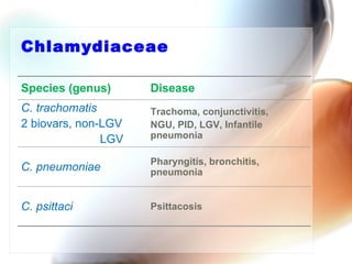
Chlamydia


     Dr Kamran Afzal
  Asst Prof Microbiology
Obligate intr acellular
bacteria
•   Rickettsia
•   Coxiella
•   Ehrlichia
•   Bartonella
•   Mycobacteria (a few species)
•   Chlamydia
    –   C. trachomatis
    –   C. pneumoniae
    –   C. psittaci
    –   C. pecorum
Char acteristics - Chlamydia
•   Family Chlamydiaceae
•   Obligate intracellular bacteria
•   Rod-shaped or coccoid
•   Aerobic
•   Gram negative but difficult to stain
•   Cell Wall – lipopolysaccharides form the outer membrane,
    not peptidoglycan
•   Infect columnar epithelial cells
•   Forms elementary bodies (EB)
•   Non-motile
•   37°C
Chlamydiaceae

Species (genus)      Disease
C. trachomatis       Trachoma, conjunctivitis,
2 biovars, non-LGV   NGU, PID, LGV, Infantile
               LGV   pneumonia

                     Pharyngitis, bronchitis,
C. pneumoniae        pneumonia


C. psittaci          Psittacosis
Chlamydia
trachomatis
Chlamydia trachomatis
• Further divided
   – 15 serovars
      • defined by serological differences in the major outer
         membrane protein (MOMP)
   – Different serovars cause different diseases
      • Trachoma (A, B, Ba, C)
      • Urogenital-STDs (D, E, F, G, H, I, J, K)
      • LGV (L1, L2, L3)
Tr achoma
• Largest single cause of preventable blindness in
  the world
   – 500 million cases worldwide
   – 5.9 million blind
   – Primarily in developing countries
• Caused by serovars A, B, Ba, C
• Clinical manifestations
   – Chronic follicular kerato-conjunctivitis
      • With conjunctival scarring and pannus formation
Pathogenesis
• Caused by repeated infections
  – Causes inflammation of the eye
  – Eyelids turn inwards so the eye lashes rub
    against the cornea causing scar tissue to
    form irreversible blindness
  – 1st infection in childhood
     • subsequently persistent infection
           or many re-infections
     • final stages 15-20 years later
  – Sensitivity to products of the organism
    causes most of the pathogenesis
• Treatment
  –   Azithromycin
  –   Topical doxycycline
  –   Improved sanitation
  –   Surgical intervention
Tr achoma
D s
S T       a rs
                 D -K

  er ov
 S
Epidemiology of STDs
• Incidence
  – 4-6 million cases/year in US
  – Increasing in some locations
• Only reservoir is human
  – Person to person transmission
  – Asymptomatic carriers important in transmission
Pathogenicity

• Chlamydia (STD)
  – Transmitted through direct contact
    between infected membranes
  – If left untreated, common cause of
    infertility
  – Newborns can contract the disease from
    infected mothers
How is it transmitted?

 Passed through an infected   Passed through an infected
 person to a partner by:      person to a partner by:

           Vaginal Sex                    Anal Sex


 Passed through an infected   Passed from an infected mother
 person to a partner by:      to her unborn child.

             Oral Sex
Infections in male
• Urethritis
   – 50-75% symptomatic
      • urethral discharge
      • pyuria
      • itching
      • dysuria
   – 25-50% asymptomatic
• Inclusion conjunctivitis
• Epididymitis
   – acute and unilateral inflammation of epididymis
   – may result in decreased fertility
• Proctitis
Male
•   Discharge from penis
•   Dysuria
•   Pain, blood, discharge from rectum
•   Signs and symptoms 1-3 weeks after exposure
Infections in female
• Urethritis (60-80%)
   – dysuria
   – pyuria
• Proctitis
   – inflammation of the rectum
• Inclusion conjunctivitis
• Peri-hepatitis
   – infection of liver capsule
Infections in neonate / infant
• Acquired from direct contact with infected cervical
  secretions of mother at delivery
• Inclusion conjunctivitis
   – most common cause of neonatal conjunctivitis
     (2-6% infants)
   – acute mucopurulent eye discharge
   – systemic therapy with erythromycin
• Pneumonia
   – 33-50% of all cases of interstitial pneumonia
   – failure to thrive
Female

•   Often no symptoms (silent injury)
•   Unusual discharge
•   Burning with urination
•   Pain, blood, discharge from rectum
•   Fever
•   Dysperunia
•   Signs and symptoms 1-3 weeks after exposure
Complications
• Reiter’s syndrome
   – A reactive arthritis secondary to an immune-mediated
     response
     o It may present as asymmetric polyarthritis, urethritis,
     inflammatory eye disease, mouth ulcers
     o 80% of affected patients are HLA-B27 positive
• Deeper pelvic complications in the female
     o PID
     o Potential infertility
     o Spread to the newborn during parturition
a
                   lom
               nu V)
         og ra
     p h        ( LG
Ly m
         re u m and L 3
     n e s L 1, L 2
  v e var
   ser o

      •Climatic or tropical bubo
      •Lymphogranuloma inguinale
Stages of infection
• Primary stage
               (untr eated)
     – 3 to 30 days after incubation
     – Small painless papule which may ulcerate at site of
     inoculation: self limiting
• Secondary    stage
     – Inguinal lymph nodes (more common in males)
      • Painful lymphadenopathy
     – Necrosis in lymph nodes – may enlarge to form abscess
     – Acute haemorrhagic proctitis (10cm of the anorectal canal)
     – Fever, myalgia and headaches
• Tertiary   stage
     – Chronic inflammatory lesions typical of chlamydial
     infection
     – Scarring in genital tract
     – Fibrosis, lymphatic obstruction, elephantiasis
     – Rectal strictures and fistulae
Risk factor s
• Adult risks
   – Non-white race
   – Multiple sexual partners
   – Age younger than 19 years
   – Non-barrier contraceptive use
   – Poor socio-economic conditions
• Neonatal risks
   – Conjunctivitis
   – Neonatal pneumonia
Dif fer ential Diagnoses
• Genital ulceration
     – Herpes simplex, syphilis, chancroid, donovanosis,
     non-venereal infections e.g; cutaneous leishmaniasis
• Inguinal lymphadenopathy
     – Herpes simplex, syphilis, chancroid, bubonic plague in
     endemic regions, carcinoma
• Proctitis
     – Ulcerative colitis, Crohn’s disease, rectal carcinoma
Chlamydia pneumoniae
• Chlamydia pneumophila
• Pneumonia, bronchitis, sinusitis, pharyngitis, laryngitis
•   Variety of mild respiratory infections w/ fever, cough
Epidemiology
•   Person to person spread by respiratory droplets
    • no animal reservoirs known
•   May be common infection among children 5-14 years old
        • 10% of pneumonia
        • 5% of bronchitis
    • 50% antibody prevalence in adults
•   Infects the lungs
    – Majority of humans are infected
    – All effects of disease not known
        • Asthma, chronic bronchitis?
•   Also, it has recently been linked to
    heart disease and
    atherosclerosis
    – organisms found
       in valve lesions
Chlamydia psittaci
• Sporadic disease in humans
  – 100-200 cases / year in US
     • generally associated with contact with an infected
       bird
  – Inhalation of respiratory secretions, droppings of
    infected birds
     • organism secreted in high numbers
• Diagnosis of Chlamydia psittaci infection
•   Primarily clinical diagnosis
    – Acute onset febrile LRTI with hepato-splenomegaly,
       history of exposure to birds
    – Symptoms
        • fever, headache, malaise, muscle aches, dry hacking
          cough, bilateral interstitial pneumonia
    – Occasional systemic symptoms
        • myocarditis, encephalitis, hepatitis
Chlamydia life cycle
Developmental cycle
•   Morphologically distinct infectious and replicative forms
• Stage 1. Attachment of elementary bodies (EBs)
   Infectious form
        • found in secretions
        • relatively resistant to environment
    Usual target cell
        • columnar epithelial cells of mucous membranes
        • not normally phagocytic
• Stage 2. Entry of EBs
•   Chlamydia-specific receptor mediated
    endocytosis
•   EBs enter cell within a phagosome
    – All development occurs here until rupture
    – Called an inclusion when visible
• Stage 3. EBs change morphologically to
  reticulate bodies (RBs)
  – 8 hours after entry
  – RB more permeable
     • metabolically active
  – not infectious at this stage
• Stage 4. Replication
   – RBs divide by binary fission for 20-24 hours
• Stage 5. RBs change back to EB form
  – DNA condenses
  – decreases in size
  – cell wall becomes more resistant
  – mature inclusion can contain hundreds of organisms
• Stage 6. Release of infectious EBs
   – both cell and inclusions lyse
T he Chlamydial Life Cycle
Dia gnosis
• Cytological diagnosis
   – By using Giemsa, iodine or papanicolaou stains to
     detect chlamydial inclusions in epithelial cells
• Isolation in cell culture
   – It is gold standard, but slow process and expensive
      • collect material containing columnar epithelial cells
        from urethra or cervix
   – Culture on monolayer of McCoy cell lines
      • incubation is for 40-72 hours
   – Stain with fluorescein-conjugated anti-Chlamydia
     monoclonal antibody after 48 hours and look for
     characteristic inclusions
Dia gnosis
• Antigen detection and nucleic acid hybridization
   – DFA staining (direct fluorescent antibody)
   – ELISA
   – Detection of chlamydial ribosomal RNA by
     hybridization with DNA probe
• Amplification techniques
   – PCR     - LCR
• Serology
   – ICT
   – CFT
   – Micro-immunofluorescence test
Tr eatment
• Azithromycin
    – treatment of choice
    – effective against C. trachomatis and N. gonorrhoeae
    – well tolerated
• Tetracycline, especially doxycycline
• Erythromycin for pregnant women and children
•   No drug resistant C. trachomatis
Pr evention

•   No vaccine
•   Safe sex
    – Barrier contraceptive
•   Topical antimicrobials
    – Silver nitrate not effective
    – topical erythromycin
•   Regular Screening
•   Educational programs
Pap smear showing Chlamydia in the vacuoles 500x H&E
Q&A

Chlamydia

  • 1.
    Chlamydia Dr Kamran Afzal Asst Prof Microbiology
  • 2.
    Obligate intr acellular bacteria • Rickettsia • Coxiella • Ehrlichia • Bartonella • Mycobacteria (a few species) • Chlamydia – C. trachomatis – C. pneumoniae – C. psittaci – C. pecorum
  • 3.
    Char acteristics -Chlamydia • Family Chlamydiaceae • Obligate intracellular bacteria • Rod-shaped or coccoid • Aerobic • Gram negative but difficult to stain • Cell Wall – lipopolysaccharides form the outer membrane, not peptidoglycan • Infect columnar epithelial cells • Forms elementary bodies (EB) • Non-motile • 37°C
  • 4.
    Chlamydiaceae Species (genus) Disease C. trachomatis Trachoma, conjunctivitis, 2 biovars, non-LGV NGU, PID, LGV, Infantile LGV pneumonia Pharyngitis, bronchitis, C. pneumoniae pneumonia C. psittaci Psittacosis
  • 5.
  • 6.
    Chlamydia trachomatis • Furtherdivided – 15 serovars • defined by serological differences in the major outer membrane protein (MOMP) – Different serovars cause different diseases • Trachoma (A, B, Ba, C) • Urogenital-STDs (D, E, F, G, H, I, J, K) • LGV (L1, L2, L3)
  • 7.
    Tr achoma • Largestsingle cause of preventable blindness in the world – 500 million cases worldwide – 5.9 million blind – Primarily in developing countries • Caused by serovars A, B, Ba, C • Clinical manifestations – Chronic follicular kerato-conjunctivitis • With conjunctival scarring and pannus formation
  • 8.
    Pathogenesis • Caused byrepeated infections – Causes inflammation of the eye – Eyelids turn inwards so the eye lashes rub against the cornea causing scar tissue to form irreversible blindness – 1st infection in childhood • subsequently persistent infection or many re-infections • final stages 15-20 years later – Sensitivity to products of the organism causes most of the pathogenesis
  • 9.
    • Treatment – Azithromycin – Topical doxycycline – Improved sanitation – Surgical intervention
  • 10.
  • 11.
    D s S T a rs D -K er ov S
  • 12.
    Epidemiology of STDs •Incidence – 4-6 million cases/year in US – Increasing in some locations • Only reservoir is human – Person to person transmission – Asymptomatic carriers important in transmission
  • 13.
    Pathogenicity • Chlamydia (STD) – Transmitted through direct contact between infected membranes – If left untreated, common cause of infertility – Newborns can contract the disease from infected mothers
  • 14.
    How is ittransmitted? Passed through an infected Passed through an infected person to a partner by: person to a partner by: Vaginal Sex Anal Sex Passed through an infected Passed from an infected mother person to a partner by: to her unborn child. Oral Sex
  • 15.
    Infections in male •Urethritis – 50-75% symptomatic • urethral discharge • pyuria • itching • dysuria – 25-50% asymptomatic • Inclusion conjunctivitis • Epididymitis – acute and unilateral inflammation of epididymis – may result in decreased fertility • Proctitis
  • 16.
    Male • Discharge from penis • Dysuria • Pain, blood, discharge from rectum • Signs and symptoms 1-3 weeks after exposure
  • 17.
    Infections in female •Urethritis (60-80%) – dysuria – pyuria • Proctitis – inflammation of the rectum • Inclusion conjunctivitis • Peri-hepatitis – infection of liver capsule
  • 18.
    Infections in neonate/ infant • Acquired from direct contact with infected cervical secretions of mother at delivery • Inclusion conjunctivitis – most common cause of neonatal conjunctivitis (2-6% infants) – acute mucopurulent eye discharge – systemic therapy with erythromycin • Pneumonia – 33-50% of all cases of interstitial pneumonia – failure to thrive
  • 19.
    Female • Often no symptoms (silent injury) • Unusual discharge • Burning with urination • Pain, blood, discharge from rectum • Fever • Dysperunia • Signs and symptoms 1-3 weeks after exposure
  • 20.
    Complications • Reiter’s syndrome – A reactive arthritis secondary to an immune-mediated response o It may present as asymmetric polyarthritis, urethritis, inflammatory eye disease, mouth ulcers o 80% of affected patients are HLA-B27 positive • Deeper pelvic complications in the female o PID o Potential infertility o Spread to the newborn during parturition
  • 21.
    a lom nu V) og ra p h ( LG Ly m re u m and L 3 n e s L 1, L 2 v e var ser o •Climatic or tropical bubo •Lymphogranuloma inguinale
  • 22.
    Stages of infection •Primary stage (untr eated) – 3 to 30 days after incubation – Small painless papule which may ulcerate at site of inoculation: self limiting • Secondary stage – Inguinal lymph nodes (more common in males) • Painful lymphadenopathy – Necrosis in lymph nodes – may enlarge to form abscess – Acute haemorrhagic proctitis (10cm of the anorectal canal) – Fever, myalgia and headaches • Tertiary stage – Chronic inflammatory lesions typical of chlamydial infection – Scarring in genital tract – Fibrosis, lymphatic obstruction, elephantiasis – Rectal strictures and fistulae
  • 23.
    Risk factor s •Adult risks – Non-white race – Multiple sexual partners – Age younger than 19 years – Non-barrier contraceptive use – Poor socio-economic conditions • Neonatal risks – Conjunctivitis – Neonatal pneumonia
  • 24.
    Dif fer entialDiagnoses • Genital ulceration – Herpes simplex, syphilis, chancroid, donovanosis, non-venereal infections e.g; cutaneous leishmaniasis • Inguinal lymphadenopathy – Herpes simplex, syphilis, chancroid, bubonic plague in endemic regions, carcinoma • Proctitis – Ulcerative colitis, Crohn’s disease, rectal carcinoma
  • 25.
  • 26.
    Chlamydia pneumoniae • Chlamydiapneumophila • Pneumonia, bronchitis, sinusitis, pharyngitis, laryngitis • Variety of mild respiratory infections w/ fever, cough
  • 27.
    Epidemiology • Person to person spread by respiratory droplets • no animal reservoirs known • May be common infection among children 5-14 years old • 10% of pneumonia • 5% of bronchitis • 50% antibody prevalence in adults
  • 28.
    Infects the lungs – Majority of humans are infected – All effects of disease not known • Asthma, chronic bronchitis? • Also, it has recently been linked to heart disease and atherosclerosis – organisms found in valve lesions
  • 29.
    Chlamydia psittaci • Sporadicdisease in humans – 100-200 cases / year in US • generally associated with contact with an infected bird – Inhalation of respiratory secretions, droppings of infected birds • organism secreted in high numbers
  • 30.
    • Diagnosis ofChlamydia psittaci infection • Primarily clinical diagnosis – Acute onset febrile LRTI with hepato-splenomegaly, history of exposure to birds – Symptoms • fever, headache, malaise, muscle aches, dry hacking cough, bilateral interstitial pneumonia – Occasional systemic symptoms • myocarditis, encephalitis, hepatitis
  • 31.
  • 32.
    Developmental cycle • Morphologically distinct infectious and replicative forms • Stage 1. Attachment of elementary bodies (EBs) Infectious form • found in secretions • relatively resistant to environment Usual target cell • columnar epithelial cells of mucous membranes • not normally phagocytic
  • 33.
    • Stage 2.Entry of EBs • Chlamydia-specific receptor mediated endocytosis • EBs enter cell within a phagosome – All development occurs here until rupture – Called an inclusion when visible
  • 34.
    • Stage 3.EBs change morphologically to reticulate bodies (RBs) – 8 hours after entry – RB more permeable • metabolically active – not infectious at this stage
  • 35.
    • Stage 4.Replication – RBs divide by binary fission for 20-24 hours
  • 36.
    • Stage 5.RBs change back to EB form – DNA condenses – decreases in size – cell wall becomes more resistant – mature inclusion can contain hundreds of organisms
  • 37.
    • Stage 6.Release of infectious EBs – both cell and inclusions lyse
  • 39.
    T he ChlamydialLife Cycle
  • 40.
    Dia gnosis • Cytologicaldiagnosis – By using Giemsa, iodine or papanicolaou stains to detect chlamydial inclusions in epithelial cells • Isolation in cell culture – It is gold standard, but slow process and expensive • collect material containing columnar epithelial cells from urethra or cervix – Culture on monolayer of McCoy cell lines • incubation is for 40-72 hours – Stain with fluorescein-conjugated anti-Chlamydia monoclonal antibody after 48 hours and look for characteristic inclusions
  • 41.
    Dia gnosis • Antigendetection and nucleic acid hybridization – DFA staining (direct fluorescent antibody) – ELISA – Detection of chlamydial ribosomal RNA by hybridization with DNA probe • Amplification techniques – PCR - LCR • Serology – ICT – CFT – Micro-immunofluorescence test
  • 42.
    Tr eatment • Azithromycin – treatment of choice – effective against C. trachomatis and N. gonorrhoeae – well tolerated • Tetracycline, especially doxycycline • Erythromycin for pregnant women and children • No drug resistant C. trachomatis
  • 43.
    Pr evention • No vaccine • Safe sex – Barrier contraceptive • Topical antimicrobials – Silver nitrate not effective – topical erythromycin • Regular Screening • Educational programs
  • 44.
    Pap smear showingChlamydia in the vacuoles 500x H&E
  • 45.

Editor's Notes

  • #4 Cell wall – one of virulence factors because, it stops white blood cells from binding to foreign organisms
  • #14 Chlamydia most common STI in world , transmitted through direct contact between infected membranes 1999 WHO estimates, 340 million new cases of curable sexually transmitted infections ages 15-49. 92 million new infections worldwide per year are chlamydia Figure: Florescent regions are the inclusion bodies on a human sperm
  • #40 Ubiquitous obligate intracellular parasites Cannot reproduce without host cells Can only be cultured in vivo Two morphological forms EB’s (elementary bodies) Infectious, small (-35 um) metabolically inactive, Do not reproduce RB’s (reticulate bodies) Form inside inclusion membrane from EB’s Metabolize, reproduce, large size (100um). Transform back into EB’s for exocytosis