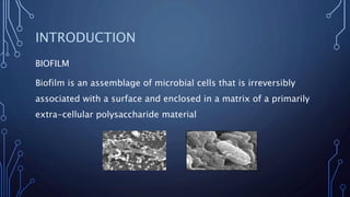
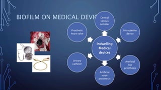
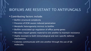
FUNGAL BIOFILM
DR KAMRAN AFZAL
MBBS, FCPS, PHD
CONSULTANT MICROBIOLOGIST
INTRODUCTION
BIOFILM
Biofilm is an assemblage of microbial cells that is irreversibly
associated with a surface and enclosed in a matrix of a primarily
extra-cellular polysaccharide material
• Biofilms are responsible for a broad spectrum of microbial
infections in human host
• Many medically important fungi produce biofilms
• Candida
• Aspergillus
• Cryptococcus
• Trichosporon
• Coccidioides
• Pneumocystis
HISTORY
• Van Leeuwenhoek credited for the discovery of microbial
biofilms
• Zobell observed number of bacteria on surface was higher than
in surrounding medium
• Jones used scanning / transmission electron microscopy to
examine biofilms
• Characklis studied microbial slimes in industrial water systems
and showed that they were highly resistant to disinfectants like
chlorine
EPIDEMIOLOGY
• Transplantation procedures, immunosuppression, use of chronic
indwelling devices, and prolonged ICU stays have increased the
prevalence of fungal disease
• The CDC estimates that over 65% of nosocomial infections are caused
by biofilms
• Indwelling medical devices (dental implants, catheters, heart valves,
vascular bypass grafts, ocular lenses, artificial joints, CNS shunts) act
as substrates for biofilm growth
• The tenacity with which Candida infects indwelling biomedical
devices necessitates their removal to effect a cure
• Mortality rate for patients with catheter-related candidemia - 41%
FUNGAL BIOFILM ARCHITECTURE
• Biofilms are complex surface-associated sessile cell
populations embedded in an ECM that possess distinct
phenotypes compared to their planktonic cell counterparts
• ECM accumulates as the biofilm matures, and contributes to
cohesion
• Contributory factors are:
• Nutrients
• Quorum-sensing molecules
• Surface contact
PHASES IN THE FORMATION OF FUNGAL
BIOFILM
• Fungal biofilm formation progresses in three distinct
developmental phases:
• Early (0-11 h)
• Initially (0 to 2 h), the majority of fungal cells appear as blastospores
(yeast forms) adhering to the surface
• At 3 to 4 h, distinct microcolonies appear on the surface, by 11 h, fungal
colonies appear as tracks of fungal growth along areas of surface
irregularities
• Intermediate (12-30 h)
• Emergence of predominantly noncellular material (12-14
h), which appear as a haze-like film covering the fungal
microcolonies
• Basal blastospore communities are covered by the matrix
• Maturation (38-72 h)
• Fungal colonies and the extracellular material in which they are
embedded constitute the biofilm
• Maturation is followed by dispersion
STAGES IN THE FORMATION OF BACTERIAL
BIOFILM
• Stage 1, initial attachment; stage 2, irreversible attachment;
stage 3, maturation I; stage 4, maturation II; stage 5, dispersion
(dispersin B)
ENVIRONMENTAL AND CULTURAL FACTORS
AFFECTING BIOFILM
BIOFILM COMMUNITY
STRUCTURE AND
EVOLUTION
ATTACHMENT
EFFICIENCY CYCLIC STAGE
ANTI EFFECTIVE
HOSTILE FORCES
PHYSIOCHEMICAL
ENVIRONMENT
MECHANICAL FACTORS
AND
SHEAR FORCES
SUBSTRATUM
GENOTYPIC
FORCES
NUTRIENT
RESOURCES
DIFFERENTIATING FEATURES
• Hyphae formation is not a uniform feature of all fungal biofilms
• Hyphal organisation is variable in the species of Aspergillus
• C. albicans can exist as yeast cells, or pseudohyphae, or both, its biofilm
has a highly heterogeneous structure
• The emerging fungal pathogen T. asahii forms biofilms comprised of yeast
and hyphal cells embedded in ECM, as do those of Coccidioides immitis
• C. neoformans forms biofilms consisting of yeast cells on many abiotic
substrates, and shed capsular polysaccharide forming the ECM
• Pneumocystis do not produce hyphal structures as part of their biofilms
ROLE OF ECM DURING BIOFILM FORMATION
• Biofilm ECM, which is also referred to as "slime“, is a self-
produced polymeric jumble of extracellular DNA, dead cells,
proteins, capsular polysaccharides and exopolysaccharides
• Facilitates attachment
• Maintains micro colonies (structural integrity)
• Protects the biofilm cells from harsh conditions and predation
• Enables the biofilms to capture nutrients (surrounding fluid
medium)
• Enhances biofilm resistance to environmental stress, fungicidal
agents
QUORUM SENSING
• Part of physiological structure
• QS (Gene expression and regulation) is a phenomenon
influencing biofilm attachment and formation
• It can occur within a single fungal species as well as between
diverse species
• It serves as a simple communication network but helps survival
of pathogens
SIGNALING IN BIOFILMS
• QS fungi release chemical signal molecules 3-oxo-C12
homoserine lactone molecules, used as signals, that increase in
concentration as a function of cell density
• These biochemicals diffuse through water channels in the
matrix
FUNGAL BIOFILM GENETICS
• Transcription factors are fundamental in both positive and
negative regulation of biofilm formation through regulation of
hyphal formation and cell surface proteins for adherence
• 124 upregulated transcription factors identified in biofilm
culture
• Transcription factors responsible for regulation of biofilm
formation:
• Bcr1 - C. albicans, C. parapsilosis and A. fumigatus
• Ace2 - C. albicans and A. fumigatus
• Efg1 - C. albicans, C. glabrata and A. fumigatus
• LAEA - A. fumigatus
GENE EXPRESSION PORTRAIT OF FUNGAL
BIOFILMS
• Biofilms of C. albicans and A. fumigatus cells have phenotypes
distinct from planktonic cells and have increased expression of
genes involved in protein synthesis, encoding:
• Ribosomal proteins, protein turnover, and translation factors
• Multi-drug resistance transporter genes:
• MDR1, MDR2, MDR4 - A. fumigatus
• MDR1, CDR1, CDR2 - C. albicans
• ERG - C. albicans
• Adherence genes:
• ALS1 - C. albicans, A. fumigatus
• Amino acid synthesis genes:
• GCN4 - C. albicans
• LAEA - A. fumigatus
• Cell wall biogenesis genes are induced:
• FKS1, BGL2, XOG1 - C. albicans
• ROD - A. fumigatus
• These features may also optimize recycling of cellular
constituents
MATING TYPE AND FUNGAL BIOFILMS
• Genetic exchange is a feature, mediated by extracellular DNA
• Main mechanism of genetic exchange involves mating and cell
fusion
• Biofilm formation of the mating-capable cell types has revealed
a regulatory pathway intimately tied to pheromone signalling
• In order to mate, C. albicans must go through a switch from the
white to opaque cell type
• Upon switching, a pheromone is released that induces a mating
response
Human
infection
s
involving
biofilms
Native
valve
endocarditi
s
Periodontiti
s
Corneal
infection
Chronic
prostatitis
Otitis
media
CLINICAL MANIFESTATIONS
BIOFILM ON MEDICAL DEVICES
Indwelling
Medical
devices
Central
venous
catheter
Intrauterine
device
Artificial
hip
prosthesis
Artificial
voice
prosthesis
Urinary
catheter
Prosthetic
heart valve
MEDICAL DEVICES AND COMMON
ORGANISMS WHICH CAUSE BIOFILM
MEDICAL DEVICES ORGANISMS
Central venous
catheter
CoNS, Staphylococcus aureus, Enterococcus
faecalis, Klebsiella pneumoniae, Pseudomonas
spp., Candida albicans
Prosthetic heart
valves
Viridans Streptococci, Staphylococcus aureus,
Enterococci
Urinary catheter Staphylococcus epidermidis, E.coli, Klebsiella,
Proteus mirabilis
Intrauterine
device
Staphylococcus epidermidis, Staphylococcus
aureus, Micrococcus spp, Enteroccocci spp,
Group B Streptococci, Candida albicans
Artificial voice
prosthesis
Candida albicans, Streptococcus spp,
Staphylococcus epidermidis.
DETECTION AND MEASUREMENT OF
BIOFILMS
• Microscopy Techniques
• Provides the best direct evidence of biofilm formation by imaging actual
cells
• Biofilms are examined by fluorescence microscopy using CW dye that
binds chitin to highlight fungal cell walls
• Most practical microscopy technique is confocal laser scanning
microscopy
• Scanning electron microscopy, a research tool
• Qualitative detection
• Quantitative detection
• Molecular detection
QUALITATIVE METHOD
Decant tubes, wash with phosphate buffer saline
Incubate at 37֯C for 24 hours
Colony inoculated into 10 ml TSB
Dried and stained with 0.1% crystal violet
RESULTS – QUALITATIVE METHODS
Positive - visible film on the wall and bottom of the tube
Negative – Ring formation at the liquid interface
QUANTITATIVE METHODS
• Maki’s Roll plate method
• Used in case of central venous catheter
• Cleri’s Quantitative method
A- High
B- Non Biofilm
Producer
C- Moderate
High
Moderate
None
• Calgary biofilm device
• Recently discovered device that can detect biofilms as in tissue culture
plate along with Antimicrobial Susceptibility testing
• Used to detect ability of an organism to form biofilm
Peg
Microtitre
plate
MOLECULAR METHODS
• Isolation of nucleic acids (DNA/RNA) and proteins provides
evidence of biological materials
• FISH, PCR, RT-PCR, RFLP, RAPD
MICROARRAYS
• Used to assess the genes present in different stages of biofilm formation
• One of the best ways to evaluate gene expression
• DNA chips are used for a solid support
RECOVERY AND MEASUREMENT OF
CLINICALLY RELEVANT BIOFILMS ON
MEDICAL DEVICES
METHOD BASIC PROTOCOL ADVANTAGE LIMITATION
Roll plate Roll the catheter tip over
surface of BA
Easy to use Examines only
outer surface .
Inaccurate
Vortex then
viable count.
Catheter section in PBS is
vortexed then cultured
on different media.
Measures
intraluminal &
extraluminal
biofilm.
Recovery
efficiency
unknown.
Sonicate ,
vortex, then
viable count.
Catheter section in TSB,
sonicate, vortex &
culture on BA.
Measures
intraluminal &
extraluminal
biofilm.
Recovery
efficiency
unknown.
Sonicate,
vortex,
homogenise,
Catheter section in
PBS/vortex repeatedly
then homogenise &
Recovery
efficiency
determined.
Measures
intraluminal
biofilms only.
Acridine orange
direct staining.
Following roll
plate, catheter
section is stained
with AO
Allows direct
examination of
catheter
Method doesn’t
allow
quantification
Endoluminal brush Brush is
introduced into
the implanted
catheter,removed,
placed in PBS,
sonicated & plated
Allows
examination of
indwelling
catheter.
Effect of
procedure on
patient & recovery
efficiency
unknown
ANTIFUNGAL SUSCEPTIBILITY TESTING
• Determination of MICs – Standard method for AST
APPARATUS ORGANISM
TESTED
FLOW
DYNAMICS
SUBSTRATUM METHOD FOR
REMOVING &
QUANTIFYING
BIOFILM
Perfused biofilm
fermentor
Candida
albicans
Continuous/op
en
system
Cellulose-
acetate filters
Shake in sterile
water, then viable
count
BIOFILMS ARE RESISTANT TO ANTIFUNGALS
• Contributing factors include:
• Biofilm structural complexity
• Presence of ECM causes reduced penetration
• Metabolic heterogeneity intrinsic to biofilm
• Biofilm-associated up-regulation of efflux pump genes
• Microbes impart genetic material to one another to maintain resistance
• Highly resistant to both immunological and non-specific defense
mechanisms
• Colonies communicate with one another through the use of QS
molecules
DIFFERENTIATING FEATURES
• C. albicans and C. parapsilosis biofilms are relatively resistant
to fluconazole, amphotericin B, nystatin and voriconazole
• A. fumigatus biofilms are relatively resistant to itraconazole
and caspofungin
• Cryptococcal biofilms are unaffected by fluconazole and
voriconazole
• Biofilms of T. asahii display elevated resistance to amphotericin
B, caspofungin, voriconazole and fluconazole
• Azole and amphotericin B therapies are ineffective against
Pneumocystis carinii biofilms
TREATMENT
• Treatment is based on MIC and MBC results
• Antifungal resistance increases during biofilm development, measurable
by increasing MICs
• Triazoles, lipid formulations of amphotericin B and echinocandins used
for Candida species
• Immune modulation
• Photodynamic therapy
• Ultrasonic wave therapy
CONTROL AND PREVENTION
• Antifungal mouth wash
• Antifungal impregnated medical devices
• Catheters impregnated with antifungals
• Catheters coated with a cationic surfactant
• Iron chelating compounds
• OMP are expressed when iron is restricted
• Enzymatic degradation, dispersing biofilms with enzymatic
bacteriophage
• Antifungal effects of chitosans and chitooligosaccharides
FUTURE RESEARCH PERSPECTIVES
• More reliable methods for detection and measurement of biofilms should be
developed
• Elucidation of the genes specifically expressed by biofilm-associated organisms
• Evaluation of various control strategies
• Development of improved imaging of biofilms in situ
• Development of improved clinically relevant in vitro and in vivo models of
biofilms under specific in vivo conditions such as flow rate, nutrient content,
and temperature
• Development of better probes (genetic, metabolic, and immunological) for
real- time analysis
• Elucidation of mechanisms of resistance of biofilms to antimicrobial agents
• Studies of host immune responses, both innate and adaptive to biofilms
• Studies on the potential of diagnostic procedures such as Broncho-alveloar
lavage and bronchoscopy to disturb local biofilm flora and inoculate distant
locations
• Development of mathematical models and computer simulations of biofilms
• Development of the methodology for the prevention and control of biofilms
from catheters, water unit lines, and other clinically important solid surfaces
CONCLUSION
• With the increasing use of prosthetic devices in the modern
practice of medicine, the prevalence of these infections is
expected to increase
• There are not many answers about how to treat chronic
infections caused by biofilm formation!!!
• However, this is something that has trickled down into the
medical field of research
Thank you

Fungal biofilm

  • 1.
    FUNGAL BIOFILM DR KAMRANAFZAL MBBS, FCPS, PHD CONSULTANT MICROBIOLOGIST
  • 2.
    INTRODUCTION BIOFILM Biofilm is anassemblage of microbial cells that is irreversibly associated with a surface and enclosed in a matrix of a primarily extra-cellular polysaccharide material
  • 3.
    • Biofilms areresponsible for a broad spectrum of microbial infections in human host • Many medically important fungi produce biofilms • Candida • Aspergillus • Cryptococcus • Trichosporon • Coccidioides • Pneumocystis
  • 4.
    HISTORY • Van Leeuwenhoekcredited for the discovery of microbial biofilms • Zobell observed number of bacteria on surface was higher than in surrounding medium • Jones used scanning / transmission electron microscopy to examine biofilms • Characklis studied microbial slimes in industrial water systems and showed that they were highly resistant to disinfectants like chlorine
  • 5.
    EPIDEMIOLOGY • Transplantation procedures,immunosuppression, use of chronic indwelling devices, and prolonged ICU stays have increased the prevalence of fungal disease • The CDC estimates that over 65% of nosocomial infections are caused by biofilms • Indwelling medical devices (dental implants, catheters, heart valves, vascular bypass grafts, ocular lenses, artificial joints, CNS shunts) act as substrates for biofilm growth • The tenacity with which Candida infects indwelling biomedical devices necessitates their removal to effect a cure • Mortality rate for patients with catheter-related candidemia - 41%
  • 6.
    FUNGAL BIOFILM ARCHITECTURE •Biofilms are complex surface-associated sessile cell populations embedded in an ECM that possess distinct phenotypes compared to their planktonic cell counterparts • ECM accumulates as the biofilm matures, and contributes to cohesion • Contributory factors are: • Nutrients • Quorum-sensing molecules • Surface contact
  • 7.
    PHASES IN THEFORMATION OF FUNGAL BIOFILM • Fungal biofilm formation progresses in three distinct developmental phases: • Early (0-11 h) • Initially (0 to 2 h), the majority of fungal cells appear as blastospores (yeast forms) adhering to the surface • At 3 to 4 h, distinct microcolonies appear on the surface, by 11 h, fungal colonies appear as tracks of fungal growth along areas of surface irregularities
  • 8.
    • Intermediate (12-30h) • Emergence of predominantly noncellular material (12-14 h), which appear as a haze-like film covering the fungal microcolonies • Basal blastospore communities are covered by the matrix
  • 9.
    • Maturation (38-72h) • Fungal colonies and the extracellular material in which they are embedded constitute the biofilm • Maturation is followed by dispersion
  • 10.
    STAGES IN THEFORMATION OF BACTERIAL BIOFILM • Stage 1, initial attachment; stage 2, irreversible attachment; stage 3, maturation I; stage 4, maturation II; stage 5, dispersion (dispersin B)
  • 11.
    ENVIRONMENTAL AND CULTURALFACTORS AFFECTING BIOFILM BIOFILM COMMUNITY STRUCTURE AND EVOLUTION ATTACHMENT EFFICIENCY CYCLIC STAGE ANTI EFFECTIVE HOSTILE FORCES PHYSIOCHEMICAL ENVIRONMENT MECHANICAL FACTORS AND SHEAR FORCES SUBSTRATUM GENOTYPIC FORCES NUTRIENT RESOURCES
  • 12.
    DIFFERENTIATING FEATURES • Hyphaeformation is not a uniform feature of all fungal biofilms • Hyphal organisation is variable in the species of Aspergillus • C. albicans can exist as yeast cells, or pseudohyphae, or both, its biofilm has a highly heterogeneous structure • The emerging fungal pathogen T. asahii forms biofilms comprised of yeast and hyphal cells embedded in ECM, as do those of Coccidioides immitis • C. neoformans forms biofilms consisting of yeast cells on many abiotic substrates, and shed capsular polysaccharide forming the ECM • Pneumocystis do not produce hyphal structures as part of their biofilms
  • 13.
    ROLE OF ECMDURING BIOFILM FORMATION • Biofilm ECM, which is also referred to as "slime“, is a self- produced polymeric jumble of extracellular DNA, dead cells, proteins, capsular polysaccharides and exopolysaccharides • Facilitates attachment • Maintains micro colonies (structural integrity) • Protects the biofilm cells from harsh conditions and predation • Enables the biofilms to capture nutrients (surrounding fluid medium) • Enhances biofilm resistance to environmental stress, fungicidal agents
  • 14.
    QUORUM SENSING • Partof physiological structure • QS (Gene expression and regulation) is a phenomenon influencing biofilm attachment and formation • It can occur within a single fungal species as well as between diverse species • It serves as a simple communication network but helps survival of pathogens
  • 15.
    SIGNALING IN BIOFILMS •QS fungi release chemical signal molecules 3-oxo-C12 homoserine lactone molecules, used as signals, that increase in concentration as a function of cell density • These biochemicals diffuse through water channels in the matrix
  • 16.
    FUNGAL BIOFILM GENETICS •Transcription factors are fundamental in both positive and negative regulation of biofilm formation through regulation of hyphal formation and cell surface proteins for adherence • 124 upregulated transcription factors identified in biofilm culture • Transcription factors responsible for regulation of biofilm formation: • Bcr1 - C. albicans, C. parapsilosis and A. fumigatus • Ace2 - C. albicans and A. fumigatus • Efg1 - C. albicans, C. glabrata and A. fumigatus • LAEA - A. fumigatus
  • 17.
    GENE EXPRESSION PORTRAITOF FUNGAL BIOFILMS • Biofilms of C. albicans and A. fumigatus cells have phenotypes distinct from planktonic cells and have increased expression of genes involved in protein synthesis, encoding: • Ribosomal proteins, protein turnover, and translation factors • Multi-drug resistance transporter genes: • MDR1, MDR2, MDR4 - A. fumigatus • MDR1, CDR1, CDR2 - C. albicans • ERG - C. albicans
  • 18.
    • Adherence genes: •ALS1 - C. albicans, A. fumigatus • Amino acid synthesis genes: • GCN4 - C. albicans • LAEA - A. fumigatus • Cell wall biogenesis genes are induced: • FKS1, BGL2, XOG1 - C. albicans • ROD - A. fumigatus • These features may also optimize recycling of cellular constituents
  • 19.
    MATING TYPE ANDFUNGAL BIOFILMS • Genetic exchange is a feature, mediated by extracellular DNA • Main mechanism of genetic exchange involves mating and cell fusion • Biofilm formation of the mating-capable cell types has revealed a regulatory pathway intimately tied to pheromone signalling • In order to mate, C. albicans must go through a switch from the white to opaque cell type • Upon switching, a pheromone is released that induces a mating response
  • 20.
  • 21.
    BIOFILM ON MEDICALDEVICES Indwelling Medical devices Central venous catheter Intrauterine device Artificial hip prosthesis Artificial voice prosthesis Urinary catheter Prosthetic heart valve
  • 22.
    MEDICAL DEVICES ANDCOMMON ORGANISMS WHICH CAUSE BIOFILM MEDICAL DEVICES ORGANISMS Central venous catheter CoNS, Staphylococcus aureus, Enterococcus faecalis, Klebsiella pneumoniae, Pseudomonas spp., Candida albicans Prosthetic heart valves Viridans Streptococci, Staphylococcus aureus, Enterococci Urinary catheter Staphylococcus epidermidis, E.coli, Klebsiella, Proteus mirabilis Intrauterine device Staphylococcus epidermidis, Staphylococcus aureus, Micrococcus spp, Enteroccocci spp, Group B Streptococci, Candida albicans Artificial voice prosthesis Candida albicans, Streptococcus spp, Staphylococcus epidermidis.
  • 23.
    DETECTION AND MEASUREMENTOF BIOFILMS • Microscopy Techniques • Provides the best direct evidence of biofilm formation by imaging actual cells • Biofilms are examined by fluorescence microscopy using CW dye that binds chitin to highlight fungal cell walls • Most practical microscopy technique is confocal laser scanning microscopy • Scanning electron microscopy, a research tool • Qualitative detection • Quantitative detection • Molecular detection
  • 24.
    QUALITATIVE METHOD Decant tubes,wash with phosphate buffer saline Incubate at 37֯C for 24 hours Colony inoculated into 10 ml TSB Dried and stained with 0.1% crystal violet
  • 25.
    RESULTS – QUALITATIVEMETHODS Positive - visible film on the wall and bottom of the tube Negative – Ring formation at the liquid interface QUANTITATIVE METHODS • Maki’s Roll plate method • Used in case of central venous catheter • Cleri’s Quantitative method A- High B- Non Biofilm Producer C- Moderate High Moderate None
  • 26.
    • Calgary biofilmdevice • Recently discovered device that can detect biofilms as in tissue culture plate along with Antimicrobial Susceptibility testing • Used to detect ability of an organism to form biofilm Peg Microtitre plate
  • 27.
    MOLECULAR METHODS • Isolationof nucleic acids (DNA/RNA) and proteins provides evidence of biological materials • FISH, PCR, RT-PCR, RFLP, RAPD MICROARRAYS • Used to assess the genes present in different stages of biofilm formation • One of the best ways to evaluate gene expression • DNA chips are used for a solid support
  • 29.
    RECOVERY AND MEASUREMENTOF CLINICALLY RELEVANT BIOFILMS ON MEDICAL DEVICES METHOD BASIC PROTOCOL ADVANTAGE LIMITATION Roll plate Roll the catheter tip over surface of BA Easy to use Examines only outer surface . Inaccurate Vortex then viable count. Catheter section in PBS is vortexed then cultured on different media. Measures intraluminal & extraluminal biofilm. Recovery efficiency unknown. Sonicate , vortex, then viable count. Catheter section in TSB, sonicate, vortex & culture on BA. Measures intraluminal & extraluminal biofilm. Recovery efficiency unknown. Sonicate, vortex, homogenise, Catheter section in PBS/vortex repeatedly then homogenise & Recovery efficiency determined. Measures intraluminal biofilms only.
  • 30.
    Acridine orange direct staining. Followingroll plate, catheter section is stained with AO Allows direct examination of catheter Method doesn’t allow quantification Endoluminal brush Brush is introduced into the implanted catheter,removed, placed in PBS, sonicated & plated Allows examination of indwelling catheter. Effect of procedure on patient & recovery efficiency unknown
  • 31.
    ANTIFUNGAL SUSCEPTIBILITY TESTING •Determination of MICs – Standard method for AST APPARATUS ORGANISM TESTED FLOW DYNAMICS SUBSTRATUM METHOD FOR REMOVING & QUANTIFYING BIOFILM Perfused biofilm fermentor Candida albicans Continuous/op en system Cellulose- acetate filters Shake in sterile water, then viable count
  • 32.
    BIOFILMS ARE RESISTANTTO ANTIFUNGALS • Contributing factors include: • Biofilm structural complexity • Presence of ECM causes reduced penetration • Metabolic heterogeneity intrinsic to biofilm • Biofilm-associated up-regulation of efflux pump genes • Microbes impart genetic material to one another to maintain resistance • Highly resistant to both immunological and non-specific defense mechanisms • Colonies communicate with one another through the use of QS molecules
  • 33.
    DIFFERENTIATING FEATURES • C.albicans and C. parapsilosis biofilms are relatively resistant to fluconazole, amphotericin B, nystatin and voriconazole • A. fumigatus biofilms are relatively resistant to itraconazole and caspofungin • Cryptococcal biofilms are unaffected by fluconazole and voriconazole • Biofilms of T. asahii display elevated resistance to amphotericin B, caspofungin, voriconazole and fluconazole • Azole and amphotericin B therapies are ineffective against Pneumocystis carinii biofilms
  • 34.
    TREATMENT • Treatment isbased on MIC and MBC results • Antifungal resistance increases during biofilm development, measurable by increasing MICs • Triazoles, lipid formulations of amphotericin B and echinocandins used for Candida species • Immune modulation • Photodynamic therapy • Ultrasonic wave therapy
  • 35.
    CONTROL AND PREVENTION •Antifungal mouth wash • Antifungal impregnated medical devices • Catheters impregnated with antifungals • Catheters coated with a cationic surfactant • Iron chelating compounds • OMP are expressed when iron is restricted • Enzymatic degradation, dispersing biofilms with enzymatic bacteriophage • Antifungal effects of chitosans and chitooligosaccharides
  • 36.
    FUTURE RESEARCH PERSPECTIVES •More reliable methods for detection and measurement of biofilms should be developed • Elucidation of the genes specifically expressed by biofilm-associated organisms • Evaluation of various control strategies • Development of improved imaging of biofilms in situ • Development of improved clinically relevant in vitro and in vivo models of biofilms under specific in vivo conditions such as flow rate, nutrient content, and temperature
  • 37.
    • Development ofbetter probes (genetic, metabolic, and immunological) for real- time analysis • Elucidation of mechanisms of resistance of biofilms to antimicrobial agents • Studies of host immune responses, both innate and adaptive to biofilms • Studies on the potential of diagnostic procedures such as Broncho-alveloar lavage and bronchoscopy to disturb local biofilm flora and inoculate distant locations • Development of mathematical models and computer simulations of biofilms • Development of the methodology for the prevention and control of biofilms from catheters, water unit lines, and other clinically important solid surfaces
  • 38.
    CONCLUSION • With theincreasing use of prosthetic devices in the modern practice of medicine, the prevalence of these infections is expected to increase • There are not many answers about how to treat chronic infections caused by biofilm formation!!! • However, this is something that has trickled down into the medical field of research
  • 39.