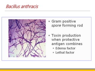
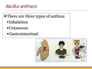
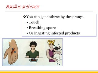
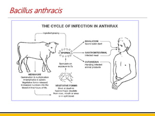
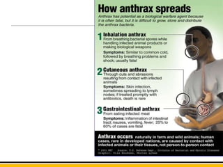
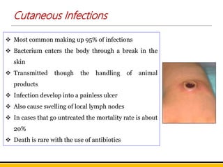
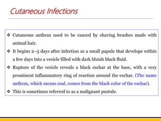
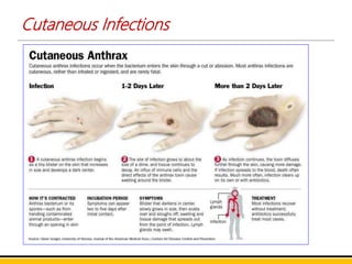
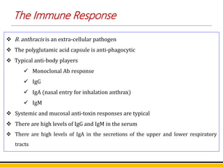
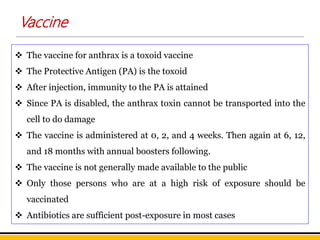
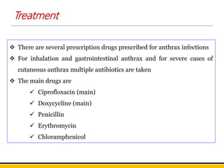
Dr. S. MEENATCHISUNDARAM
ASSOCIATE PROFESSOR
DEPARTMENT OF MICROBIOLOGY
SNMV COLLEGE OF ARTS AND SCIENCE
COIMBATORE
https://orcid.org/0000-0002-8691-449X
95496
https://scholar.google.com/citations?user=IkdZ5XsAAAAJ&hl=en
Bacillus anthracis
Anthrax
• Caused by spore forming
bacterium called Bacillus
anthracis
• Anthrax is an acute, febrile
disease of virtually all
worm blooded animals,
including man
Bacillus anthracis
• Anthrax is usually
transmitted to
humans secondary
• We usually get it
from animals such as
sheep, cattle, horses,
goats, and pigs; pigs
and horses rarely get
anthrax
Bacillus anthracis
 It was the first pathogenic bacterium to be observed under the microscope
(Pollender, 1849).
 The first communicable disease shown to be transmitted by inoculation of
infected blood (Davaine, 1850) was anthrax.
 B. anthracis was first bacillus to be isolated in pure culture and shown to
possess spores (Koch, 1876).
 It was in studies on anthrax that Koch demonstrated for the first time a set of
criteria or postulates.
 The first bacterium used for the preparation of an attenuated vaccine by
Pasteur.
 Nobel Prize winner Metchnikoff studied virulent and attenuated strains of
B. anthracis, in his pioneering work on phagocytosis.
Bacillus anthracis
Bacillus anthracis
 B. anthracis is one of the largest of pathogenic bacteria.
 3–8 by 1–1.3 μm and is gram-positive nonacid fast, non-motile
straight, sporing bacillus.
 It is rectangular in shape and arranged in filamentous chains in
culture.
 In cultures, the bacilli are arranged end to end in long chains.
 The ends of the bacilli are truncated or often concave and
somewhat swollen so that a chain of bacilli presents a ‘bamboo
stick’ appearance.
Bacillus anthracis
 The spore is oval (ellipsoidal), refractile,
central in position and of the same
diameter as the Bacillus and not swelling
the mother cell
 Spores are formed in culture, in the soil,
and in the tissue and exudates of dead
animals but never in the blood or tissues
of living animals.
 Spores seen as unstained spaces in Gram-
stained bacilli and, when free, faintly
outlined with Gram counterstain.
 The anthrax bacillus is nonmotile, unlike
most other members of this genus
Bacillus anthracis
 It is found singly, in pairs or in short chains in tissues.
 The entire chain being surrounded by a capsule which is
polypeptide in nature, being composed of a polymer of d (–)
glutamic acid.
 Capsules are formed in the animal body but in culture only if
the media contain added bicarbonate or are incubated under
10–25% CO2.
 They are gram-positive
Bacillus anthracis
 When blood films containing anthrax bacilli are stained with
polychrome methylene blue for a few seconds and examined
under the microscope, an amorphous purplish material is
noticed around the blue bacilli.
 This represents the capsular material and is characteristic of the
anthrax bacillus. This is called the M’Fadyean’s reaction and
is employed for the presumptive diagnosis of anthrax in
animals.
 Purple bacillus with red capsule is seen with Giemsa’s stain.
 Fat globules may be made out within the bacilli when stained
with Sudan black
 B. Spores seen as unstained spaces in Gram stained bacilli and,
when free, faintly outlined with Gram counterstain.
Cultural Characteristics
 It is aerobe and facultative anaerobe.
 Temperature range for growth is 12–45°C (optimum 37°C).
 Good growth occurs on ordinary media.
Nutrient agar:
 On nutrient agar, colonies are irregularly round, 2–3 mm in
diameter, raised, dull, opaque, greyish white, with a frosted
glass appearance.
 The edge of the colony is composed of long, interlacing chains of
bacilli, resembling locks of matted hair under the low power
microscope.
 This is called the ‘Medusa head appearance’
Cultural Characteristics
Blood agar:
Colonies on horse or sheep blood agar are virtually nonhemolytic
In a gelatin stab, there is growth down the stab line with lateral
spikes, longer near the surface, giving an ‘inverted fir tree’
appearance
Selective medium:
A selective medium (PLET medium), consisting of polymyxin,
lysozyme, ethylene diamine tetra acetic acid (EDTA) and
thallous acetate added to heart infusion agar, has been
devised to isolate B. antracis from mixtures containing other spore-
bearing bacilli.
Cultural Characteristics
Solid medium containing penicillin:
 When B. anthracis is grown on a solid medium containing 0.05–
0.5 units penicillin per mL, in 3–4 hours the cells become large,
spherical, and occurs in chains on the surface of the agar,
resembling a string of pearls.
 This string of pearls reaction useful in differentiation of B.
anthracis from B. cereus and other aerobic spore bearers.
Resistance
 The spores are resistant to chemical disinfectants and heat.
 With moist heat, the vegetative bacilli are killed at 60°C in 30
minutes and the spores at 100°C in 10 minutes.
 With dry heat the spores are killed at 150°C in 60 minutes.
 The spores are also killed by 4% formaldehyde or 4% potassium
permanganate in a few minutes.
 The bacilli are sensitive to benzylpenicillin, streptomycin,
tetracyclines, chloramphenicol, ciprofloxacin, the
cephalosporins and sulfonamides
Bacillus anthracis
There are three types of anthrax
Inhalation
Cutaneous
Gastrointestinal
Bacillus anthracis
You can get anthrax by three ways
• Touch
• Breathing spores
• Or ingesting infected products
Bacillus anthracis
Cutaneous Infections
 Most common making up 95% of infections
 Bacterium enters the body through a break in the
skin
 Transmitted though the handling of animal
products
 Infection develop into a painless ulcer
 Also cause swelling of local lymph nodes
 In cases that go untreated the mortality rate is about
20%
 Death is rare with the use of antibiotics
Cutaneous Infections
 Cutaneous anthrax used to be caused by shaving brushes made with
animal hair.
 It begins 2–5 days after infection as a small papule that develops within
a few days into a vesicle filled with dark bluish black fluid.
 Rupture of the vesicle reveals a black eschar at the base, with a very
prominent inflammatory ring of reaction around the eschar. (The name
anthrax, which means coal, comes from the black color of the eschar).
 This is sometimes referred to as a malignant pustule.
Cutaneous Infections
 The lesion is classically found on the hands, forearms, or head and is
painless.
 The disease used to be common in dock workers carrying loads of hides
and skins on their bare backs and hence was known as the ‘hide porter’s
disease.’
 Cutaneous anthrax generally resolves spontaneously, but 10–20% of
untreated patients may develop fatal septicemia or meningitis.
Cutaneous Infections
 Transmission is through the inhalation of anthrax
spores
 This is the type that was used in the 2001
bioterrorist attack
 Starts with common cold and progresses to severe
breathing problems and shock
 Inhalation anthrax is usually fatal
 This picture shows the lung tissue of a patient
infected with inhalation anthrax
 The red arrow points to the infective spores
Inhalation Anthrax
 Pulmonary anthrax, known as ‘wool-sorter’s disease, because it used to
be common in workers in wool factories, due to inhalation of dust from
infected wool.
 It occurs in patients who handle raw wool, hides, or horsehair and
acquire the disease by the inhalation of spores.
 This is a hemorrhagic pneumonia with a high fatality rate.
 Hemorrhagic meningitis may occur as a complication
Inhalation Anthrax
 Transmitted through eating of undercooked
contaminated meat or animal products
 Results in acute inflammation of the gut
 Causes nausea, vomiting, and diarrhea which
become more severe with time
 The mortality rate of patients infected with
Gastrointestinal Anthrax is anywhere from 25-60%
 This is a picture of the intestines of a patient that
died from Gastrointestinal Anthrax
 There is severe edema and hemorrhage
Gastrointestinal Anthrax
 Intestinal anthrax, is rare and occurs mainly in primitive communities
who eat the carcasses of animals dying of anthrax.
 An individual may suffer after a day or so from hemorrhagic diarrhea,
and dies rapidly from septicemia.
Gastrointestinal Anthrax
 The lethal affects of a B. anthracis infection are mediated by a toxin
 The toxin has 3 parts
 A lethal factor (LF)
 An edema factor (EF)
 A protective antigen (PA)
 PA transports LF and EF to the cytosol of cells where they do their
damage
 The protective antigen is used to induce immunity
Toxin
 B. anthracis is an extra-cellular pathogen
 The polyglutamic acid capsule is anti-phagocytic
 Typical anti-body players
 Monoclonal Ab response
 IgG
 IgA (nasal entry for inhalation anthrax)
 IgM
 Systemic and mucosal anti-toxin responses are typical
 There are high levels of IgG and IgM in the serum
 There are high levels of IgA in the secretions of the upper and lower respiratory
tracts
The Immune Response
 The vaccine for anthrax is a toxoid vaccine
 The Protective Antigen (PA) is the toxoid
 After injection, immunity to the PA is attained
 Since PA is disabled, the anthrax toxin cannot be transported into the
cell to do damage
 The vaccine is administered at 0, 2, and 4 weeks. Then again at 6, 12,
and 18 months with annual boosters following.
 The vaccine is not generally made available to the public
 Only those persons who are at a high risk of exposure should be
vaccinated
 Antibiotics are sufficient post-exposure in most cases
Vaccine
 Military personnel
 Laboratory workers who come into contact with the organism
 Anyone who handles animal products that are at a high potential of
contamination
Who is at risk?
 There are several prescription drugs prescribed for anthrax infections
 For inhalation and gastrointestinal anthrax and for severe cases of
cutaneous anthrax multiple antibiotics are taken
 The main drugs are
 Ciprofloxacin (main)
 Doxycycline (main)
 Penicillin
 Erythromycin
 Chloramphenicol
Treatment
Treatment
Penicillin
Tetracyclines
Amoxicillin
Chloramphenicol
Ciprofloxacin
Doxycycline
Erythromycin
Gentamicin
Streptomycin
Sulfonamides

BACILLUS ANTHRACIS

  • 1.
    Dr. S. MEENATCHISUNDARAM ASSOCIATEPROFESSOR DEPARTMENT OF MICROBIOLOGY SNMV COLLEGE OF ARTS AND SCIENCE COIMBATORE https://orcid.org/0000-0002-8691-449X 95496 https://scholar.google.com/citations?user=IkdZ5XsAAAAJ&hl=en Bacillus anthracis
  • 2.
    Anthrax • Caused byspore forming bacterium called Bacillus anthracis • Anthrax is an acute, febrile disease of virtually all worm blooded animals, including man
  • 3.
    Bacillus anthracis • Anthraxis usually transmitted to humans secondary • We usually get it from animals such as sheep, cattle, horses, goats, and pigs; pigs and horses rarely get anthrax
  • 4.
    Bacillus anthracis  Itwas the first pathogenic bacterium to be observed under the microscope (Pollender, 1849).  The first communicable disease shown to be transmitted by inoculation of infected blood (Davaine, 1850) was anthrax.  B. anthracis was first bacillus to be isolated in pure culture and shown to possess spores (Koch, 1876).  It was in studies on anthrax that Koch demonstrated for the first time a set of criteria or postulates.  The first bacterium used for the preparation of an attenuated vaccine by Pasteur.  Nobel Prize winner Metchnikoff studied virulent and attenuated strains of B. anthracis, in his pioneering work on phagocytosis.
  • 5.
  • 6.
    Bacillus anthracis  B.anthracis is one of the largest of pathogenic bacteria.  3–8 by 1–1.3 μm and is gram-positive nonacid fast, non-motile straight, sporing bacillus.  It is rectangular in shape and arranged in filamentous chains in culture.  In cultures, the bacilli are arranged end to end in long chains.  The ends of the bacilli are truncated or often concave and somewhat swollen so that a chain of bacilli presents a ‘bamboo stick’ appearance.
  • 7.
    Bacillus anthracis  Thespore is oval (ellipsoidal), refractile, central in position and of the same diameter as the Bacillus and not swelling the mother cell  Spores are formed in culture, in the soil, and in the tissue and exudates of dead animals but never in the blood or tissues of living animals.  Spores seen as unstained spaces in Gram- stained bacilli and, when free, faintly outlined with Gram counterstain.  The anthrax bacillus is nonmotile, unlike most other members of this genus
  • 8.
    Bacillus anthracis  Itis found singly, in pairs or in short chains in tissues.  The entire chain being surrounded by a capsule which is polypeptide in nature, being composed of a polymer of d (–) glutamic acid.  Capsules are formed in the animal body but in culture only if the media contain added bicarbonate or are incubated under 10–25% CO2.  They are gram-positive
  • 9.
    Bacillus anthracis  Whenblood films containing anthrax bacilli are stained with polychrome methylene blue for a few seconds and examined under the microscope, an amorphous purplish material is noticed around the blue bacilli.  This represents the capsular material and is characteristic of the anthrax bacillus. This is called the M’Fadyean’s reaction and is employed for the presumptive diagnosis of anthrax in animals.  Purple bacillus with red capsule is seen with Giemsa’s stain.  Fat globules may be made out within the bacilli when stained with Sudan black  B. Spores seen as unstained spaces in Gram stained bacilli and, when free, faintly outlined with Gram counterstain.
  • 10.
    Cultural Characteristics  Itis aerobe and facultative anaerobe.  Temperature range for growth is 12–45°C (optimum 37°C).  Good growth occurs on ordinary media. Nutrient agar:  On nutrient agar, colonies are irregularly round, 2–3 mm in diameter, raised, dull, opaque, greyish white, with a frosted glass appearance.  The edge of the colony is composed of long, interlacing chains of bacilli, resembling locks of matted hair under the low power microscope.  This is called the ‘Medusa head appearance’
  • 11.
    Cultural Characteristics Blood agar: Colonieson horse or sheep blood agar are virtually nonhemolytic In a gelatin stab, there is growth down the stab line with lateral spikes, longer near the surface, giving an ‘inverted fir tree’ appearance Selective medium: A selective medium (PLET medium), consisting of polymyxin, lysozyme, ethylene diamine tetra acetic acid (EDTA) and thallous acetate added to heart infusion agar, has been devised to isolate B. antracis from mixtures containing other spore- bearing bacilli.
  • 12.
    Cultural Characteristics Solid mediumcontaining penicillin:  When B. anthracis is grown on a solid medium containing 0.05– 0.5 units penicillin per mL, in 3–4 hours the cells become large, spherical, and occurs in chains on the surface of the agar, resembling a string of pearls.  This string of pearls reaction useful in differentiation of B. anthracis from B. cereus and other aerobic spore bearers.
  • 13.
    Resistance  The sporesare resistant to chemical disinfectants and heat.  With moist heat, the vegetative bacilli are killed at 60°C in 30 minutes and the spores at 100°C in 10 minutes.  With dry heat the spores are killed at 150°C in 60 minutes.  The spores are also killed by 4% formaldehyde or 4% potassium permanganate in a few minutes.  The bacilli are sensitive to benzylpenicillin, streptomycin, tetracyclines, chloramphenicol, ciprofloxacin, the cephalosporins and sulfonamides
  • 14.
    Bacillus anthracis There arethree types of anthrax Inhalation Cutaneous Gastrointestinal
  • 15.
    Bacillus anthracis You canget anthrax by three ways • Touch • Breathing spores • Or ingesting infected products
  • 16.
  • 18.
    Cutaneous Infections  Mostcommon making up 95% of infections  Bacterium enters the body through a break in the skin  Transmitted though the handling of animal products  Infection develop into a painless ulcer  Also cause swelling of local lymph nodes  In cases that go untreated the mortality rate is about 20%  Death is rare with the use of antibiotics
  • 19.
    Cutaneous Infections  Cutaneousanthrax used to be caused by shaving brushes made with animal hair.  It begins 2–5 days after infection as a small papule that develops within a few days into a vesicle filled with dark bluish black fluid.  Rupture of the vesicle reveals a black eschar at the base, with a very prominent inflammatory ring of reaction around the eschar. (The name anthrax, which means coal, comes from the black color of the eschar).  This is sometimes referred to as a malignant pustule.
  • 20.
    Cutaneous Infections  Thelesion is classically found on the hands, forearms, or head and is painless.  The disease used to be common in dock workers carrying loads of hides and skins on their bare backs and hence was known as the ‘hide porter’s disease.’  Cutaneous anthrax generally resolves spontaneously, but 10–20% of untreated patients may develop fatal septicemia or meningitis.
  • 21.
  • 22.
     Transmission isthrough the inhalation of anthrax spores  This is the type that was used in the 2001 bioterrorist attack  Starts with common cold and progresses to severe breathing problems and shock  Inhalation anthrax is usually fatal  This picture shows the lung tissue of a patient infected with inhalation anthrax  The red arrow points to the infective spores Inhalation Anthrax
  • 23.
     Pulmonary anthrax,known as ‘wool-sorter’s disease, because it used to be common in workers in wool factories, due to inhalation of dust from infected wool.  It occurs in patients who handle raw wool, hides, or horsehair and acquire the disease by the inhalation of spores.  This is a hemorrhagic pneumonia with a high fatality rate.  Hemorrhagic meningitis may occur as a complication Inhalation Anthrax
  • 24.
     Transmitted througheating of undercooked contaminated meat or animal products  Results in acute inflammation of the gut  Causes nausea, vomiting, and diarrhea which become more severe with time  The mortality rate of patients infected with Gastrointestinal Anthrax is anywhere from 25-60%  This is a picture of the intestines of a patient that died from Gastrointestinal Anthrax  There is severe edema and hemorrhage Gastrointestinal Anthrax
  • 25.
     Intestinal anthrax,is rare and occurs mainly in primitive communities who eat the carcasses of animals dying of anthrax.  An individual may suffer after a day or so from hemorrhagic diarrhea, and dies rapidly from septicemia. Gastrointestinal Anthrax
  • 26.
     The lethalaffects of a B. anthracis infection are mediated by a toxin  The toxin has 3 parts  A lethal factor (LF)  An edema factor (EF)  A protective antigen (PA)  PA transports LF and EF to the cytosol of cells where they do their damage  The protective antigen is used to induce immunity Toxin
  • 27.
     B. anthracisis an extra-cellular pathogen  The polyglutamic acid capsule is anti-phagocytic  Typical anti-body players  Monoclonal Ab response  IgG  IgA (nasal entry for inhalation anthrax)  IgM  Systemic and mucosal anti-toxin responses are typical  There are high levels of IgG and IgM in the serum  There are high levels of IgA in the secretions of the upper and lower respiratory tracts The Immune Response
  • 28.
     The vaccinefor anthrax is a toxoid vaccine  The Protective Antigen (PA) is the toxoid  After injection, immunity to the PA is attained  Since PA is disabled, the anthrax toxin cannot be transported into the cell to do damage  The vaccine is administered at 0, 2, and 4 weeks. Then again at 6, 12, and 18 months with annual boosters following.  The vaccine is not generally made available to the public  Only those persons who are at a high risk of exposure should be vaccinated  Antibiotics are sufficient post-exposure in most cases Vaccine
  • 29.
     Military personnel Laboratory workers who come into contact with the organism  Anyone who handles animal products that are at a high potential of contamination Who is at risk?
  • 30.
     There areseveral prescription drugs prescribed for anthrax infections  For inhalation and gastrointestinal anthrax and for severe cases of cutaneous anthrax multiple antibiotics are taken  The main drugs are  Ciprofloxacin (main)  Doxycycline (main)  Penicillin  Erythromycin  Chloramphenicol Treatment
  • 31.

Editor's Notes

  • #2 This template was inserted from Power-user, the productivity add-in for PowerPoint and Excel. Get thousands of templates, icons, maps, diagrams and charts with Power-user. Visit www.powerusersoftwares.com!