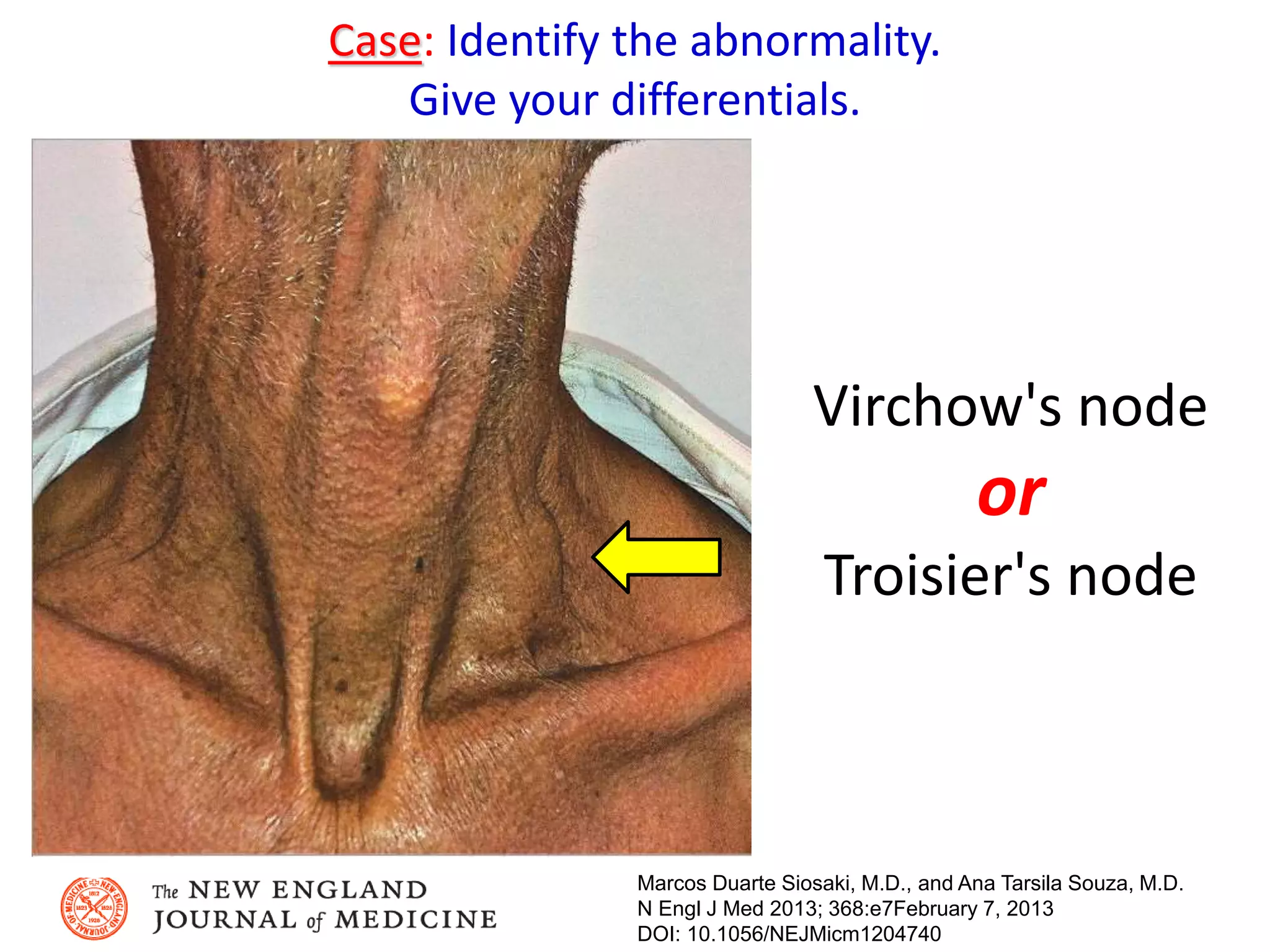
Downloaded 49 times

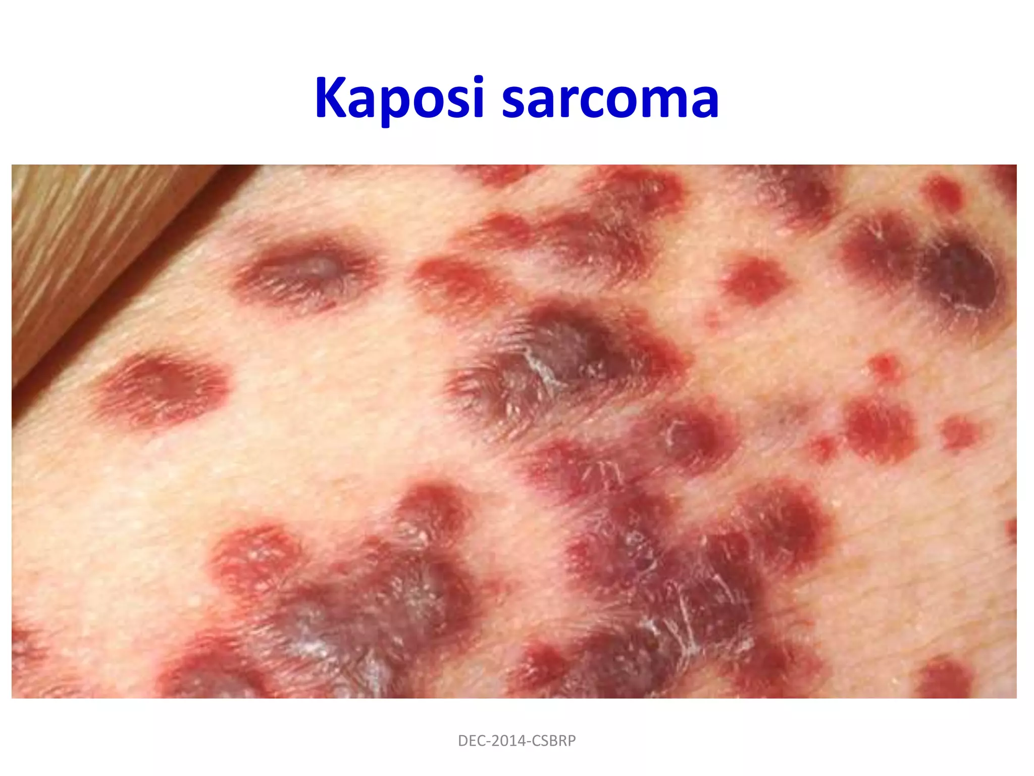
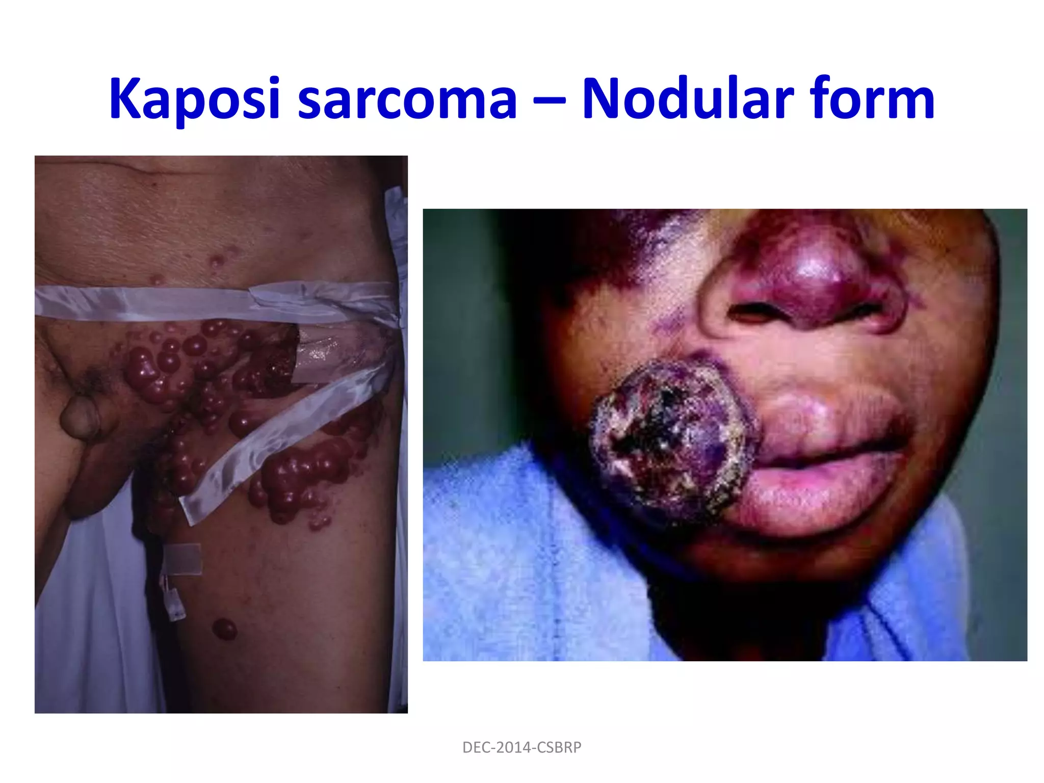
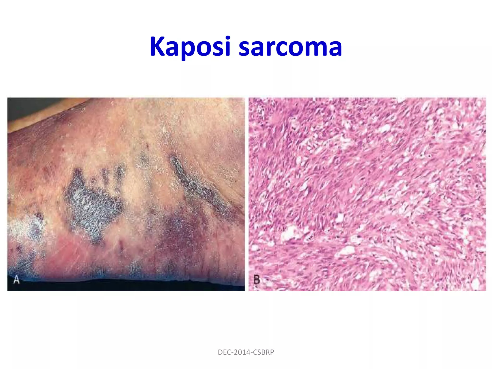
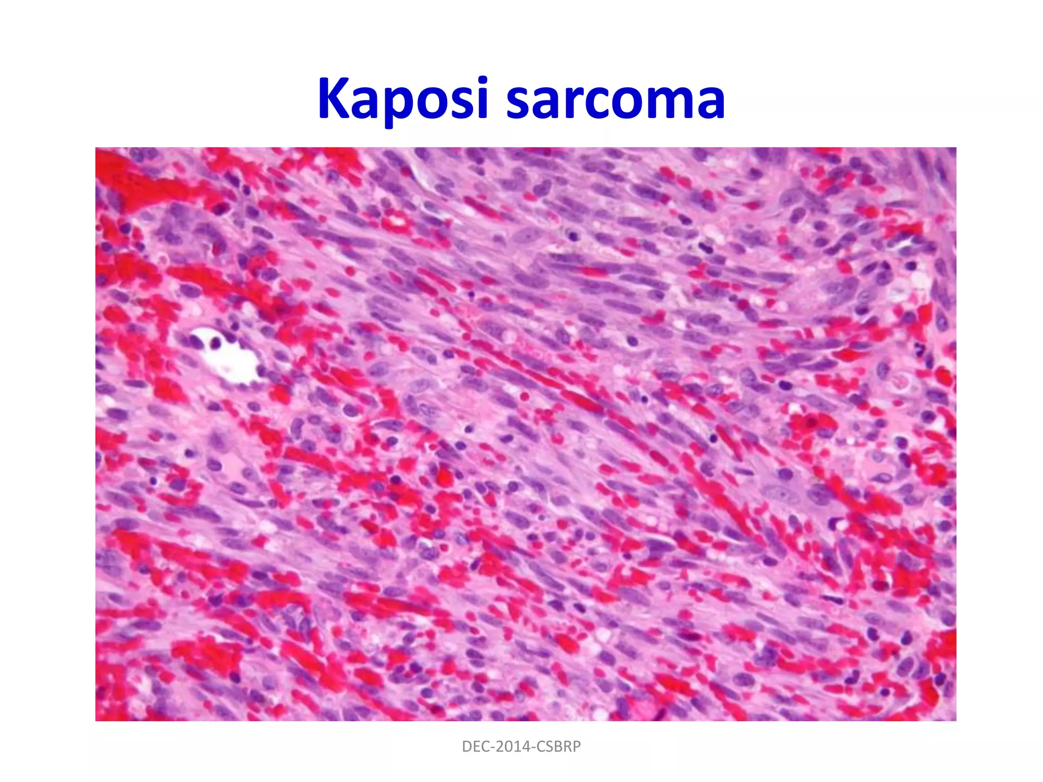
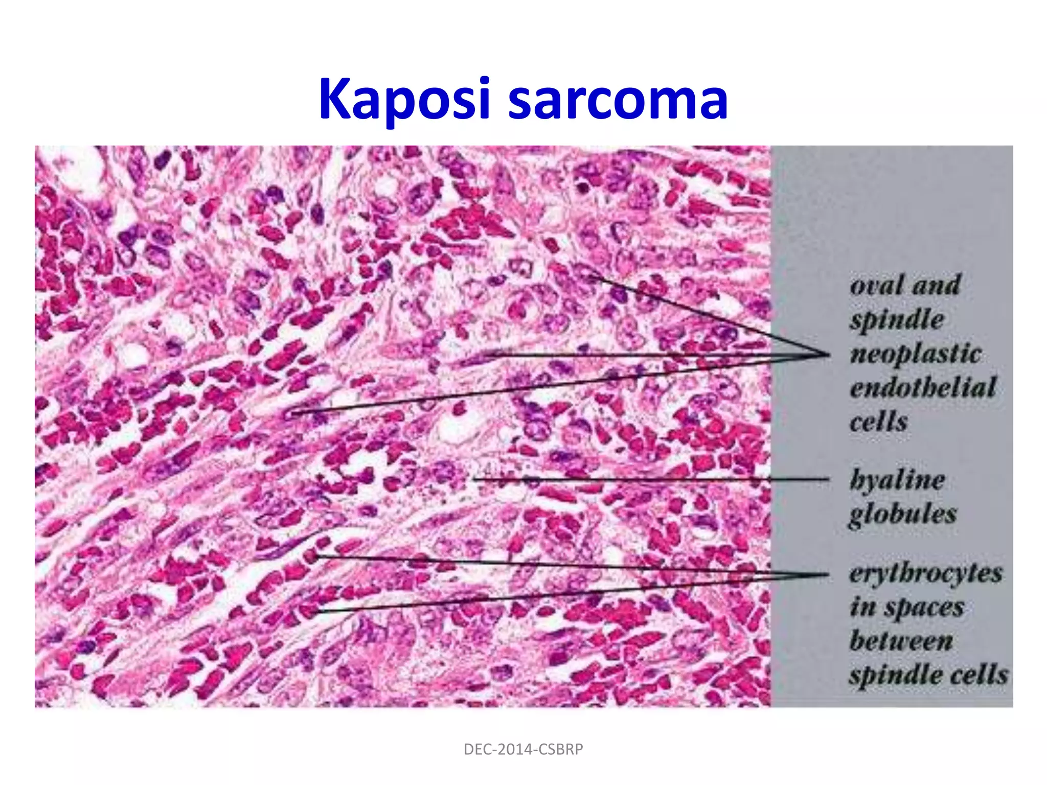
![Kaposi sarcoma (KS)
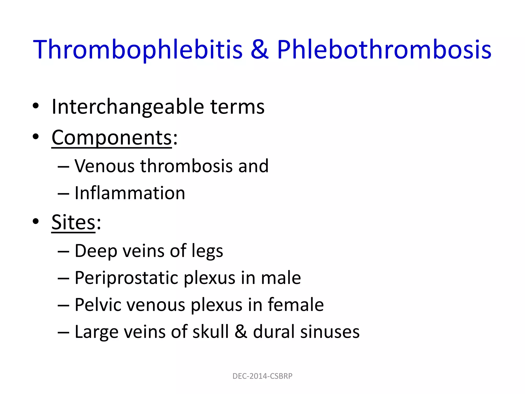
Forms of KS:
• Chronic KS (Classic)
• Lymphadenopathic KS
• Transplant associated KS
• AIDS associated KS
Pathogenesis:
• HHV8 (KSHV)
• Latent infection in endothelial cells > [p53
inhibitors + Cyclin-D equivalent + viral G protein
which induces VEGF] > Cellular proliferation
DEC-2014-CSBRP](https://image.slidesharecdn.com/cvs-misc-1-csbrp-151029115933-lva1-app6891/75/Cvs-misc-1-csbrp-41-2048.jpg)
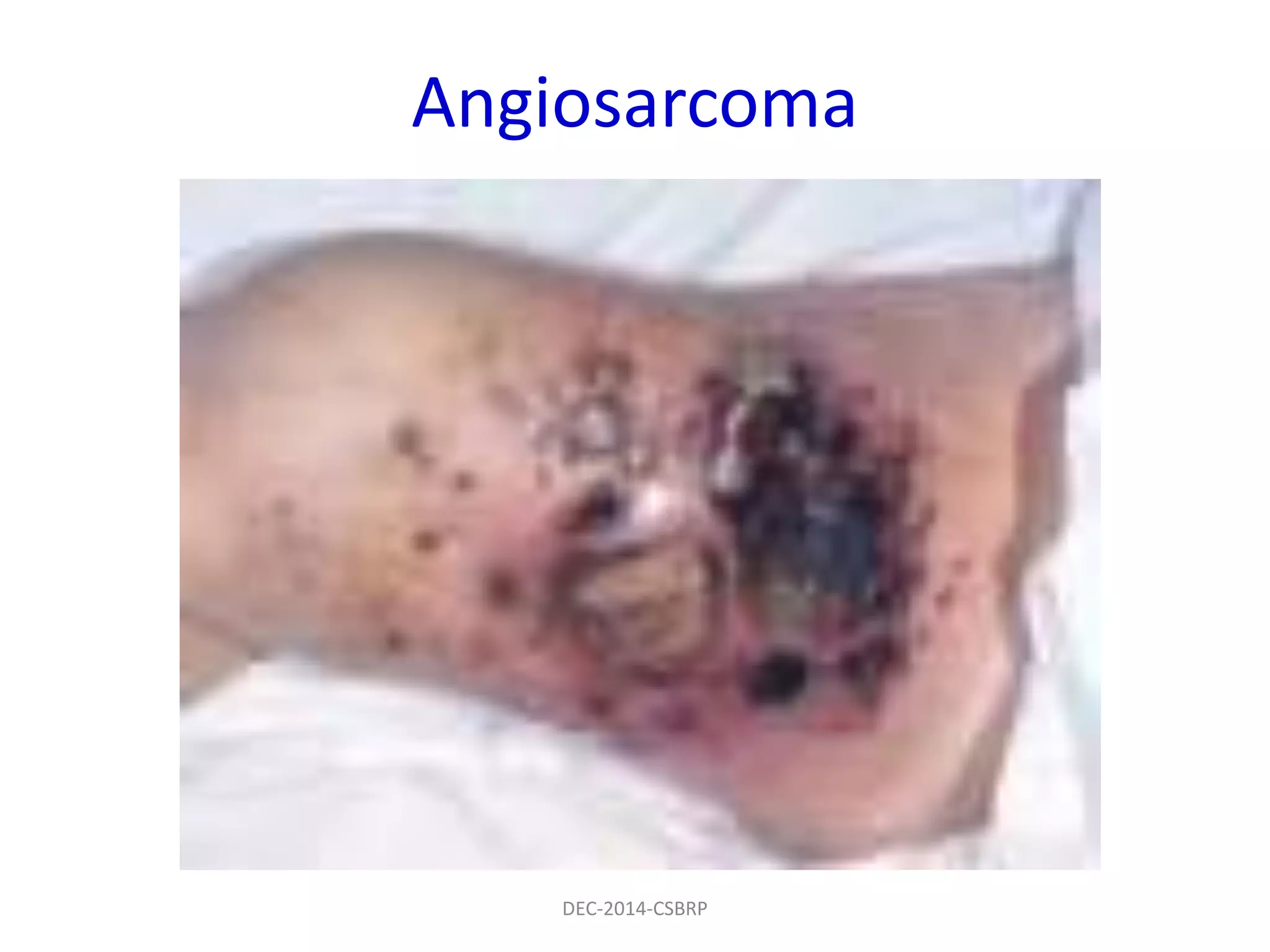
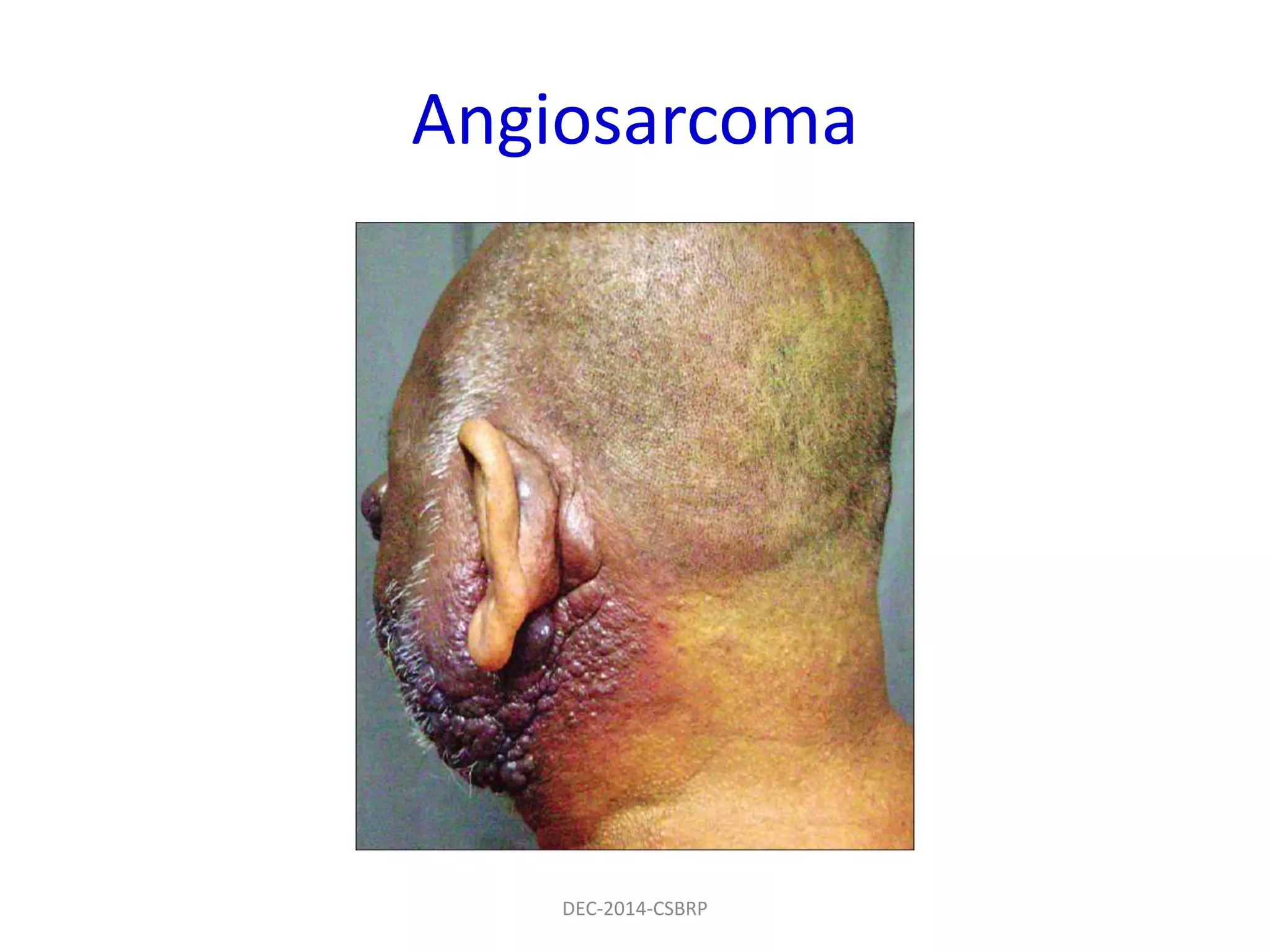


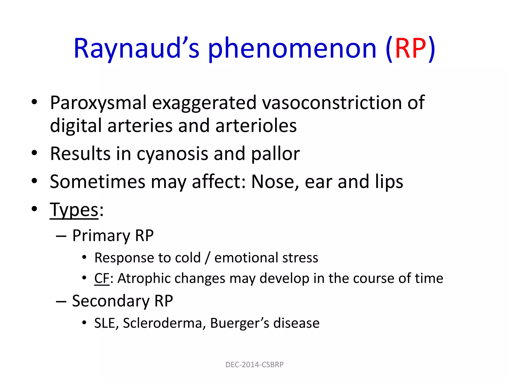
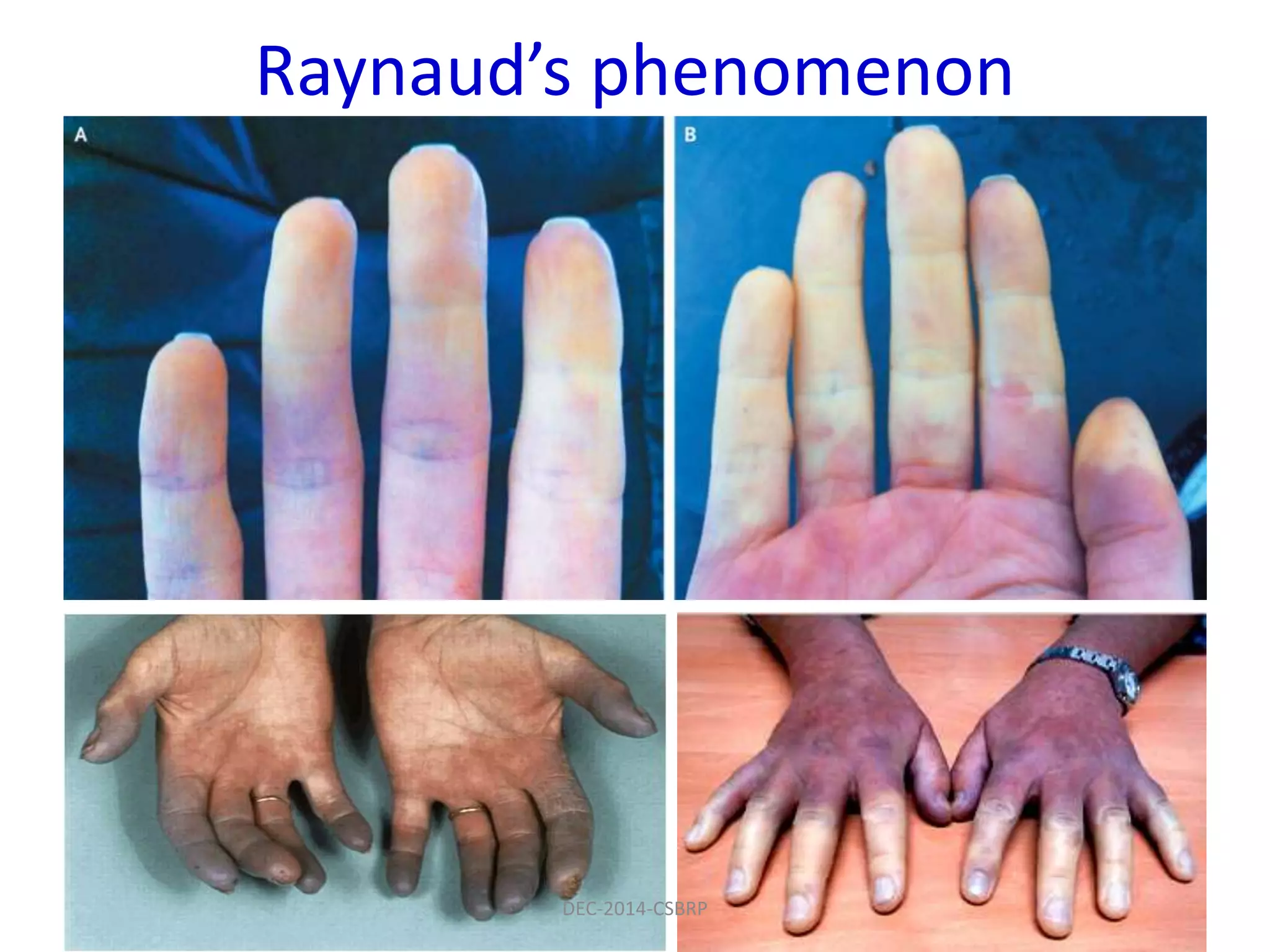
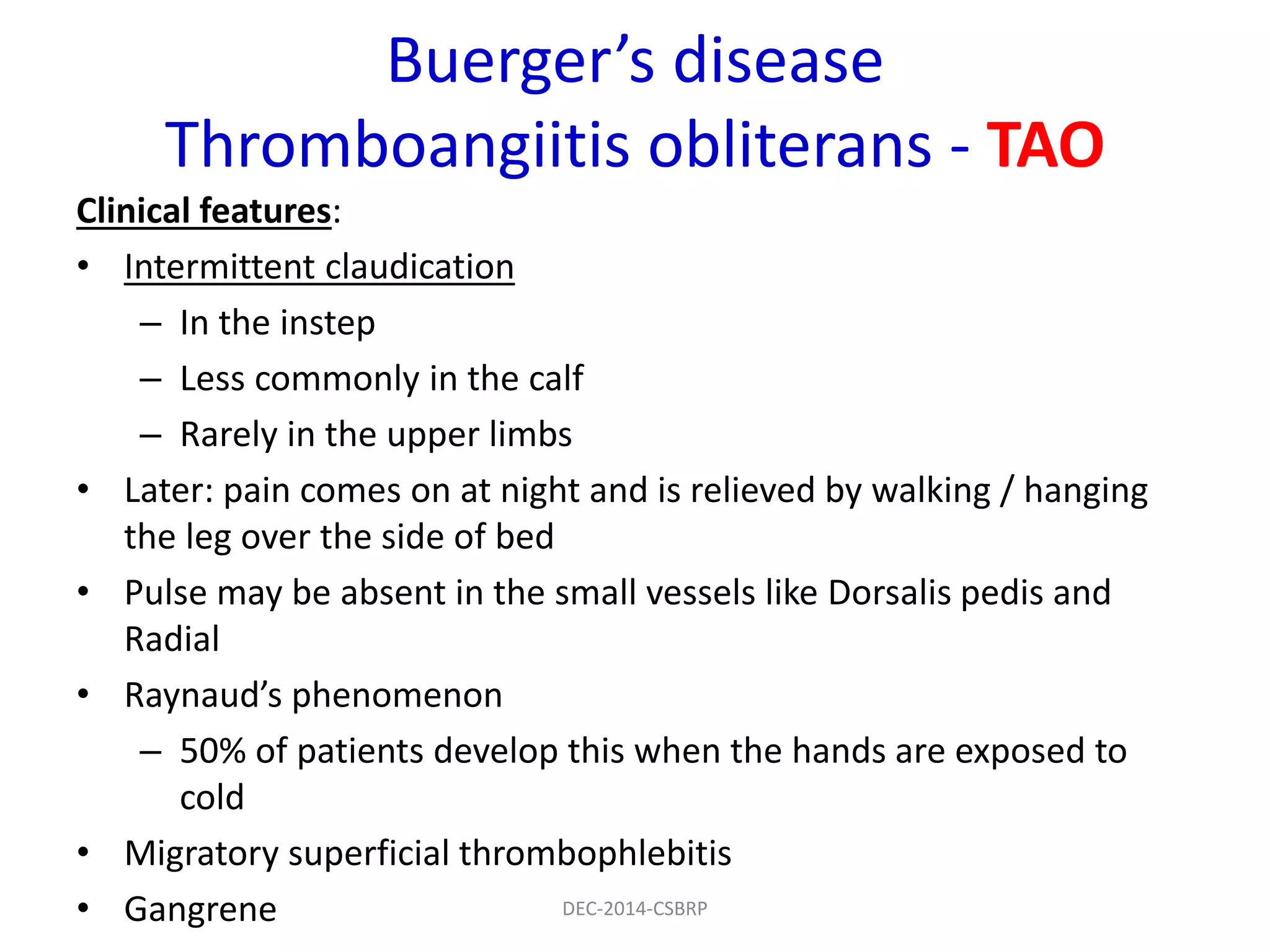
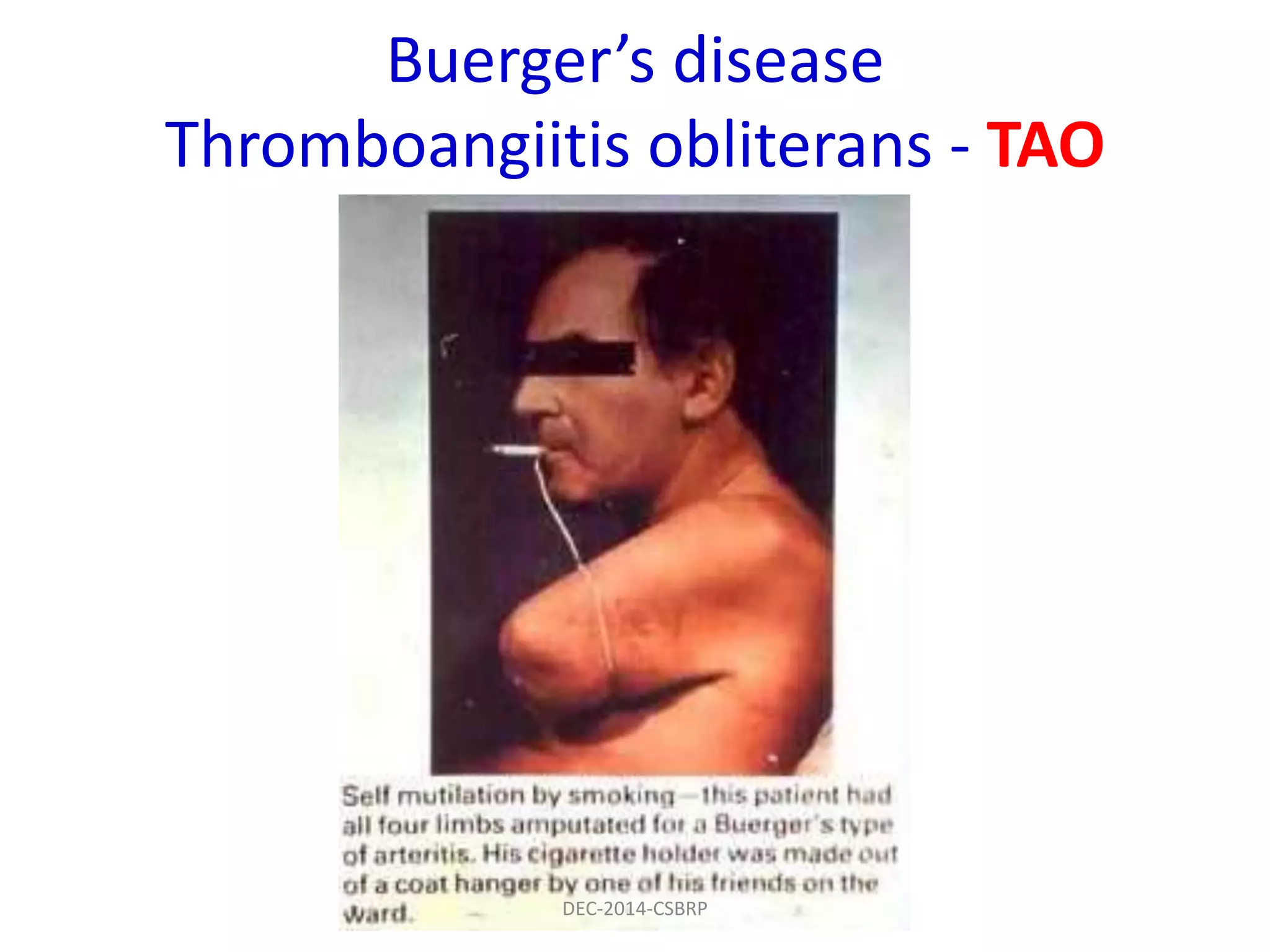
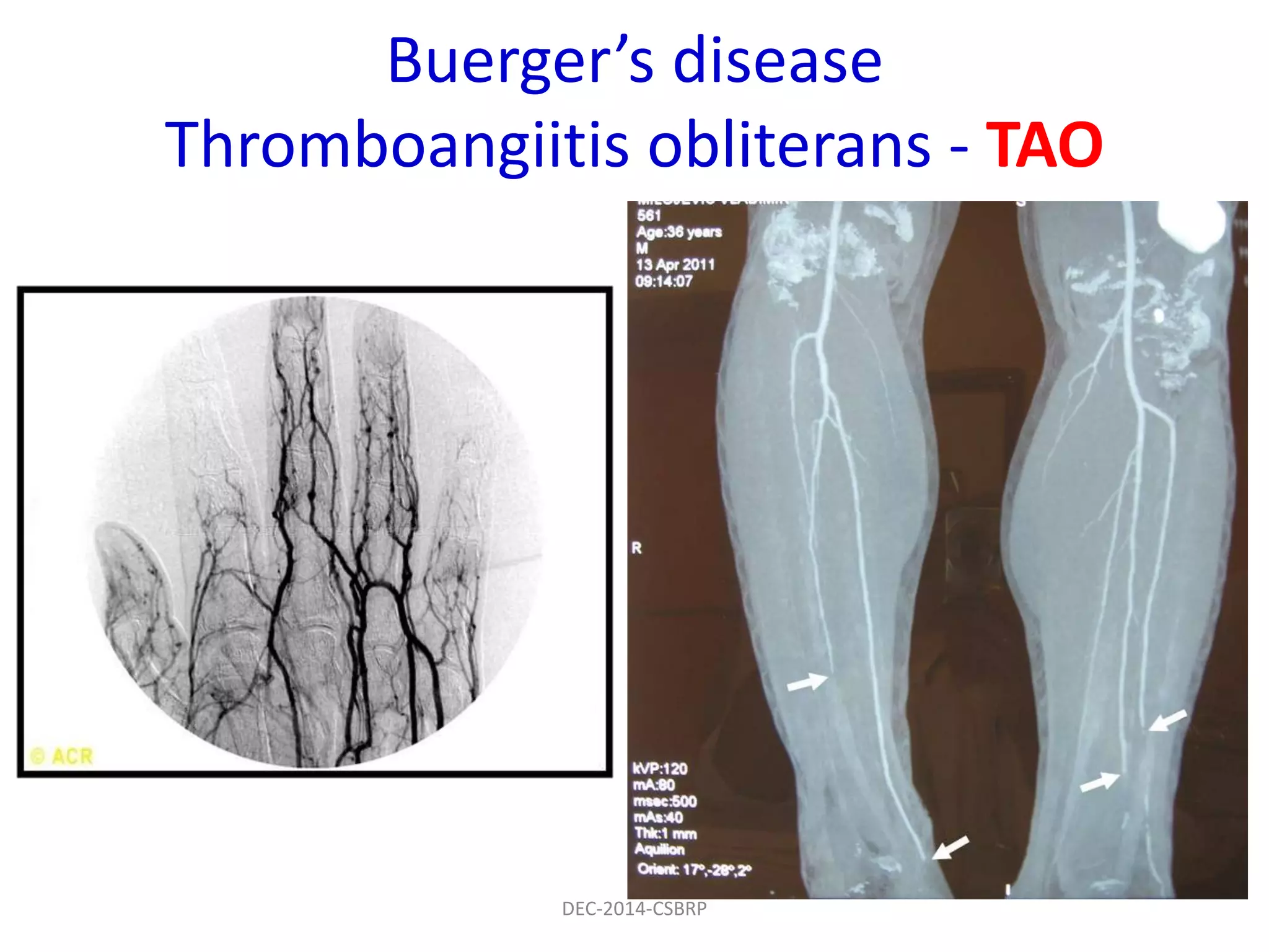
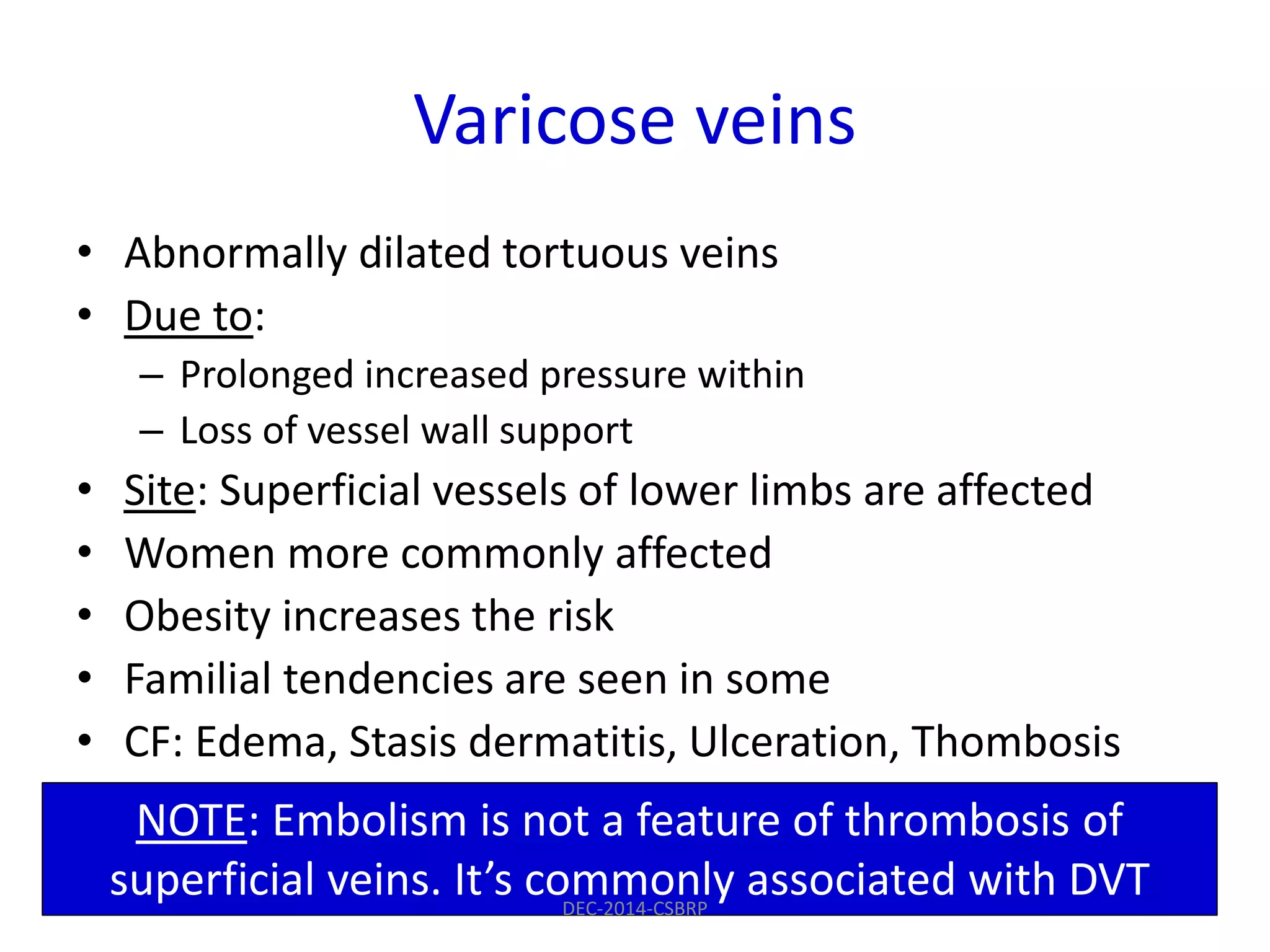
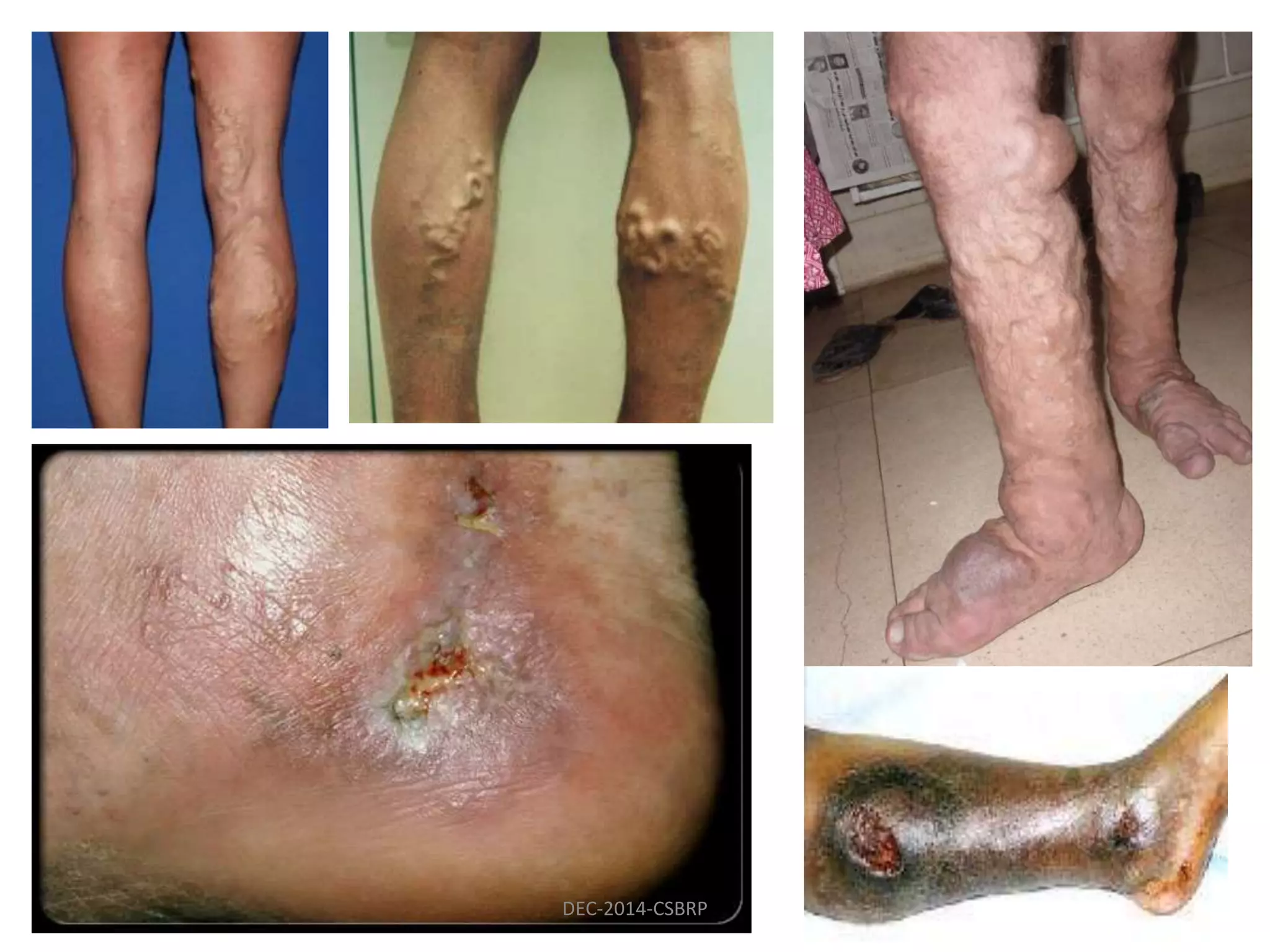
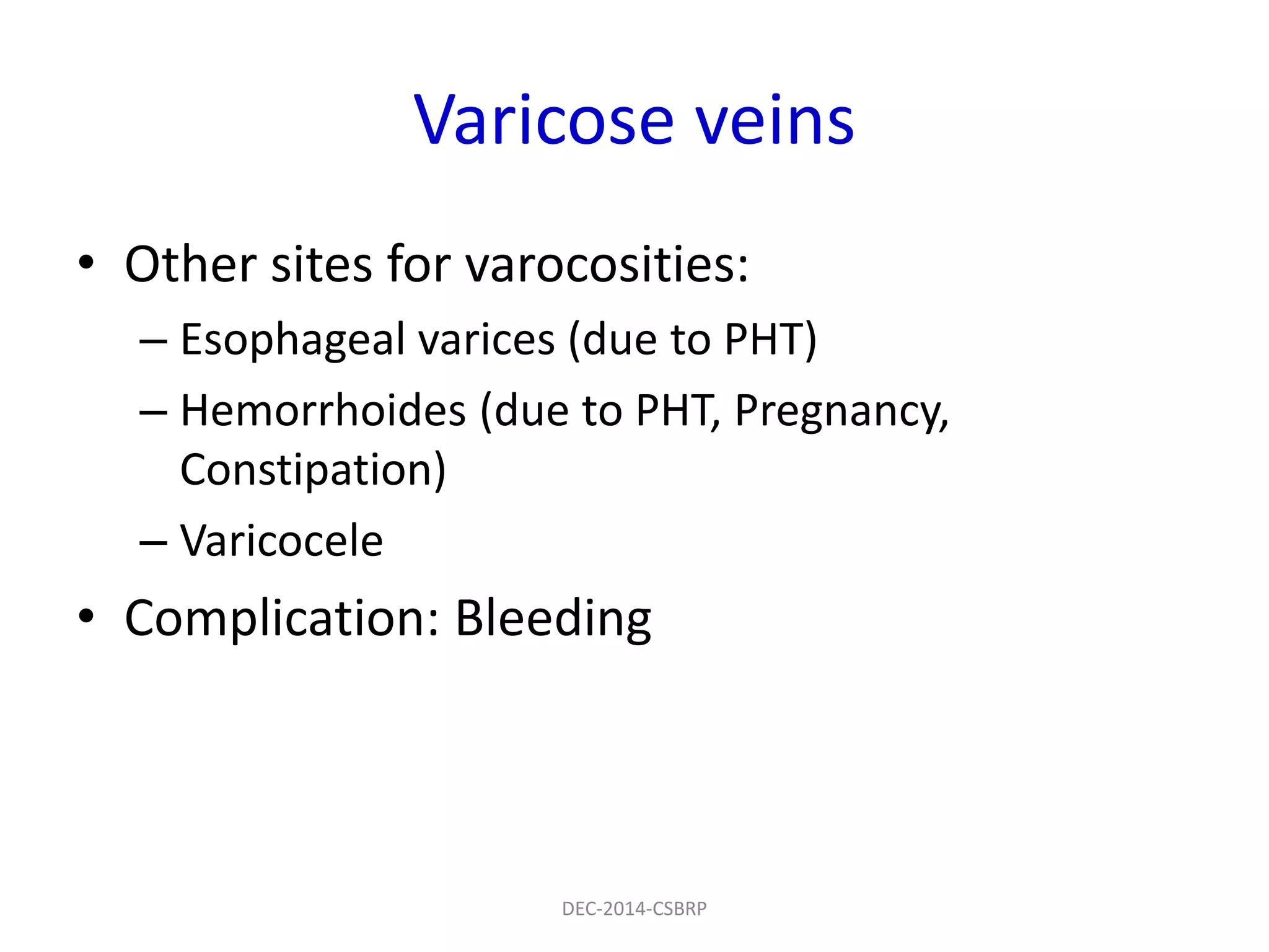
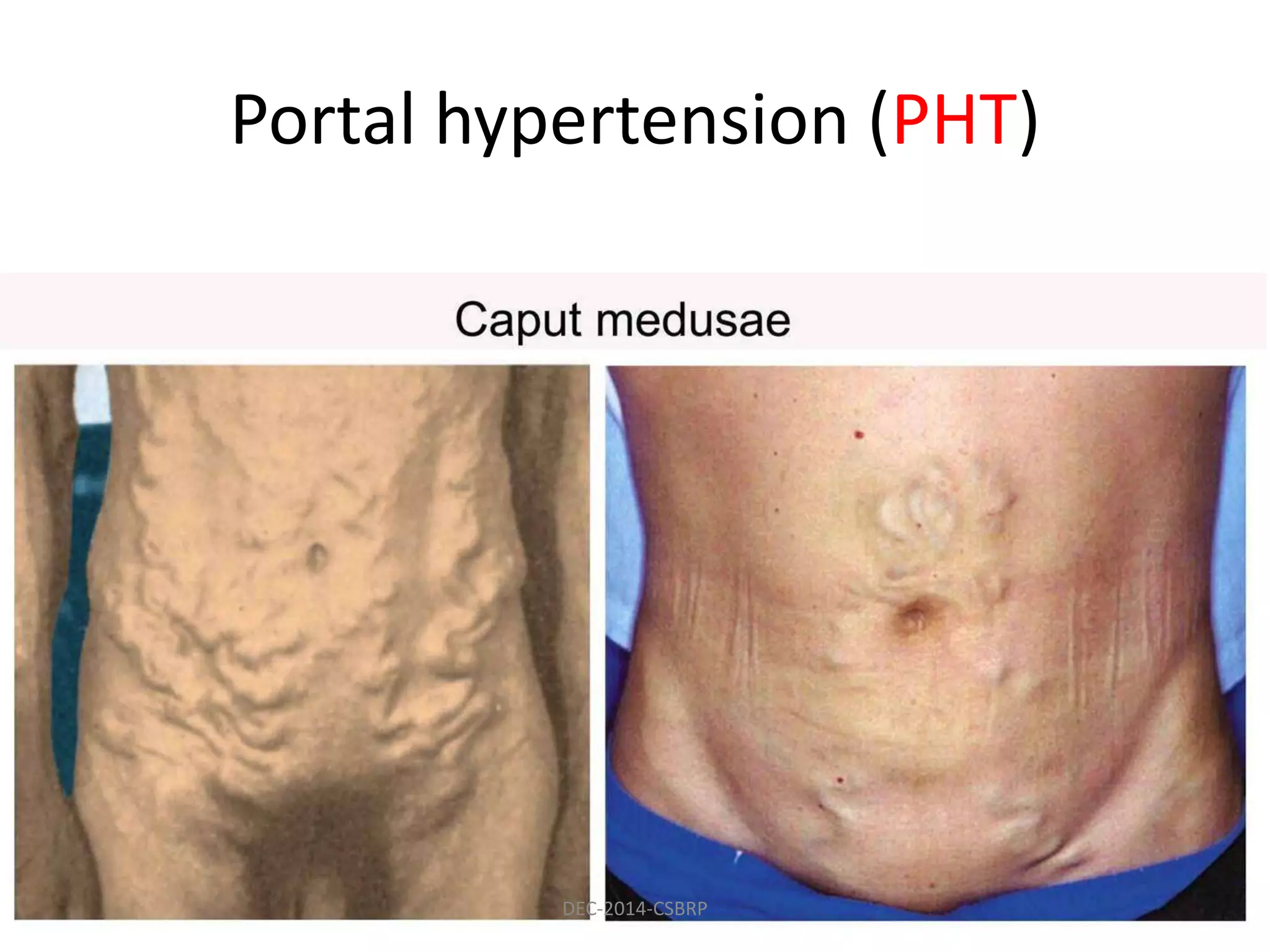
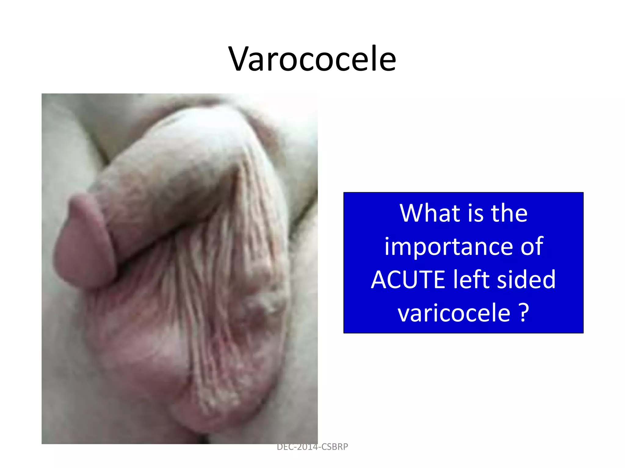
1. Raynaud's phenomenon is characterized by paroxysmal vasoconstriction of the digital arteries, typically in response to cold or stress, and results in color changes in the fingers. It can occur primary or secondary to conditions like SLE or scleroderma. 2. Buerger's disease (thromboangiitis obliterans) is a non-atherosclerotic inflammatory disease affecting small and medium arteries of the limbs. It mainly affects young male smokers and causes symptoms like claudication, pain relieved by hanging the leg over the bed, and Raynaud's phenomenon. 3. Varicose veins are abnormally dilated and tortuous veins most commonly seen


