SlideShare a Scribd company logo
BACHELOR OF SCIENCE IN NURSING:
HEALTH ASSESSMENT
COURSE MODULE COURSE UNIT WEEK
3 13 13
Adult Physical Assessment: Peripheral Vascular System
✓ Read course and unit objectives
✓ Read and comprehend study guide prior to
class attendance
✓ Read and comprehend required learning
resources
✓ Engage in classroom discussions
✓ Participate in weekly discussion board
(Canvas)
✓ Answer and submit course unit tasks
At the end of this unit, the students are expected to:
Cognitive:
1. Describe the structure and the function of the blood vessels, including capillaries and
lymphatic circulation.
2. Discuss risk factors associated with peripheral vascular disease across the cultures and ways
to reduce one’s risks.
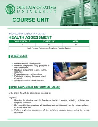
3. Perform a physical assessment of the peripheral vascular system using the correct
techniques.
4. Differentiate between normal and abnormal findings of the peripheral vascular system.
5. Analyze the data from the interview and physical assessment of the peripheral vascular
system to formulate valid nursing diagnosis, collaborative problems, and/or referrals.
Affective:
1. Listen attentively during class discussions
2. Demonstrate tact and respect when challenging other people’s opinions and ideas
3. Accept comments and reactions of classmates on one’s opinions openly and graciously.
Psychomotor:
1. Participate actively during class discussions
2. Confidently express personal opinion and thoughts in front of the class
Weber, J.R., and Kelley, J. H., (2018) Health Assessment in Nursing 6th Edition, Philadelphia:
Wolters Kluwer
ASSESSMENT OF THE PERIPHERAL VASCULAR SYSTEM
Structure and Function
Arteries
• These are blood vessels that carry oxygenated, nutrient rich blood from the heart to the
capillaries
• A high-pressure system
• Arterial pulse
o The surge of blood as a result from a heartbeat which forces blood through the arterial
vessels under high pressure
• Major Arteries of the Arm
o Brachial artery
▪ Major artery that supplies the arm
▪ Palpated medial to the biceps tendon in and above the bend of the elbow
▪ Divides near the elbow to become the radial artery (extending down the thumb
side of the arm) and the ulnar artery (extending down the little-finger side of the
arm)
• Both arteries provide blood to the hand
o Radial artery
▪ Palpated on the lateral aspect of the wrist
o Ulnar artery
▪ Located on the medial aspect of the wrist
▪ A deeper pulse, not easily palpated
• Major Arteries of the Leg
o Femoral artery
▪ Major supplier of blood to the legs
▪ Pulse palpated under the inguinal ligament
o Popliteal artery
▪ Pulse palpated behind the knee
o Dorsalis pedis artery
▪ Pulse palpated on the great-toe side of the top of the foot
o Posterior tibial artery
▪ Palpated behind the medial malleolus of the ankle
Veins
• Carry deoxygenated, nutrient depleted, waste-laden blood from the tissues back to the heart
• The veins of the arms, upper trunk, head, and neck carry blood to the superior vena cava,
where it passes into the right atrium
• Blood from the lower trunk and legs drains upward into the inferior vena cava
• Contain nearly 70% of the body’s blood volume
• Walls are much thinner, low-pressure system
• 3 types
o Deep veins
▪ Femoral veins
▪ Popliteal veins
o Superficial veins
▪ Great and small saphenous veins
o Perforator (or communicator) veins
▪ Connect the superficial veins with the deep veins
• 3 mechanisms of venous function
o 1st mechanism
▪ Structure of the veins
o 2nd mechanism
▪ Muscular contraction
o 3rd mechanism
▪ Creation of a pressure gradient through the act of breathing
Capillaries
• Small blood vessels that form the connection between the arterioles and venules
• Allow the circulatory system to maintain the vital equilibrium between the vascular and
interstitial spaces
Collecting Objective Data
Physical Examination
• The purpose is to identify any signs or symptoms of PVD including arterial insufficiency,
venous insufficiency, or lymphatic involvement
• Useful in acute care, extended care, and home health care settings
• A complete peripheral vascular examination involves inspection, palpation, and
auscultation
• When performing PE, the nurse should:
o Discuss risk factors for PVD with the client.
o Accurately inspect arms and legs for edema and venous patterning
o Observe carefully for signs of arterial and venous insufficiency (skin color, venous
pattern, hair distribution, lesions or ulcers) and inadequate lymphatic drainage
o Recognize characteristic clubbing
o Palpate pulse points correctly
o Use the Doppler ultrasound instrument correctly
Assessment Procedure
Arms
Inspection
• Observe coloration of the hands and arms
o Normal findings
▪ Color varies depending on the client’s skin tone, although color should be the
same bilaterally
o Abnormal findings
▪ Raynaud disorder
• A vascular disorder caused by vasoconstriction or vasospasm of the
fingers or toes, characterized by rapid changes of color (pallor,
cyanosis, and redness), swelling, pain, numbness, tingling, burning,
throbbing, and coldness
Palpation
• Palpate the client’s fingers, hands, and arms, and note the temperature
o Normal findings
▪ Skin is warm to the touch bilaterally from fingertips to upper arms
o Abnormal findings
▪ A cool extremity may be a sign of arterial insufficiency.
▪ Cold fingers and hands, for example, are common findings with Raynaud’s
• Palpate to assess capillary refill time.
✓ Compress the nailbed until it blanches. Release the pressure and calculate the time it takes
for color to return. This test indicates peripheral perfusion and reflects cardiac output.
o Normal findings
▪ Capillary beds refill (and, therefore, color returns) in 2 seconds or less
o Abnormal findings
▪ Capillary refill time exceeding 2 seconds may indicate vasoconstriction,
decreased cardiac output, shock, arterial occlusion, or hypothermia
• Palpate the radial pulse.
✓ Gently press the radial artery against the radius.
Note elasticity and strength.
o Normal findings
▪ Radial pulses are bilaterally strong (2+). Artery walls have a resilient quality
(bounce).
o Abnormal findings
▪ Increased radial pulse volume indicates a hyperkinetic state (3+ or bounding
pulse). Diminished (1+) or absent (0) pulse suggests partial or complete arterial
occlusion (which is more common in the legs than the arms). The pulse could
also be decreased from Buerger’s disease or scleroderma.
• Palpate the ulnar pulses.
✓ Apply pressure with your first three fingertips to the medial
aspects of the inner wrists. The ulnar pulses are not
routinely assessed because they are located deeper than
the radial pulses and are difficult to detect. Palpate the
ulnar arteries if you suspect arterial insufficiency.
o Normal findings
▪ The ulnar pulses may not be detectable
o Abnormal findings
▪ Obliteration of the pulse may result from compression by external sources, as
in compartment syndrome. Lack of resilience or inelasticity of the artery wall
may indicate arteriosclerosis.
• Palpate the brachial pulses.
✓ Do this by placing the first three fingertips of each hand at
the client’s right and left medial antecubital creases.
Alternatively, palpate the brachial pulse in the groove
between the biceps and triceps.
o Normal findings
▪ Brachial pulses have equal strength
bilaterally
o Abnormal findings
▪ Brachial pulses are increased, diminished, or absent
• Perform the Allen test.
✓ Evaluates patency of the radial or ulnar arteries. It is done when patency is questionable or
before such procedures as a radial artery puncture.
✓ The test begins by assessing ulnar patency. Have the
client rest the hand palm side up on the examination table
and make a fist. Then use your thumbs to occlude the
radial and ulnar arteries.
✓ Continue pressure to keep both arteries occluded and have
the client release the fist.
✓ Note that the palm remains pale. Release the pressure on
the ulnar artery and watch for color to return to the hand.
To assess radial patency, repeat the procedure as before,
but at the last step, release pressure on the radial artery.
o Normal findings
▪ Pink coloration returns to the palms within 3–
5 seconds if the ulnar artery is patent.
▪ Pink coloration returns within 3–5 seconds if the radial artery is patent.
o Abnormal findings
▪ With arterial insufficiency or occlusion of the ulnar artery, pallor persists. With
arterial insufficiency or occlusion of the radial artery, pallor persists.
Legs
Inspection, Palpation, and Auscultation
✓ Ask the client to lie supine. Then drape the groin area and place a pillow under the client’s
head for comfort. Observe skin color while inspecting both legs from the toes to the groin.
• Observe skin color while inspecting both legs from the toes to the groin
o Normal findings
▪ Pink color for lighter-skinned clients and pink or red tones visible under darker-
pigmented skin. There should be no changes in pigmentation.
o Abnormal findings
▪ Pallor, especially when elevated, and rubor, when dependent, suggests arterial
insufficiency.
▪ Cyanosis when dependent suggests venous insufficiency.
▪ A rusty or brownish pigmentation around the ankles indicates venous
insufficiency
• Inspect distribution of hair on legs
o Normal findings
▪ Hair covers the skin on the legs and appears on the dorsal surface of the toes.
o Abnormal findings
▪ Loss of hair on the legs suggests arterial insufficiency. Often thin, shiny skin is
noted as well.
• Inspect for lesions or ulcers.
o Normal findings
▪ Legs are free of lesions or ulcerations.
o Abnormal findings
▪ Ulcers with smooth, even margins that occur at pressure areas, such as the
toes and lateral ankle, result from arterial insufficiency.
▪ Ulcers with irregular edges, bleeding, and possible bacterial infection that occur
on the medial ankle result from venous insufficiency.
• Inspect for edema.
✓ Inspect the legs for unilateral or bilateral edema. Note
veins, tendons, and bony prominences. If the legs appear
asymmetric, use a centimeter tape to measure in four
different areas: circumference at mid-thigh, largest
circumference at the calf, smallest circumference above the
ankle, and across the forefoot. Compare both extremities at
the same locations.
o Normal findings
▪ Identical size and shape bilaterally; no swelling or atrophy.
o Abnormal findings
▪ May be detected by the absence of visible veins, tendons, or bony
prominences.
▪ Bilateral edema usually indicates a systemic problem
▪ Unilateral edema is characterized by a 1-cm difference in measurement at the
ankles or a 2-cm difference at the calf, and a swollen extremity.
▪ A difference in measurement between legs may also be due to muscular
atrophy.
• Palpate edema.
✓ Determine if it is pitting or nonpitting. Press the edematous area with the tips of your fingers,
hold for a few seconds, then release. If the depression does not rapidly refill and the skin
remains indented on release, pitting edema is present.
o Normal findings
▪ No edema (pitting or nonpitting) present in the legs.
o Abnormal findings
▪ Pitting edema is associated with systemic problems
▪ A 1+ to 4+ scale is used to grade the severity of pitting edema, with 4+ being
most severe.
• Palpate bilaterally for temperature of the feet and legs.
✓ Use the backs of your fingers. Compare your findings in the same areas bilaterally. Note
location of any changes in temperature.
o Normal findings
▪ Toes, feet, and legs are equally warm bilaterally.
o Abnormal findings
▪ Generalized coolness in one leg or change in temperature from warm to cool
as you move down the leg suggests arterial insufficiency.
▪ Increased warmth in the leg may be caused by superficial thrombophlebitis
resulting from a secondary inflammation in the tissue around the vein.
• Palpate the femoral pulses.
✓ Ask the client to bend the knee and move it out to the side.
Press deeply and slowly below and medial to the inguinal
ligament. Use two hands if necessary. Release pressure
until you feel the pulse. Repeat palpation on the opposite
leg. Compare amplitude bilaterally
o Normal findings
▪ Femoral pulses strong and equal bilaterally.
o Abnormal findings
▪ Weak or absent femoral pulses indicate partial or complete arterial occlusion.
• Auscultate the femoral pulses.
✓ If arterial occlusion is suspected in the femoral pulse,
position the stethoscope over the femoral artery and listen
for bruits. Repeat for other artery.
o Normal findings
▪ No sounds auscultated over the femoral
arteries.
o Abnormal findings
▪ Bruits over one or both femoral arteries
suggest partial obstruction of the vessel and diminished blood flow to the lower
extremities.
• Palpate the popliteal pulses.
✓ Ask the client to raise (flex) the knee partially. Place your thumbs on the knee while positioning
your fingers deep in the bend of the knee. Apply pressure to locate the pulse. It is usually
detected lateral to the medial tendon.
o Normal findings
▪ It is not unusual for the popliteal pulse to be difficult or impossible to detect,
and yet for circulation to be normal.
o Abnormal findings
▪ Although normal popliteal arteries may be nonpalpable, an absent pulse may
also be the result of an occluded artery.
• Palpate the dorsalis pedis pulses.
✓ Dorsiflex the client’s foot and apply light pressure lateral to
and along the side of the extensor tendon of the big toe.
The pulses of both feet may be assessed at the same time
to aid in making comparisons. Assess amplitude bilaterally.
o Normal findings
▪ Dorsalis pedis pulses are bilaterally strong.
o Abnormal findings
▪ A weak or absent pulse may indicate impaired arterial circulation.
• Palpate the posterior tibial pulses.
✓ Palpate behind and just below the medial malleolus (in the
groove between the ankle and the Achilles tendon).
Palpating both posterior tibial pulses at the same time aids
in making comparisons. Assess amplitude bilaterally.
o Normal findings
▪ The posterior tibial pulses should be strong
bilaterally.
o Abnormal findings
▪ A weak or absent pulse indicates partial or complete arterial occlusion.
• Inspect for varicosities and thrombophlebitis.
✓ Ask the client to stand because varicose veins may not be visible when the client is supine
and not as pronounced when the client is sitting. As the client is standing, inspect for
superficial vein thrombophlebitis. To fully assess for a suspected phlebitis, lightly palpate for
tenderness. If superficial vein thrombophlebitis is present, note redness or discoloration on
the skin surface over the vein.
o Normal findings
▪ Veins are flat and barely seen under the surface of the skin.
o Abnormal findings
▪ Varicose veins may appear as distended, nodular, bulging, and tortuous,
depending on severity.
▪ Varicosities are common in the anterior lateral thigh and lower leg, the posterior
lateral calf, or anus (known as hemorrhoids).
▪ Superficial vein thrombophlebitis is marked by redness, thickening, and
tenderness along the vein.
▪ Aching or cramping may occur with walking.
Characteristics of Arterial and Venous Insufficiency
Arterial Insufficiency
• Pain: Intermittent claudication to sharp, unrelenting, constant
• Pulses: Diminished or absent
• Skin Characteristics: Dependent rubor
o Elevation pallor of foot
o Dry, shiny skin
o Cool-to-cold temperature
o Loss of hair over toes and dorsum of foot
o Nails thickened and ridged
• Ulcer Characteristics:
o Location: Tips of toes, toe webs, heel or other
pressure areas if confined to bed
o Pain: Very painful
o Depth of ulcer: Deep, often involving joint space
o Shape: Circular
o Ulcer base: Pale black to dry and gangrene
o Leg edema: Minimal unless extremity kept in
dependent position constantly to relieve pain
Venous Insufficiency
• Pain: Aching, cramping
• Pulses: Present but may be difficult to palpate through
edema
• Skin Characteristics:
o Pigmentation in gaiter area (area of medial and
lateral malleolus)
o Skin thickened and tough
o May be reddish-blue in color
o Frequently associated with dermatitis
• Ulcer Characteristics:
o Location: Medial malleolus or anterior tibial area
o Pain: If superficial, minimal pain; but may be very
painful
o Depth of ulcer: Superficial
o Shape: Irregular border
o Ulcer base: Granulation tissue—beefy red to yellow fibrinous in chronic long-term ulcer
o Leg edema: Moderate to severe
Types of Peripheral Edema
Edema Associated with Lymphedema
• Caused by abnormal or blocked lymph vessels
• Nonpitting
• Usually bilateral; may be unilateral
• No skin ulceration or pigmentation
Edema Associated with Chronic Venous Insufficiency
• Caused by obstruction or insufficiency of deep veins
• Pitting, documented as:
o 1+ = slight pitting
o 2+ = deeper than 1+
o 3+ = noticeably deep pit; extremity looks larger
o 4+ = very deep pit; gross edema in extremity
• Usually unilateral; may be bilateral
• Skin ulceration and pigmentation may be present
Abnormal Arterial Findings
• Necrotic great toes with blisters on toes and foot
• Raynaud Disease
o Blanching of fingers on both hands
Abnormal Venous Findings
• Superficial thrombophlebitis
o Often seen with unilateral localized pain, achiness, edema, redness, and warmth to
touch
• Lymphedema
• Varicose veins
D’Amico, D., and Barbarito, C., (2019) Health & Physical Assessment in Nursing 3rd Edition,
Singapore: Pearson Education, Inc.
Weber, J.R., and Kelley, J. H., (2018) Health Assessment in Nursing
6th Edition, Philadelphia: Wolters Kluwer

More Related Content

What's hot

Bowel elimination
Bowel eliminationBowel elimination
Bowel elimination
NEHA BHARTI
 
Nursing Case study potts disease
Nursing Case study potts diseaseNursing Case study potts disease
Nursing Case study potts disease
pinoy nurze
 
Assessing the breasts and axillae
Assessing the breasts and axillae Assessing the breasts and axillae
Assessing the breasts and axillae
chrissie argana
 
Stages of dying
Stages of dyingStages of dying
Stages of dying
rajnee shrestha
 
Assessment of the anus & rectum
Assessment of the anus & rectum Assessment of the anus & rectum
Assessment of the anus & rectum
Carmela Domocmat
 
Adult physical assessment
Adult physical assessmentAdult physical assessment
Adult physical assessment
Chinna Chadayan
 
Assessment of the musclo skletal system
Assessment of the musclo skletal systemAssessment of the musclo skletal system
Assessment of the musclo skletal system
Mohamed Mansor
 
Note on assessment of renal or urinary system
Note on assessment of renal or urinary systemNote on assessment of renal or urinary system
Note on assessment of renal or urinary system
Babitha Devu
 
Fracture,types and its management-MSN...
Fracture,types and its management-MSN...Fracture,types and its management-MSN...
Fracture,types and its management-MSN...
RNRM Shalini Rana
 
Gatrointestinal assessment
Gatrointestinal assessmentGatrointestinal assessment
Gatrointestinal assessment
CHETAN RSANGATI
 
Vital signs Lecture
Vital signs LectureVital signs Lecture
Vital signs Lecture
enrico baldoviso
 

What's hot (11)

Bowel elimination
Bowel eliminationBowel elimination
Bowel elimination
 
Nursing Case study potts disease
Nursing Case study potts diseaseNursing Case study potts disease
Nursing Case study potts disease
 
Assessing the breasts and axillae
Assessing the breasts and axillae Assessing the breasts and axillae
Assessing the breasts and axillae
 
Stages of dying
Stages of dyingStages of dying
Stages of dying
 
Assessment of the anus & rectum
Assessment of the anus & rectum Assessment of the anus & rectum
Assessment of the anus & rectum
 
Adult physical assessment
Adult physical assessmentAdult physical assessment
Adult physical assessment
 
Assessment of the musclo skletal system
Assessment of the musclo skletal systemAssessment of the musclo skletal system
Assessment of the musclo skletal system
 
Note on assessment of renal or urinary system
Note on assessment of renal or urinary systemNote on assessment of renal or urinary system
Note on assessment of renal or urinary system
 
Fracture,types and its management-MSN...
Fracture,types and its management-MSN...Fracture,types and its management-MSN...
Fracture,types and its management-MSN...
 
Gatrointestinal assessment
Gatrointestinal assessmentGatrointestinal assessment
Gatrointestinal assessment
 
Vital signs Lecture
Vital signs LectureVital signs Lecture
Vital signs Lecture
 

Similar to CM3 - CU13 ASSESSMENT OF PERIPHERAL VASCULAR SYSTEM.pdf

CU13-assessment-of-perivascular-system.pptx
CU13-assessment-of-perivascular-system.pptxCU13-assessment-of-perivascular-system.pptx
CU13-assessment-of-perivascular-system.pptx
ZyraPascual1
 
Assessing the Thorax and Lungs presentation
Assessing the  Thorax  and  Lungs presentationAssessing the  Thorax  and  Lungs presentation
Assessing the Thorax and Lungs presentation
srslytrd
 
12. Peripheral Vascular System-bates.pptx
12. Peripheral Vascular System-bates.pptx12. Peripheral Vascular System-bates.pptx
12. Peripheral Vascular System-bates.pptx
MuSic402834
 
Surgery 6th year, Tutorial (Dr. Aram Baram)
Surgery 6th year, Tutorial (Dr. Aram Baram)Surgery 6th year, Tutorial (Dr. Aram Baram)
Surgery 6th year, Tutorial (Dr. Aram Baram)
College of Medicine, Sulaymaniyah
 
general physical examination .pptx
general physical examination .pptxgeneral physical examination .pptx
general physical examination .pptx
NaeemShehzad18
 
Detailed first and second year OSCE stations
Detailed first and second year OSCE stationsDetailed first and second year OSCE stations
Detailed first and second year OSCE stations
meducationdotnet
 
CM3 - CU12 ASSESSMENT OF HEART & NECK VESSELS.pdf
CM3 - CU12 ASSESSMENT OF HEART & NECK VESSELS.pdfCM3 - CU12 ASSESSMENT OF HEART & NECK VESSELS.pdf
CM3 - CU12 ASSESSMENT OF HEART & NECK VESSELS.pdf
ZyraPascual1
 
Cardiovascular examination
Cardiovascular examinationCardiovascular examination
Cardiovascular examination
abeerabdulkareem
 
Peripheral Vascular Examination
Peripheral  Vascular  ExaminationPeripheral  Vascular  Examination
Peripheral Vascular Examination
Shkar Ahmed
 
Basics of nursing of patient with heart disease 1.pptx
Basics of nursing of patient with heart disease 1.pptxBasics of nursing of patient with heart disease 1.pptx
Basics of nursing of patient with heart disease 1.pptx
sneha334357
 
GENERAL PHYSICAL EXAMINATION
GENERAL PHYSICAL EXAMINATIONGENERAL PHYSICAL EXAMINATION
GENERAL PHYSICAL EXAMINATION
NiteshKumar1150
 
Cardiovascular System
Cardiovascular SystemCardiovascular System
Cardiovascular assessment aser
Cardiovascular assessment aser Cardiovascular assessment aser
Cardiovascular assessment aser
AlAhly sporting club
 
Cardiovascular assessment
Cardiovascular assessmentCardiovascular assessment
Cardiovascular assessment
Gianne Gregorio
 
Chapter 6
Chapter 6Chapter 6
Chapter 6
Reach Na
 
Cardiovascular assessment
Cardiovascular assessment Cardiovascular assessment
Cardiovascular assessment
faculty of nursing Tanta University
 
Peripheral pulsations and blood pressure measurement
Peripheral pulsations and blood pressure measurementPeripheral pulsations and blood pressure measurement
Peripheral pulsations and blood pressure measurement
abeerabdulkareem
 
vasospastic.pptx
vasospastic.pptxvasospastic.pptx
vasospastic.pptx
AvinashYakkaliAvin
 
Bood pressure .ppt
Bood pressure .pptBood pressure .ppt
Bood pressure .ppt
JarallahAlghobari
 
Heart
HeartHeart

Similar to CM3 - CU13 ASSESSMENT OF PERIPHERAL VASCULAR SYSTEM.pdf (20)

CU13-assessment-of-perivascular-system.pptx
CU13-assessment-of-perivascular-system.pptxCU13-assessment-of-perivascular-system.pptx
CU13-assessment-of-perivascular-system.pptx
 
Assessing the Thorax and Lungs presentation
Assessing the  Thorax  and  Lungs presentationAssessing the  Thorax  and  Lungs presentation
Assessing the Thorax and Lungs presentation
 
12. Peripheral Vascular System-bates.pptx
12. Peripheral Vascular System-bates.pptx12. Peripheral Vascular System-bates.pptx
12. Peripheral Vascular System-bates.pptx
 
Surgery 6th year, Tutorial (Dr. Aram Baram)
Surgery 6th year, Tutorial (Dr. Aram Baram)Surgery 6th year, Tutorial (Dr. Aram Baram)
Surgery 6th year, Tutorial (Dr. Aram Baram)
 
general physical examination .pptx
general physical examination .pptxgeneral physical examination .pptx
general physical examination .pptx
 
Detailed first and second year OSCE stations
Detailed first and second year OSCE stationsDetailed first and second year OSCE stations
Detailed first and second year OSCE stations
 
CM3 - CU12 ASSESSMENT OF HEART & NECK VESSELS.pdf
CM3 - CU12 ASSESSMENT OF HEART & NECK VESSELS.pdfCM3 - CU12 ASSESSMENT OF HEART & NECK VESSELS.pdf
CM3 - CU12 ASSESSMENT OF HEART & NECK VESSELS.pdf
 
Cardiovascular examination
Cardiovascular examinationCardiovascular examination
Cardiovascular examination
 
Peripheral Vascular Examination
Peripheral  Vascular  ExaminationPeripheral  Vascular  Examination
Peripheral Vascular Examination
 
Basics of nursing of patient with heart disease 1.pptx
Basics of nursing of patient with heart disease 1.pptxBasics of nursing of patient with heart disease 1.pptx
Basics of nursing of patient with heart disease 1.pptx
 
GENERAL PHYSICAL EXAMINATION
GENERAL PHYSICAL EXAMINATIONGENERAL PHYSICAL EXAMINATION
GENERAL PHYSICAL EXAMINATION
 
Cardiovascular System
Cardiovascular SystemCardiovascular System
Cardiovascular System
 
Cardiovascular assessment aser
Cardiovascular assessment aser Cardiovascular assessment aser
Cardiovascular assessment aser
 
Cardiovascular assessment
Cardiovascular assessmentCardiovascular assessment
Cardiovascular assessment
 
Chapter 6
Chapter 6Chapter 6
Chapter 6
 
Cardiovascular assessment
Cardiovascular assessment Cardiovascular assessment
Cardiovascular assessment
 
Peripheral pulsations and blood pressure measurement
Peripheral pulsations and blood pressure measurementPeripheral pulsations and blood pressure measurement
Peripheral pulsations and blood pressure measurement
 
vasospastic.pptx
vasospastic.pptxvasospastic.pptx
vasospastic.pptx
 
Bood pressure .ppt
Bood pressure .pptBood pressure .ppt
Bood pressure .ppt
 
Heart
HeartHeart
Heart
 

Recently uploaded

Basavarajeeyam - Ayurvedic heritage book of Andhra pradesh
Basavarajeeyam - Ayurvedic heritage book of Andhra pradeshBasavarajeeyam - Ayurvedic heritage book of Andhra pradesh
Basavarajeeyam - Ayurvedic heritage book of Andhra pradesh
Dr. Madduru Muni Haritha
 
A Classical Text Review on Basavarajeeyam
A Classical Text Review on BasavarajeeyamA Classical Text Review on Basavarajeeyam
A Classical Text Review on Basavarajeeyam
Dr. Jyothirmai Paindla
 
The Best Ayurvedic Antacid Tablets in India
The Best Ayurvedic Antacid Tablets in IndiaThe Best Ayurvedic Antacid Tablets in India
The Best Ayurvedic Antacid Tablets in India
Swastik Ayurveda
 
CHEMOTHERAPY_RDP_CHAPTER 6_Anti Malarial Drugs.pdf
CHEMOTHERAPY_RDP_CHAPTER 6_Anti Malarial Drugs.pdfCHEMOTHERAPY_RDP_CHAPTER 6_Anti Malarial Drugs.pdf
CHEMOTHERAPY_RDP_CHAPTER 6_Anti Malarial Drugs.pdf
rishi2789
 
Local Advanced Lung Cancer: Artificial Intelligence, Synergetics, Complex Sys...
Local Advanced Lung Cancer: Artificial Intelligence, Synergetics, Complex Sys...Local Advanced Lung Cancer: Artificial Intelligence, Synergetics, Complex Sys...
Local Advanced Lung Cancer: Artificial Intelligence, Synergetics, Complex Sys...
Oleg Kshivets
 
CHEMOTHERAPY_RDP_CHAPTER 4_ANTI VIRAL DRUGS.pdf
CHEMOTHERAPY_RDP_CHAPTER 4_ANTI VIRAL DRUGS.pdfCHEMOTHERAPY_RDP_CHAPTER 4_ANTI VIRAL DRUGS.pdf
CHEMOTHERAPY_RDP_CHAPTER 4_ANTI VIRAL DRUGS.pdf
rishi2789
 
Dehradun #ℂall #gIRLS Oyo Hotel 8107221448 #ℂall #gIRL in Dehradun
Dehradun #ℂall #gIRLS Oyo Hotel 8107221448 #ℂall #gIRL in DehradunDehradun #ℂall #gIRLS Oyo Hotel 8107221448 #ℂall #gIRL in Dehradun
Dehradun #ℂall #gIRLS Oyo Hotel 8107221448 #ℂall #gIRL in Dehradun
chandankumarsmartiso
 
Thyroid Gland- Gross Anatomy by Dr. Rabia Inam Gandapore.pptx
Thyroid Gland- Gross Anatomy by Dr. Rabia Inam Gandapore.pptxThyroid Gland- Gross Anatomy by Dr. Rabia Inam Gandapore.pptx
Thyroid Gland- Gross Anatomy by Dr. Rabia Inam Gandapore.pptx
Dr. Rabia Inam Gandapore
 
Novas diretrizes da OMS para os cuidados perinatais de mais qualidade
Novas diretrizes da OMS para os cuidados perinatais de mais qualidadeNovas diretrizes da OMS para os cuidados perinatais de mais qualidade
Novas diretrizes da OMS para os cuidados perinatais de mais qualidade
Prof. Marcus Renato de Carvalho
 
#cALL# #gIRLS# In Dehradun ꧁❤8107221448❤꧂#cALL# #gIRLS# Service In Dehradun W...
#cALL# #gIRLS# In Dehradun ꧁❤8107221448❤꧂#cALL# #gIRLS# Service In Dehradun W...#cALL# #gIRLS# In Dehradun ꧁❤8107221448❤꧂#cALL# #gIRLS# Service In Dehradun W...
#cALL# #gIRLS# In Dehradun ꧁❤8107221448❤꧂#cALL# #gIRLS# Service In Dehradun W...
chandankumarsmartiso
 
TEST BANK For Community Health Nursing A Canadian Perspective, 5th Edition by...
TEST BANK For Community Health Nursing A Canadian Perspective, 5th Edition by...TEST BANK For Community Health Nursing A Canadian Perspective, 5th Edition by...
TEST BANK For Community Health Nursing A Canadian Perspective, 5th Edition by...
Donc Test
 
Adhd Medication Shortage Uk - trinexpharmacy.com
Adhd Medication Shortage Uk - trinexpharmacy.comAdhd Medication Shortage Uk - trinexpharmacy.com
Adhd Medication Shortage Uk - trinexpharmacy.com
reignlana06
 
Tests for analysis of different pharmaceutical.pptx
Tests for analysis of different pharmaceutical.pptxTests for analysis of different pharmaceutical.pptx
Tests for analysis of different pharmaceutical.pptx
taiba qazi
 
Part II - Body Grief: Losing parts of ourselves and our identity before, duri...
Part II - Body Grief: Losing parts of ourselves and our identity before, duri...Part II - Body Grief: Losing parts of ourselves and our identity before, duri...
Part II - Body Grief: Losing parts of ourselves and our identity before, duri...
bkling
 
share - Lions, tigers, AI and health misinformation, oh my!.pptx
share - Lions, tigers, AI and health misinformation, oh my!.pptxshare - Lions, tigers, AI and health misinformation, oh my!.pptx
share - Lions, tigers, AI and health misinformation, oh my!.pptx
Tina Purnat
 
Vestibulocochlear Nerve by Dr. Rabia Inam Gandapore.pptx
Vestibulocochlear Nerve by Dr. Rabia Inam Gandapore.pptxVestibulocochlear Nerve by Dr. Rabia Inam Gandapore.pptx
Vestibulocochlear Nerve by Dr. Rabia Inam Gandapore.pptx
Dr. Rabia Inam Gandapore
 
Promoting Wellbeing - Applied Social Psychology - Psychology SuperNotes
Promoting Wellbeing - Applied Social Psychology - Psychology SuperNotesPromoting Wellbeing - Applied Social Psychology - Psychology SuperNotes
Promoting Wellbeing - Applied Social Psychology - Psychology SuperNotes
PsychoTech Services
 
THERAPEUTIC ANTISENSE MOLECULES .pptx
THERAPEUTIC ANTISENSE MOLECULES    .pptxTHERAPEUTIC ANTISENSE MOLECULES    .pptx
THERAPEUTIC ANTISENSE MOLECULES .pptx
70KRISHPATEL
 
How STIs Influence the Development of Pelvic Inflammatory Disease.pptx
How STIs Influence the Development of Pelvic Inflammatory Disease.pptxHow STIs Influence the Development of Pelvic Inflammatory Disease.pptx
How STIs Influence the Development of Pelvic Inflammatory Disease.pptx
FFragrant
 
NVBDCP.pptx Nation vector borne disease control program
NVBDCP.pptx Nation vector borne disease control programNVBDCP.pptx Nation vector borne disease control program
NVBDCP.pptx Nation vector borne disease control program
Sapna Thakur
 

Recently uploaded (20)

Basavarajeeyam - Ayurvedic heritage book of Andhra pradesh
Basavarajeeyam - Ayurvedic heritage book of Andhra pradeshBasavarajeeyam - Ayurvedic heritage book of Andhra pradesh
Basavarajeeyam - Ayurvedic heritage book of Andhra pradesh
 
A Classical Text Review on Basavarajeeyam
A Classical Text Review on BasavarajeeyamA Classical Text Review on Basavarajeeyam
A Classical Text Review on Basavarajeeyam
 
The Best Ayurvedic Antacid Tablets in India
The Best Ayurvedic Antacid Tablets in IndiaThe Best Ayurvedic Antacid Tablets in India
The Best Ayurvedic Antacid Tablets in India
 
CHEMOTHERAPY_RDP_CHAPTER 6_Anti Malarial Drugs.pdf
CHEMOTHERAPY_RDP_CHAPTER 6_Anti Malarial Drugs.pdfCHEMOTHERAPY_RDP_CHAPTER 6_Anti Malarial Drugs.pdf
CHEMOTHERAPY_RDP_CHAPTER 6_Anti Malarial Drugs.pdf
 
Local Advanced Lung Cancer: Artificial Intelligence, Synergetics, Complex Sys...
Local Advanced Lung Cancer: Artificial Intelligence, Synergetics, Complex Sys...Local Advanced Lung Cancer: Artificial Intelligence, Synergetics, Complex Sys...
Local Advanced Lung Cancer: Artificial Intelligence, Synergetics, Complex Sys...
 
CHEMOTHERAPY_RDP_CHAPTER 4_ANTI VIRAL DRUGS.pdf
CHEMOTHERAPY_RDP_CHAPTER 4_ANTI VIRAL DRUGS.pdfCHEMOTHERAPY_RDP_CHAPTER 4_ANTI VIRAL DRUGS.pdf
CHEMOTHERAPY_RDP_CHAPTER 4_ANTI VIRAL DRUGS.pdf
 
Dehradun #ℂall #gIRLS Oyo Hotel 8107221448 #ℂall #gIRL in Dehradun
Dehradun #ℂall #gIRLS Oyo Hotel 8107221448 #ℂall #gIRL in DehradunDehradun #ℂall #gIRLS Oyo Hotel 8107221448 #ℂall #gIRL in Dehradun
Dehradun #ℂall #gIRLS Oyo Hotel 8107221448 #ℂall #gIRL in Dehradun
 
Thyroid Gland- Gross Anatomy by Dr. Rabia Inam Gandapore.pptx
Thyroid Gland- Gross Anatomy by Dr. Rabia Inam Gandapore.pptxThyroid Gland- Gross Anatomy by Dr. Rabia Inam Gandapore.pptx
Thyroid Gland- Gross Anatomy by Dr. Rabia Inam Gandapore.pptx
 
Novas diretrizes da OMS para os cuidados perinatais de mais qualidade
Novas diretrizes da OMS para os cuidados perinatais de mais qualidadeNovas diretrizes da OMS para os cuidados perinatais de mais qualidade
Novas diretrizes da OMS para os cuidados perinatais de mais qualidade
 
#cALL# #gIRLS# In Dehradun ꧁❤8107221448❤꧂#cALL# #gIRLS# Service In Dehradun W...
#cALL# #gIRLS# In Dehradun ꧁❤8107221448❤꧂#cALL# #gIRLS# Service In Dehradun W...#cALL# #gIRLS# In Dehradun ꧁❤8107221448❤꧂#cALL# #gIRLS# Service In Dehradun W...
#cALL# #gIRLS# In Dehradun ꧁❤8107221448❤꧂#cALL# #gIRLS# Service In Dehradun W...
 
TEST BANK For Community Health Nursing A Canadian Perspective, 5th Edition by...
TEST BANK For Community Health Nursing A Canadian Perspective, 5th Edition by...TEST BANK For Community Health Nursing A Canadian Perspective, 5th Edition by...
TEST BANK For Community Health Nursing A Canadian Perspective, 5th Edition by...
 
Adhd Medication Shortage Uk - trinexpharmacy.com
Adhd Medication Shortage Uk - trinexpharmacy.comAdhd Medication Shortage Uk - trinexpharmacy.com
Adhd Medication Shortage Uk - trinexpharmacy.com
 
Tests for analysis of different pharmaceutical.pptx
Tests for analysis of different pharmaceutical.pptxTests for analysis of different pharmaceutical.pptx
Tests for analysis of different pharmaceutical.pptx
 
Part II - Body Grief: Losing parts of ourselves and our identity before, duri...
Part II - Body Grief: Losing parts of ourselves and our identity before, duri...Part II - Body Grief: Losing parts of ourselves and our identity before, duri...
Part II - Body Grief: Losing parts of ourselves and our identity before, duri...
 
share - Lions, tigers, AI and health misinformation, oh my!.pptx
share - Lions, tigers, AI and health misinformation, oh my!.pptxshare - Lions, tigers, AI and health misinformation, oh my!.pptx
share - Lions, tigers, AI and health misinformation, oh my!.pptx
 
Vestibulocochlear Nerve by Dr. Rabia Inam Gandapore.pptx
Vestibulocochlear Nerve by Dr. Rabia Inam Gandapore.pptxVestibulocochlear Nerve by Dr. Rabia Inam Gandapore.pptx
Vestibulocochlear Nerve by Dr. Rabia Inam Gandapore.pptx
 
Promoting Wellbeing - Applied Social Psychology - Psychology SuperNotes
Promoting Wellbeing - Applied Social Psychology - Psychology SuperNotesPromoting Wellbeing - Applied Social Psychology - Psychology SuperNotes
Promoting Wellbeing - Applied Social Psychology - Psychology SuperNotes
 
THERAPEUTIC ANTISENSE MOLECULES .pptx
THERAPEUTIC ANTISENSE MOLECULES    .pptxTHERAPEUTIC ANTISENSE MOLECULES    .pptx
THERAPEUTIC ANTISENSE MOLECULES .pptx
 
How STIs Influence the Development of Pelvic Inflammatory Disease.pptx
How STIs Influence the Development of Pelvic Inflammatory Disease.pptxHow STIs Influence the Development of Pelvic Inflammatory Disease.pptx
How STIs Influence the Development of Pelvic Inflammatory Disease.pptx
 
NVBDCP.pptx Nation vector borne disease control program
NVBDCP.pptx Nation vector borne disease control programNVBDCP.pptx Nation vector borne disease control program
NVBDCP.pptx Nation vector borne disease control program
 

CM3 - CU13 ASSESSMENT OF PERIPHERAL VASCULAR SYSTEM.pdf

  • 1. BACHELOR OF SCIENCE IN NURSING: HEALTH ASSESSMENT COURSE MODULE COURSE UNIT WEEK 3 13 13 Adult Physical Assessment: Peripheral Vascular System ✓ Read course and unit objectives ✓ Read and comprehend study guide prior to class attendance ✓ Read and comprehend required learning resources ✓ Engage in classroom discussions ✓ Participate in weekly discussion board (Canvas) ✓ Answer and submit course unit tasks At the end of this unit, the students are expected to: Cognitive: 1. Describe the structure and the function of the blood vessels, including capillaries and lymphatic circulation. 2. Discuss risk factors associated with peripheral vascular disease across the cultures and ways to reduce one’s risks. 3. Perform a physical assessment of the peripheral vascular system using the correct techniques.
  • 2. 4. Differentiate between normal and abnormal findings of the peripheral vascular system. 5. Analyze the data from the interview and physical assessment of the peripheral vascular system to formulate valid nursing diagnosis, collaborative problems, and/or referrals. Affective: 1. Listen attentively during class discussions 2. Demonstrate tact and respect when challenging other people’s opinions and ideas 3. Accept comments and reactions of classmates on one’s opinions openly and graciously. Psychomotor: 1. Participate actively during class discussions 2. Confidently express personal opinion and thoughts in front of the class Weber, J.R., and Kelley, J. H., (2018) Health Assessment in Nursing 6th Edition, Philadelphia: Wolters Kluwer ASSESSMENT OF THE PERIPHERAL VASCULAR SYSTEM Structure and Function Arteries • These are blood vessels that carry oxygenated, nutrient rich blood from the heart to the capillaries • A high-pressure system • Arterial pulse o The surge of blood as a result from a heartbeat which forces blood through the arterial vessels under high pressure • Major Arteries of the Arm o Brachial artery ▪ Major artery that supplies the arm ▪ Palpated medial to the biceps tendon in and above the bend of the elbow ▪ Divides near the elbow to become the radial artery (extending down the thumb side of the arm) and the ulnar artery (extending down the little-finger side of the arm) • Both arteries provide blood to the hand o Radial artery
  • 3. ▪ Palpated on the lateral aspect of the wrist o Ulnar artery ▪ Located on the medial aspect of the wrist ▪ A deeper pulse, not easily palpated • Major Arteries of the Leg o Femoral artery ▪ Major supplier of blood to the legs ▪ Pulse palpated under the inguinal ligament o Popliteal artery ▪ Pulse palpated behind the knee o Dorsalis pedis artery ▪ Pulse palpated on the great-toe side of the top of the foot o Posterior tibial artery ▪ Palpated behind the medial malleolus of the ankle Veins • Carry deoxygenated, nutrient depleted, waste-laden blood from the tissues back to the heart • The veins of the arms, upper trunk, head, and neck carry blood to the superior vena cava, where it passes into the right atrium • Blood from the lower trunk and legs drains upward into the inferior vena cava • Contain nearly 70% of the body’s blood volume • Walls are much thinner, low-pressure system • 3 types o Deep veins ▪ Femoral veins ▪ Popliteal veins o Superficial veins ▪ Great and small saphenous veins o Perforator (or communicator) veins ▪ Connect the superficial veins with the deep veins • 3 mechanisms of venous function o 1st mechanism ▪ Structure of the veins o 2nd mechanism ▪ Muscular contraction o 3rd mechanism ▪ Creation of a pressure gradient through the act of breathing
  • 4. Capillaries • Small blood vessels that form the connection between the arterioles and venules • Allow the circulatory system to maintain the vital equilibrium between the vascular and interstitial spaces Collecting Objective Data Physical Examination • The purpose is to identify any signs or symptoms of PVD including arterial insufficiency, venous insufficiency, or lymphatic involvement • Useful in acute care, extended care, and home health care settings • A complete peripheral vascular examination involves inspection, palpation, and auscultation • When performing PE, the nurse should: o Discuss risk factors for PVD with the client. o Accurately inspect arms and legs for edema and venous patterning o Observe carefully for signs of arterial and venous insufficiency (skin color, venous pattern, hair distribution, lesions or ulcers) and inadequate lymphatic drainage o Recognize characteristic clubbing o Palpate pulse points correctly o Use the Doppler ultrasound instrument correctly Assessment Procedure Arms Inspection • Observe coloration of the hands and arms
  • 5. o Normal findings ▪ Color varies depending on the client’s skin tone, although color should be the same bilaterally o Abnormal findings ▪ Raynaud disorder • A vascular disorder caused by vasoconstriction or vasospasm of the fingers or toes, characterized by rapid changes of color (pallor, cyanosis, and redness), swelling, pain, numbness, tingling, burning, throbbing, and coldness Palpation • Palpate the client’s fingers, hands, and arms, and note the temperature o Normal findings ▪ Skin is warm to the touch bilaterally from fingertips to upper arms o Abnormal findings ▪ A cool extremity may be a sign of arterial insufficiency. ▪ Cold fingers and hands, for example, are common findings with Raynaud’s • Palpate to assess capillary refill time. ✓ Compress the nailbed until it blanches. Release the pressure and calculate the time it takes for color to return. This test indicates peripheral perfusion and reflects cardiac output. o Normal findings ▪ Capillary beds refill (and, therefore, color returns) in 2 seconds or less o Abnormal findings ▪ Capillary refill time exceeding 2 seconds may indicate vasoconstriction, decreased cardiac output, shock, arterial occlusion, or hypothermia • Palpate the radial pulse. ✓ Gently press the radial artery against the radius. Note elasticity and strength.
  • 6. o Normal findings ▪ Radial pulses are bilaterally strong (2+). Artery walls have a resilient quality (bounce). o Abnormal findings ▪ Increased radial pulse volume indicates a hyperkinetic state (3+ or bounding pulse). Diminished (1+) or absent (0) pulse suggests partial or complete arterial occlusion (which is more common in the legs than the arms). The pulse could also be decreased from Buerger’s disease or scleroderma. • Palpate the ulnar pulses. ✓ Apply pressure with your first three fingertips to the medial aspects of the inner wrists. The ulnar pulses are not routinely assessed because they are located deeper than the radial pulses and are difficult to detect. Palpate the ulnar arteries if you suspect arterial insufficiency. o Normal findings ▪ The ulnar pulses may not be detectable o Abnormal findings ▪ Obliteration of the pulse may result from compression by external sources, as in compartment syndrome. Lack of resilience or inelasticity of the artery wall may indicate arteriosclerosis. • Palpate the brachial pulses. ✓ Do this by placing the first three fingertips of each hand at the client’s right and left medial antecubital creases. Alternatively, palpate the brachial pulse in the groove between the biceps and triceps. o Normal findings ▪ Brachial pulses have equal strength bilaterally o Abnormal findings ▪ Brachial pulses are increased, diminished, or absent • Perform the Allen test. ✓ Evaluates patency of the radial or ulnar arteries. It is done when patency is questionable or before such procedures as a radial artery puncture. ✓ The test begins by assessing ulnar patency. Have the client rest the hand palm side up on the examination table and make a fist. Then use your thumbs to occlude the radial and ulnar arteries.
  • 7. ✓ Continue pressure to keep both arteries occluded and have the client release the fist. ✓ Note that the palm remains pale. Release the pressure on the ulnar artery and watch for color to return to the hand. To assess radial patency, repeat the procedure as before, but at the last step, release pressure on the radial artery. o Normal findings ▪ Pink coloration returns to the palms within 3– 5 seconds if the ulnar artery is patent. ▪ Pink coloration returns within 3–5 seconds if the radial artery is patent. o Abnormal findings ▪ With arterial insufficiency or occlusion of the ulnar artery, pallor persists. With arterial insufficiency or occlusion of the radial artery, pallor persists. Legs Inspection, Palpation, and Auscultation ✓ Ask the client to lie supine. Then drape the groin area and place a pillow under the client’s head for comfort. Observe skin color while inspecting both legs from the toes to the groin. • Observe skin color while inspecting both legs from the toes to the groin o Normal findings ▪ Pink color for lighter-skinned clients and pink or red tones visible under darker- pigmented skin. There should be no changes in pigmentation. o Abnormal findings ▪ Pallor, especially when elevated, and rubor, when dependent, suggests arterial insufficiency. ▪ Cyanosis when dependent suggests venous insufficiency. ▪ A rusty or brownish pigmentation around the ankles indicates venous insufficiency • Inspect distribution of hair on legs o Normal findings ▪ Hair covers the skin on the legs and appears on the dorsal surface of the toes. o Abnormal findings ▪ Loss of hair on the legs suggests arterial insufficiency. Often thin, shiny skin is noted as well. • Inspect for lesions or ulcers. o Normal findings ▪ Legs are free of lesions or ulcerations.
  • 8. o Abnormal findings ▪ Ulcers with smooth, even margins that occur at pressure areas, such as the toes and lateral ankle, result from arterial insufficiency. ▪ Ulcers with irregular edges, bleeding, and possible bacterial infection that occur on the medial ankle result from venous insufficiency. • Inspect for edema. ✓ Inspect the legs for unilateral or bilateral edema. Note veins, tendons, and bony prominences. If the legs appear asymmetric, use a centimeter tape to measure in four different areas: circumference at mid-thigh, largest circumference at the calf, smallest circumference above the ankle, and across the forefoot. Compare both extremities at the same locations. o Normal findings ▪ Identical size and shape bilaterally; no swelling or atrophy. o Abnormal findings ▪ May be detected by the absence of visible veins, tendons, or bony prominences. ▪ Bilateral edema usually indicates a systemic problem ▪ Unilateral edema is characterized by a 1-cm difference in measurement at the ankles or a 2-cm difference at the calf, and a swollen extremity. ▪ A difference in measurement between legs may also be due to muscular atrophy. • Palpate edema. ✓ Determine if it is pitting or nonpitting. Press the edematous area with the tips of your fingers, hold for a few seconds, then release. If the depression does not rapidly refill and the skin remains indented on release, pitting edema is present. o Normal findings ▪ No edema (pitting or nonpitting) present in the legs. o Abnormal findings ▪ Pitting edema is associated with systemic problems ▪ A 1+ to 4+ scale is used to grade the severity of pitting edema, with 4+ being most severe.
  • 9. • Palpate bilaterally for temperature of the feet and legs. ✓ Use the backs of your fingers. Compare your findings in the same areas bilaterally. Note location of any changes in temperature. o Normal findings ▪ Toes, feet, and legs are equally warm bilaterally. o Abnormal findings ▪ Generalized coolness in one leg or change in temperature from warm to cool as you move down the leg suggests arterial insufficiency. ▪ Increased warmth in the leg may be caused by superficial thrombophlebitis resulting from a secondary inflammation in the tissue around the vein. • Palpate the femoral pulses. ✓ Ask the client to bend the knee and move it out to the side. Press deeply and slowly below and medial to the inguinal ligament. Use two hands if necessary. Release pressure until you feel the pulse. Repeat palpation on the opposite leg. Compare amplitude bilaterally o Normal findings ▪ Femoral pulses strong and equal bilaterally. o Abnormal findings ▪ Weak or absent femoral pulses indicate partial or complete arterial occlusion. • Auscultate the femoral pulses. ✓ If arterial occlusion is suspected in the femoral pulse, position the stethoscope over the femoral artery and listen for bruits. Repeat for other artery. o Normal findings ▪ No sounds auscultated over the femoral arteries. o Abnormal findings ▪ Bruits over one or both femoral arteries suggest partial obstruction of the vessel and diminished blood flow to the lower extremities. • Palpate the popliteal pulses. ✓ Ask the client to raise (flex) the knee partially. Place your thumbs on the knee while positioning your fingers deep in the bend of the knee. Apply pressure to locate the pulse. It is usually detected lateral to the medial tendon. o Normal findings ▪ It is not unusual for the popliteal pulse to be difficult or impossible to detect, and yet for circulation to be normal. o Abnormal findings ▪ Although normal popliteal arteries may be nonpalpable, an absent pulse may also be the result of an occluded artery.
  • 10. • Palpate the dorsalis pedis pulses. ✓ Dorsiflex the client’s foot and apply light pressure lateral to and along the side of the extensor tendon of the big toe. The pulses of both feet may be assessed at the same time to aid in making comparisons. Assess amplitude bilaterally. o Normal findings ▪ Dorsalis pedis pulses are bilaterally strong. o Abnormal findings ▪ A weak or absent pulse may indicate impaired arterial circulation. • Palpate the posterior tibial pulses. ✓ Palpate behind and just below the medial malleolus (in the groove between the ankle and the Achilles tendon). Palpating both posterior tibial pulses at the same time aids in making comparisons. Assess amplitude bilaterally. o Normal findings ▪ The posterior tibial pulses should be strong bilaterally. o Abnormal findings ▪ A weak or absent pulse indicates partial or complete arterial occlusion. • Inspect for varicosities and thrombophlebitis. ✓ Ask the client to stand because varicose veins may not be visible when the client is supine and not as pronounced when the client is sitting. As the client is standing, inspect for superficial vein thrombophlebitis. To fully assess for a suspected phlebitis, lightly palpate for tenderness. If superficial vein thrombophlebitis is present, note redness or discoloration on the skin surface over the vein. o Normal findings ▪ Veins are flat and barely seen under the surface of the skin. o Abnormal findings ▪ Varicose veins may appear as distended, nodular, bulging, and tortuous, depending on severity. ▪ Varicosities are common in the anterior lateral thigh and lower leg, the posterior lateral calf, or anus (known as hemorrhoids). ▪ Superficial vein thrombophlebitis is marked by redness, thickening, and tenderness along the vein. ▪ Aching or cramping may occur with walking. Characteristics of Arterial and Venous Insufficiency Arterial Insufficiency • Pain: Intermittent claudication to sharp, unrelenting, constant • Pulses: Diminished or absent • Skin Characteristics: Dependent rubor
  • 11. o Elevation pallor of foot o Dry, shiny skin o Cool-to-cold temperature o Loss of hair over toes and dorsum of foot o Nails thickened and ridged • Ulcer Characteristics: o Location: Tips of toes, toe webs, heel or other pressure areas if confined to bed o Pain: Very painful o Depth of ulcer: Deep, often involving joint space o Shape: Circular o Ulcer base: Pale black to dry and gangrene o Leg edema: Minimal unless extremity kept in dependent position constantly to relieve pain Venous Insufficiency • Pain: Aching, cramping • Pulses: Present but may be difficult to palpate through edema • Skin Characteristics: o Pigmentation in gaiter area (area of medial and lateral malleolus) o Skin thickened and tough o May be reddish-blue in color o Frequently associated with dermatitis • Ulcer Characteristics: o Location: Medial malleolus or anterior tibial area o Pain: If superficial, minimal pain; but may be very painful o Depth of ulcer: Superficial o Shape: Irregular border o Ulcer base: Granulation tissue—beefy red to yellow fibrinous in chronic long-term ulcer o Leg edema: Moderate to severe Types of Peripheral Edema Edema Associated with Lymphedema • Caused by abnormal or blocked lymph vessels • Nonpitting • Usually bilateral; may be unilateral • No skin ulceration or pigmentation
  • 12. Edema Associated with Chronic Venous Insufficiency • Caused by obstruction or insufficiency of deep veins • Pitting, documented as: o 1+ = slight pitting o 2+ = deeper than 1+ o 3+ = noticeably deep pit; extremity looks larger o 4+ = very deep pit; gross edema in extremity • Usually unilateral; may be bilateral • Skin ulceration and pigmentation may be present Abnormal Arterial Findings • Necrotic great toes with blisters on toes and foot • Raynaud Disease o Blanching of fingers on both hands Abnormal Venous Findings • Superficial thrombophlebitis o Often seen with unilateral localized pain, achiness, edema, redness, and warmth to touch • Lymphedema • Varicose veins D’Amico, D., and Barbarito, C., (2019) Health & Physical Assessment in Nursing 3rd Edition, Singapore: Pearson Education, Inc. Weber, J.R., and Kelley, J. H., (2018) Health Assessment in Nursing 6th Edition, Philadelphia: Wolters Kluwer