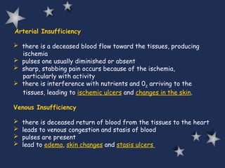
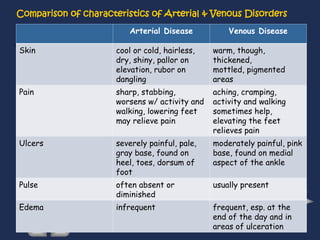
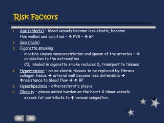
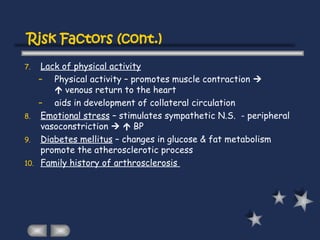
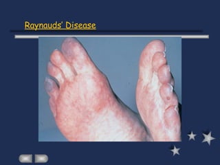
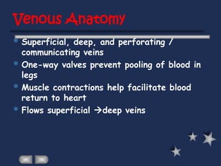
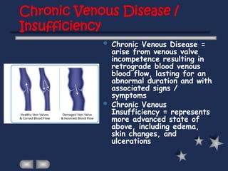
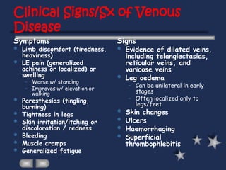
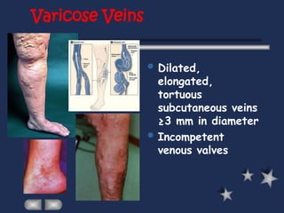
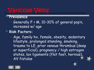
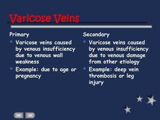
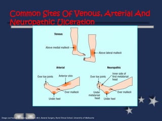
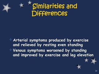
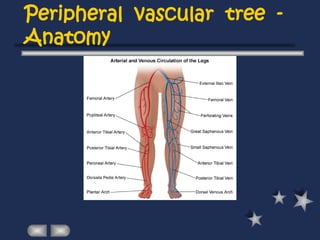
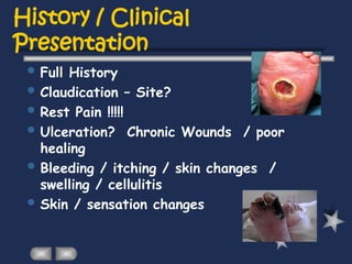
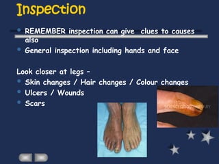
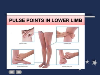
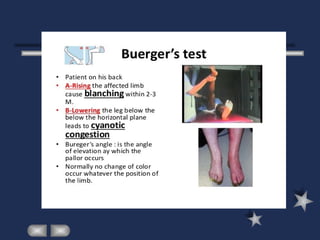
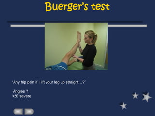
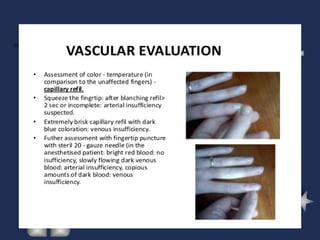
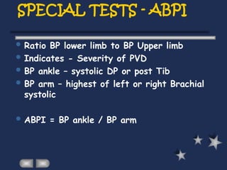
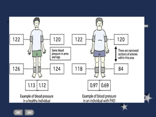
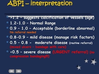
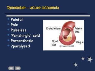
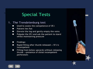
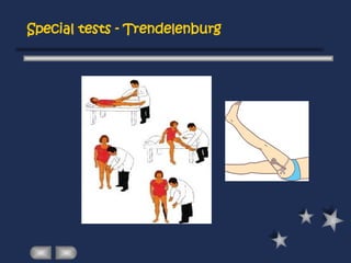
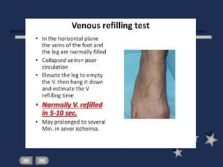
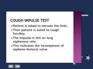
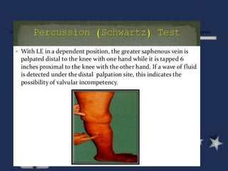
Peripheral Vascular
Diseases
Arteries
 are thick-walled vessels that transport 02
and blood via
the aorta from the heart to the tissues
3 Layers of Arteries
1. inner layer of endothelium (intima)
2. middle layer of connective tissue, smooth muscle and elastic
fibers (media)
3.outer layer of connective tissue (adventitia)
 have smooth muscles that contracts & relaxes to respond
changes in blood volume.
Veins
 are thin-walled vessels that transport deoxygenated blood
from the capillaries back to the right side of the heart
3 Layers – intima, media, adventitia
 there is little smooth muscle &
connective tissue  makes
the veins more distensible 
they accumulate large volumes of blood
 Major veins, particularly in the lower
extremities, have one-way valves
---allow blood flow against gravity
 Valves allow blood to be pumped back
to the heart but prevent it from
draining back into the periphery
Peripheral Vascular Diseases
 charac. by a reduction in blood flow and hence 02 through the
peripheral vessels
 when the need of the tissues for 02 exceeds the supply, areas of
ischemia and necrosis will develop
Factors that can contribute to the development of peripheral
vascular disorders :
 atherosclerotic changes
 thrombus formation
 embolization
  coagulability of blood
 hypertension
 inflammatory process/infection
Arterial Insufficiency
 there is a deceased blood flow toward the tissues, producing
ischemia
 pulses one usually diminished or absent
 sharp, stabbing pain occurs because of the ischemia,
particularly with activity
 there is interference with nutrients and 02
arriving to the
tissues, leading to ischemic ulcers and changes in the skin.
Venous Insufficiency
 there is deceased return of blood from the tissues to the heart
 leads to venous congestion and stasis of blood
 pulses are present
 lead to edema, skin changes and stasis ulcers
Comparison of characteristics of Arterial & Venous Disorders
Arterial Disease Venous Disease
Skin cool or cold, hairless,
dry, shiny, pallor on
elevation, rubor on
dangling
warm, though,
thickened,
mottled, pigmented
areas
Pain sharp, stabbing,
worsens w/ activity and
walking, lowering feet
may relieve pain
aching, cramping,
activity and walking
sometimes help,
elevating the feet
relieves pain
Ulcers severely painful, pale,
gray base, found on
heel, toes, dorsum of
foot
moderately painful, pink
base, found on medial
aspect of the ankle
Pulse often absent or
diminished
usually present
Edema infrequent frequent, esp. at the
end of the day and in
areas of ulceration
Risk Factors
1. Age (elderly) – blood vessels become less elastic, become
thin walled and calcified –  PVR –  BP
2. Sex (male)
3. Cigarette smoking
– nicotine causes vasoconstriction and spasm of the arteries – 
circulation to the extremities
– C02 inhaled in cigarette smoke reduces 02 transport to tissues
4. Hypertension – cause elastic tissues to be replaced by fibrous
collagen tissue  arterial wall become less distensible 
resistance to blood flow   BP
5. Hyperlipedimia – atherosclerotic plaque
6. Obesity – places added burden on the heart & blood vessels
– excess fat contribute to  venous congestion
7. Lack of physical activity
– Physical activity – promotes muscle contraction 
 venous return to the heart
– aids in development of collateral circulation
8. Emotional stress – stimulates sympathetic N.S. - peripheral
vasoconstriction   BP
9. Diabetes mellitus – changes in glucose & fat metabolism
promote the atherosclerotic process
10. Family history of arthrosclerosis
Risk Factors (cont.)
Arteriosclerosis Obliterans
 is a disorder in which there is an arteriosclerotic narrowing
or obstruction of the inner & middle layer of the artery
 most common cause of arterial obstructive disease in the
extremities
 the lower extremities are involved more than upper
extremities
 common site of disease – femoral artery, iliac arteries,
popliteal arteries
 in a diabetic, the disease becomes more progressive, affects
the smaller arteries and often involves vessels below the
knee
Thromboangitis Obliterans ( Buerger’s
Disease)
 characterized by acute inflammatory lesions and occlusive
thrombosis of the arteries & veins
 has a very strong assoc. with cigarette smoking
 commonly occurs in male – bet. 20-40 y.o
 may involve the arteries of the upper extremities (wrists)
 usually affect the lower leg. toes, feet
Thromboangitis Obliterans
Raynaud’s phenomenon
 refers to intermittent episodes during which small arteries or
arterioles of L and R arm constrict (spasm) causing changes in skin
color and temperature
 generally unilateral and may affect only 1 or 2 fingers
 may occur after trauma, neurogenic lesions, occlusive arterial
disease, connective tissues disease
 charac. by reduction of blood flow to the fingers manifested by
cutaneous vessel constriction and resulting in blanching (pallor)
Raynauds’ Disease
 unknown etiology, may be due to immunologic abnormalities
 common in women 20-40 y.o
 maybe stimulated by emotional stress, hypersensitivity to cold,
alteration in sympathetic innervation
Raynauds’ Disease
Aneurysm
 is a localized or diffuse enlargement of an artery at some point
along its course
 can occur when the vessel becomes weakened from trauma,
congenital vascular disease, infection or atherosclerosis
Pathophysiology
 enlargement of a segment of an artery  the tunica media
(middle layer composed of smooth muscle & elastic tissue) is
damaged  progressive dilation, degeneration  risk of rupture
 * most common site is the aorta
 may develop in any blood vessel
Arterial Embolism
 blood clots floating in the circulating arterial blood.
 the embolus is frequently a fragment of arterioscherotic plaque
loosened from the aorta
 emboli will tend to lodge in femoral or popliteal arteries, blood flow
is impaired and ischemia develops
Clinical manifestations:
 S/Sx depends on the size of the embolus, the presence of collateral
circulation and if it is close to a major organ
 abrupt onset of severe pain from the sudden cessation of
circulation
 muscular weakness and burning, aching pain occur
 distal pulses are absent and extremity becomes cold, numb and pale
 symptoms of shock may develop if the embolus blocks a large artery
 Venous disease
Venous Anatomy
 Superficial, deep, and perforating /
communicating veins
 One-way valves prevent pooling of blood in
legs
 Muscle contractions help facilitate blood
return to heart
 Flows superficial deep veins
Chronic Venous Disease /
Insufficiency
 Chronic Venous Disease =
arise from venous valve
incompetence resulting in
retrograde blood venous
blood flow, lasting for an
abnormal duration and with
associated signs /
symptoms
 Chronic Venous
Insufficiency = represents
more advanced state of
above, including edema,
skin changes, and
ulcerations
Clinical Signs/Sx of Venous
Disease
Symptoms Signs
 Limb discomfort (tiredness,
heaviness)
 LE pain (generalized
achiness or localized) or
swelling
– Worse w/ standing
– Improves w/ elevation or
walking
 Paresthesias (tingling,
burning)
 Tightness in legs
 Skin irritation/itching or
discoloration / redness
 Bleeding
 Muscle cramps
 Generalized fatigue
 Evidence of dilated veins,
including telangiectasias,
reticular veins, and
varicose veins
 Leg oedema
– Can be unilateral in early
stages
– Often localized only to
legs/feet
 Skin changes
 Ulcers
 Haemorrhaging
 Superficial
thrombophlebitis
Varicose Veins
 Dilated,
elongated,
tortuous
subcutaneous veins
≥3 mm in diameter
 Incompetent
venous valves
Varicose Veins
 Prevalence
– Generally F > M, 10-30% of general pop’n,
increases w/ age
 Risk Factors:
– Age, family hx, female, obesity, sedentary
lifestyle, prolonged standing, smoking,
trauma to LE, prior venous thrombus (deep
or superficial), pregnancy / high estrogen
states, lax ligaments (flat feet, hernias),
AV fistulas
Varicose Veins
Primary Secondary
 Varicose veins caused
by venous insufficiency
due to venous wall
weakness
 Example: due to age or
pregnancy
 Varicose veins caused
by venous insufficiency
due to venous damage
from other etiology
 Example: deep vein
thrombosis or leg
injury
Common Sites Of Venous, Arterial And
Neuropathic Ulceration
Image courtesy of Mr. Nikolas Kosanovic M.D, General Surgery, Rural Clinical School, University of Melbourne
 Arterial symptoms produced by exercise
and relieved by resting even standing
 Venous symptoms worsened by standing
and improved by exercise and leg elevation
24
Similarities and
Differences
 In the arterial system, treatment is
aimed at opening and restoring blood flow
 In the venous system, treatment is aimed
at obstructing and eliminating
dysfunctional blood vessels
25
Treatment Differences
Peripheral Vascular
system examination
Assessment
 Anatomy
 History / clinical presentation
 Aetiology or Causes / Risk Factors
 Examination – special tests
Peripheral vascular tree -
Anatomy
History / Clinical
Presentation
 Full History
 Claudication – Site?
 Rest Pain !!!!!
 Ulceration? Chronic Wounds / poor
healing
 Bleeding / itching / skin changes /
swelling / cellulitis
 Skin / sensation changes
RISK FACTORS
SMOKING! (even a few
cigarettes/day)
Diabetes (esp poorly controlled)
Dyslipidaemia
Hypertension
Family Hx (CVS/PVD/dyslipidaemias)
Age
Ethnicity
Obesity
(Vasculitides – active inflammation, hyperhomocysteinaemias)
Arterial System
Inspection
 REMEMBER inspection can give clues to causes
also
 General inspection including hands and face
Look closer at legs –
 Skin changes / Hair changes / Colour changes
 Ulcers / Wounds
 Scars
Palpation
 SUPINE – expose abdomen and legs
Feel –
Start distally – temperature / scars (compare L Vs R)
Tibial shaft for oedema (pitting or non) – note upper
level (severity)
scrotum /abdominal wall maybe involved
unilateral disease – DVT or compression of large veins
by node/tumour
Non pitting – lymphatic diseases / hypothyroidism
Palpation (and auscultation)
 Feel PULSES (and auscultate each for bruits)
DP / Post Tib / Pop / Femoral
Abdo– MUST complete exam by assessing Abdominal
Aorta, bruits and assess for sacral oedema…
Full abdo exam: ascites (severe CF), tender liver-
capsule/veins, splenomegaly, full CVS exam
PULSES POPLITEAL
Relax the fossa
Use both hands to feel behind fat pad – almost like
bimanual palp
PULSES ANT tibial /
D.PEDIS / POST TIB
Buerger’s test
“Any hip pain if I lift your leg up straight…?”
Angles ?
<20 severe
SPECIAL TESTS - ABPI
 Ratio BP lower limb to BP Upper limb
 Indicates - Severity of PVD
 BP ankle – systolic DP or post Tib
 BP arm – highest of left or right Brachial
systolic
 ABPI = BP ankle / BP arm
ABPI – interpretation
 >1.2 – suggests calcification of vessels (age)
 1.2-1.0 – Normal Range
 0.9 – 1.0 – Acceptable (borderline abnormal)
No referral needed
 0.8-0.9 – mild disease (manage risk factors)
 0.5 – 0.8 - moderate disease (routine referral)
(mixed ulcers – bandage with care)
 <0.5 – severe disease (URGENT referral) (no
compression bandaging!!!)
Remember – acute ischaemia
 Painful
 Pale
 Pulseless
 ‘Perishingly’ cold
 Paraesthetic
 ?paralysed
Venous System
Venous system
 Position – this time STANDING
 Remainder the same – Inspect /
Palpate /
 Special tests
Examination
Inspect WHOLE leg – long saph / short saph regions
If unilateral swelling –measure circumference
(Use bony landmark as point of reference)
Palpate – hard veins = Thrombosis, Tenderness =
phlebitis
Cough Impulse test: saphenofemoral valve (thrill –
incompetence)
Trendelenburg – incompetence of saphenofemoral valve
(POSITIVE)
If veins still fill up – incompetence lower - Perthes
Test
Varicose veins
Trendelenberg tests
Special tests - Trendelenburg
Perthes test
Same as Trendelenburg – but on standing release small vol
blood into veins
Ask patient to pump calves (stand up and down on tip toes)
Veins become less tense if perforators have competent valves
More tense if incompetence
Note Pt will feel pain when veins fill up!!!!
So BE VIGILANT and ready to STOP IMMEDIATELY
 Do systematically
Thank you!

peripheral vascular diseases overview and physiology

  • 1.
  • 2.
    Arteries  are thick-walledvessels that transport 02 and blood via the aorta from the heart to the tissues 3 Layers of Arteries 1. inner layer of endothelium (intima) 2. middle layer of connective tissue, smooth muscle and elastic fibers (media) 3.outer layer of connective tissue (adventitia)  have smooth muscles that contracts & relaxes to respond changes in blood volume.
  • 3.
    Veins  are thin-walledvessels that transport deoxygenated blood from the capillaries back to the right side of the heart 3 Layers – intima, media, adventitia  there is little smooth muscle & connective tissue  makes the veins more distensible  they accumulate large volumes of blood  Major veins, particularly in the lower extremities, have one-way valves ---allow blood flow against gravity  Valves allow blood to be pumped back to the heart but prevent it from draining back into the periphery
  • 4.
    Peripheral Vascular Diseases charac. by a reduction in blood flow and hence 02 through the peripheral vessels  when the need of the tissues for 02 exceeds the supply, areas of ischemia and necrosis will develop Factors that can contribute to the development of peripheral vascular disorders :  atherosclerotic changes  thrombus formation  embolization   coagulability of blood  hypertension  inflammatory process/infection
  • 5.
    Arterial Insufficiency  thereis a deceased blood flow toward the tissues, producing ischemia  pulses one usually diminished or absent  sharp, stabbing pain occurs because of the ischemia, particularly with activity  there is interference with nutrients and 02 arriving to the tissues, leading to ischemic ulcers and changes in the skin. Venous Insufficiency  there is deceased return of blood from the tissues to the heart  leads to venous congestion and stasis of blood  pulses are present  lead to edema, skin changes and stasis ulcers
  • 6.
    Comparison of characteristicsof Arterial & Venous Disorders Arterial Disease Venous Disease Skin cool or cold, hairless, dry, shiny, pallor on elevation, rubor on dangling warm, though, thickened, mottled, pigmented areas Pain sharp, stabbing, worsens w/ activity and walking, lowering feet may relieve pain aching, cramping, activity and walking sometimes help, elevating the feet relieves pain Ulcers severely painful, pale, gray base, found on heel, toes, dorsum of foot moderately painful, pink base, found on medial aspect of the ankle Pulse often absent or diminished usually present Edema infrequent frequent, esp. at the end of the day and in areas of ulceration
  • 7.
    Risk Factors 1. Age(elderly) – blood vessels become less elastic, become thin walled and calcified –  PVR –  BP 2. Sex (male) 3. Cigarette smoking – nicotine causes vasoconstriction and spasm of the arteries –  circulation to the extremities – C02 inhaled in cigarette smoke reduces 02 transport to tissues 4. Hypertension – cause elastic tissues to be replaced by fibrous collagen tissue  arterial wall become less distensible  resistance to blood flow   BP 5. Hyperlipedimia – atherosclerotic plaque 6. Obesity – places added burden on the heart & blood vessels – excess fat contribute to  venous congestion
  • 8.
    7. Lack ofphysical activity – Physical activity – promotes muscle contraction   venous return to the heart – aids in development of collateral circulation 8. Emotional stress – stimulates sympathetic N.S. - peripheral vasoconstriction   BP 9. Diabetes mellitus – changes in glucose & fat metabolism promote the atherosclerotic process 10. Family history of arthrosclerosis Risk Factors (cont.)
  • 9.
    Arteriosclerosis Obliterans  isa disorder in which there is an arteriosclerotic narrowing or obstruction of the inner & middle layer of the artery  most common cause of arterial obstructive disease in the extremities  the lower extremities are involved more than upper extremities  common site of disease – femoral artery, iliac arteries, popliteal arteries  in a diabetic, the disease becomes more progressive, affects the smaller arteries and often involves vessels below the knee
  • 10.
    Thromboangitis Obliterans (Buerger’s Disease)  characterized by acute inflammatory lesions and occlusive thrombosis of the arteries & veins  has a very strong assoc. with cigarette smoking  commonly occurs in male – bet. 20-40 y.o  may involve the arteries of the upper extremities (wrists)  usually affect the lower leg. toes, feet
  • 11.
  • 12.
    Raynaud’s phenomenon  refersto intermittent episodes during which small arteries or arterioles of L and R arm constrict (spasm) causing changes in skin color and temperature  generally unilateral and may affect only 1 or 2 fingers  may occur after trauma, neurogenic lesions, occlusive arterial disease, connective tissues disease  charac. by reduction of blood flow to the fingers manifested by cutaneous vessel constriction and resulting in blanching (pallor) Raynauds’ Disease  unknown etiology, may be due to immunologic abnormalities  common in women 20-40 y.o  maybe stimulated by emotional stress, hypersensitivity to cold, alteration in sympathetic innervation
  • 13.
  • 14.
    Aneurysm  is alocalized or diffuse enlargement of an artery at some point along its course  can occur when the vessel becomes weakened from trauma, congenital vascular disease, infection or atherosclerosis Pathophysiology  enlargement of a segment of an artery  the tunica media (middle layer composed of smooth muscle & elastic tissue) is damaged  progressive dilation, degeneration  risk of rupture  * most common site is the aorta  may develop in any blood vessel
  • 15.
    Arterial Embolism  bloodclots floating in the circulating arterial blood.  the embolus is frequently a fragment of arterioscherotic plaque loosened from the aorta  emboli will tend to lodge in femoral or popliteal arteries, blood flow is impaired and ischemia develops Clinical manifestations:  S/Sx depends on the size of the embolus, the presence of collateral circulation and if it is close to a major organ  abrupt onset of severe pain from the sudden cessation of circulation  muscular weakness and burning, aching pain occur  distal pulses are absent and extremity becomes cold, numb and pale  symptoms of shock may develop if the embolus blocks a large artery
  • 16.
  • 17.
    Venous Anatomy  Superficial,deep, and perforating / communicating veins  One-way valves prevent pooling of blood in legs  Muscle contractions help facilitate blood return to heart  Flows superficial deep veins
  • 18.
    Chronic Venous Disease/ Insufficiency  Chronic Venous Disease = arise from venous valve incompetence resulting in retrograde blood venous blood flow, lasting for an abnormal duration and with associated signs / symptoms  Chronic Venous Insufficiency = represents more advanced state of above, including edema, skin changes, and ulcerations
  • 19.
    Clinical Signs/Sx ofVenous Disease Symptoms Signs  Limb discomfort (tiredness, heaviness)  LE pain (generalized achiness or localized) or swelling – Worse w/ standing – Improves w/ elevation or walking  Paresthesias (tingling, burning)  Tightness in legs  Skin irritation/itching or discoloration / redness  Bleeding  Muscle cramps  Generalized fatigue  Evidence of dilated veins, including telangiectasias, reticular veins, and varicose veins  Leg oedema – Can be unilateral in early stages – Often localized only to legs/feet  Skin changes  Ulcers  Haemorrhaging  Superficial thrombophlebitis
  • 20.
    Varicose Veins  Dilated, elongated, tortuous subcutaneousveins ≥3 mm in diameter  Incompetent venous valves
  • 21.
    Varicose Veins  Prevalence –Generally F > M, 10-30% of general pop’n, increases w/ age  Risk Factors: – Age, family hx, female, obesity, sedentary lifestyle, prolonged standing, smoking, trauma to LE, prior venous thrombus (deep or superficial), pregnancy / high estrogen states, lax ligaments (flat feet, hernias), AV fistulas
  • 22.
    Varicose Veins Primary Secondary Varicose veins caused by venous insufficiency due to venous wall weakness  Example: due to age or pregnancy  Varicose veins caused by venous insufficiency due to venous damage from other etiology  Example: deep vein thrombosis or leg injury
  • 23.
    Common Sites OfVenous, Arterial And Neuropathic Ulceration Image courtesy of Mr. Nikolas Kosanovic M.D, General Surgery, Rural Clinical School, University of Melbourne
  • 24.
     Arterial symptomsproduced by exercise and relieved by resting even standing  Venous symptoms worsened by standing and improved by exercise and leg elevation 24 Similarities and Differences
  • 25.
     In thearterial system, treatment is aimed at opening and restoring blood flow  In the venous system, treatment is aimed at obstructing and eliminating dysfunctional blood vessels 25 Treatment Differences
  • 26.
  • 27.
    Assessment  Anatomy  History/ clinical presentation  Aetiology or Causes / Risk Factors  Examination – special tests
  • 28.
  • 29.
    History / Clinical Presentation Full History  Claudication – Site?  Rest Pain !!!!!  Ulceration? Chronic Wounds / poor healing  Bleeding / itching / skin changes / swelling / cellulitis  Skin / sensation changes
  • 30.
    RISK FACTORS SMOKING! (evena few cigarettes/day) Diabetes (esp poorly controlled) Dyslipidaemia Hypertension Family Hx (CVS/PVD/dyslipidaemias) Age Ethnicity Obesity (Vasculitides – active inflammation, hyperhomocysteinaemias)
  • 31.
  • 32.
    Inspection  REMEMBER inspectioncan give clues to causes also  General inspection including hands and face Look closer at legs –  Skin changes / Hair changes / Colour changes  Ulcers / Wounds  Scars
  • 33.
    Palpation  SUPINE –expose abdomen and legs Feel – Start distally – temperature / scars (compare L Vs R) Tibial shaft for oedema (pitting or non) – note upper level (severity) scrotum /abdominal wall maybe involved unilateral disease – DVT or compression of large veins by node/tumour Non pitting – lymphatic diseases / hypothyroidism
  • 34.
    Palpation (and auscultation) Feel PULSES (and auscultate each for bruits) DP / Post Tib / Pop / Femoral Abdo– MUST complete exam by assessing Abdominal Aorta, bruits and assess for sacral oedema… Full abdo exam: ascites (severe CF), tender liver- capsule/veins, splenomegaly, full CVS exam
  • 35.
    PULSES POPLITEAL Relax thefossa Use both hands to feel behind fat pad – almost like bimanual palp
  • 36.
    PULSES ANT tibial/ D.PEDIS / POST TIB
  • 39.
    Buerger’s test “Any hippain if I lift your leg up straight…?” Angles ? <20 severe
  • 41.
    SPECIAL TESTS -ABPI  Ratio BP lower limb to BP Upper limb  Indicates - Severity of PVD  BP ankle – systolic DP or post Tib  BP arm – highest of left or right Brachial systolic  ABPI = BP ankle / BP arm
  • 43.
    ABPI – interpretation >1.2 – suggests calcification of vessels (age)  1.2-1.0 – Normal Range  0.9 – 1.0 – Acceptable (borderline abnormal) No referral needed  0.8-0.9 – mild disease (manage risk factors)  0.5 – 0.8 - moderate disease (routine referral) (mixed ulcers – bandage with care)  <0.5 – severe disease (URGENT referral) (no compression bandaging!!!)
  • 44.
    Remember – acuteischaemia  Painful  Pale  Pulseless  ‘Perishingly’ cold  Paraesthetic  ?paralysed
  • 45.
  • 46.
    Venous system  Position– this time STANDING  Remainder the same – Inspect / Palpate /  Special tests
  • 47.
    Examination Inspect WHOLE leg– long saph / short saph regions If unilateral swelling –measure circumference (Use bony landmark as point of reference) Palpate – hard veins = Thrombosis, Tenderness = phlebitis Cough Impulse test: saphenofemoral valve (thrill – incompetence) Trendelenburg – incompetence of saphenofemoral valve (POSITIVE) If veins still fill up – incompetence lower - Perthes Test
  • 48.
  • 49.
  • 51.
    Special tests -Trendelenburg
  • 52.
    Perthes test Same asTrendelenburg – but on standing release small vol blood into veins Ask patient to pump calves (stand up and down on tip toes) Veins become less tense if perforators have competent valves More tense if incompetence Note Pt will feel pain when veins fill up!!!! So BE VIGILANT and ready to STOP IMMEDIATELY
  • 56.
  • 57.