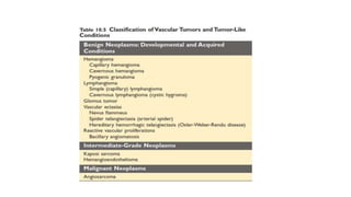
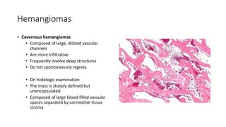
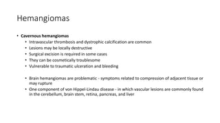
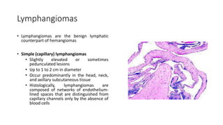
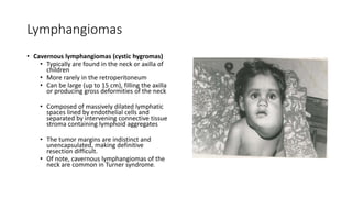
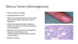
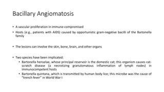
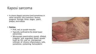
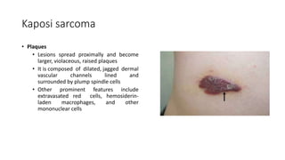
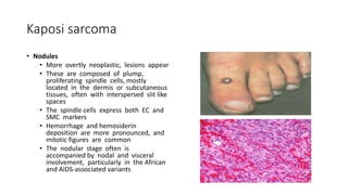
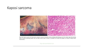
This document discusses various types of vascular tumors, including benign tumors like hemangiomas and lymphangiomas, intermediate tumors like hemangioendotheliomas, and malignant tumors such as Kaposi sarcoma and angiosarcomas. It covers the histological and clinical features used to classify and diagnose these different vascular neoplasms. Key information includes that benign tumors form well-organized vessels, while malignant tumors are more cellular and proliferative without such organization. Immunohistochemistry can help identify the endothelial cell origin of these proliferations.
This document discusses various vascular diseases and tumors. It covers topics such as varicose veins, thrombophlebitis, hemorrhoids, risk factors for deep vein thrombosis, and assessments for DVT. It also discusses lymphangitis, lymphedema, and various benign and malignant vascular tumors including hemangiomas, lymphangiomas, glomus tumors, Kaposi sarcoma, and angiosarcomas.
Vasoformative disorders are distinct type of lesions that covers benign, lesions of intermediate malignancy and malignant disorders along with syndromes. very useful for oral path, oral medicine and as well as general pathology and residents.
What are Vascular Anomalies?
Hemangioma
Rendu Osler Weber Disease
Sturge Weber syndrome
Lymphangioma
Cystic Hygroma
Hemangiomas: lesions demonstrating endothelial hyperplasia.
Vascular Malformations : lesions with normal endothelial turnover.
A hemangioma is a benign and usually self-involuting tumor of the endothelial cells that line blood vessels, and is characterised by increased number of normal or abnormal vessels filled with blood.
May be present at Birth or arise during early childhood.
Hemangiomas and vascular malformations are benign vascular proliferations that can occur in the oral cavity. Hemangiomas are considered benign tumors that grow rapidly in infancy through endothelial cell proliferation, then slowly involute over time. In contrast, vascular malformations are structural anomalies present at birth that do not involute. The document discusses several subtypes of hemangiomas including capillary hemangioma, tufted angioma, verrucous hemangioma and lobular capillary hemangioma. It also covers histologic features that can help distinguish hemangiomas from vascular malformations and malignant lesions.
This document discusses cardiac tumors, beginning with an introduction stating that primary heart tumors are rare but the most common ones are benign. The five most common benign tumors collectively account for 50-90% of primary cardiac tumors. The heart can also be a site of metastatic tumors in about 5% of cancer cases.
The document then discusses specific tumor types in more detail, including myxomas (the most common primary adult cardiac tumor), lipomas, fibroelastomas, rhabdomyomas, and malignant primary tumors such as angiosarcomas, rhabdomyosarcomas, and mesotheliomas. Clinical manifestations, morphology, histology, and prognosis are described for each tumor type. Secondary
The document provides an overview of mediastinal tumors and lesions. It discusses inflammatory diseases like acute and chronic mediastinitis. It also covers cysts, thymic lesions including thymoma, lymphomas, neurogenic tumors, and metastatic tumors. Specific conditions are described such as fibrosing mediastinitis, thymic cysts, and different non-neoplastic thymic diseases. The anatomy of the mediastinum and different tumor types are outlined in detail.
This document discusses various non-odontogenic tumors classified according to their origin. It covers giant cell lesions including central giant cell granuloma and brown tumor of hyperparathyroidism. Vascular lesions such as hemangiomas, vascular malformations, and neurogenic tumors including neurofibromas are also discussed. For each type of lesion, the document provides information on classification, clinical features, radiographic findings, diagnosis, and treatment options.
Based on the information provided about Mr. Gamal:
- CT angiography would likely reveal an abdominal aortic aneurysm, showing markedly increased aortic diameter below the renal arteries.
- The most probable cause in his case is atherosclerosis, given his risk factors of diabetes, hypertension, smoking history and hyperlipidemia.
- His risk factors are diabetes, hypertension, smoking history, obesity (BMI of 30), and hyperlipidemia.
- His condition would be classified as an atherosclerotic abdominal aortic aneurysm.
- Possible complications include rupture of the aneurysm, thrombosis/embolism, compression of adjacent structures, or occlusion of branches supplying organs like the kidneys or intestines
This document provides information on various neck masses including branchial cysts, branchial fistulas, cystic hygromas, carotid body tumors, and cervical lymphadenopathy. It describes the anatomy, etiology, clinical presentation, diagnosis and management of each condition. Branchial cysts are the most common congenital neck masses and usually present as soft, fluctuant swellings in the neck. Cystic hygromas are lymphangiomas that occur in the neck of newborns. Carotid body tumors develop in the carotid artery bifurcation and may cause cranial nerve palsies or Horner's syndrome. Surgical excision is the main treatment for branchial cysts, cystic hyg
This document discusses various non-tumoral spinal cord lesions including vascular, infectious, inflammatory, and demyelinating causes. It provides details on imaging features of common conditions such as spinal cord ischemia seen as "owl's eyes" or "butterfly" patterns on MRI. Arteriovenous malformations appear as flow voids and cord expansion on MRI while cavernous angiomas have a "popcorn" appearance. Infections like tuberculosis may cause cord enhancement and abscesses. Demyelinating diseases like multiple sclerosis can cause multiple spinal cord lesions seen as hyperintensities on MRI.
This document discusses vascular malformations, which are collections of abnormal vessels with a normal endothelial turnover rate and thin basement membrane. They can involve arteries, veins, or capillaries. Vascular malformations result from defects during embryonic vascular development and are classified based on the type of involved vessel and blood flow. Common types include venous, capillary, lymphatic, and arteriovenous malformations. Signs and symptoms depend on the location and size of the lesion. Diagnosis involves physical examination, imaging studies, and sometimes biopsy. Hematologic evaluation is also important due to potential coagulation disorders.
imaging of scrotum [Repaired] [Repaired].pptxdypradio
The scrotum contains the testes and epididymides. On ultrasound, the normal anatomy includes the oval testes with homogeneous echotexture and color flow. Potential pathological findings include infections like epididymitis, tumors such as seminomas which appear hypoechoic and well-defined, and traumatic injuries or torsion which may demonstrate absent flow. Malignancies require evaluation for metastases while infections require treatment with antibiotics. Imaging guides diagnosis and management of scrotal pathologies.
1. An aneurysm is an abnormal dilation of a blood vessel that can be congenital or acquired due to weakening of the vessel wall.
2. Aneurysms are classified based on composition, shape, location, and pathogenetic mechanism. The most common type is atherosclerotic aneurysms, which often affect the abdominal aorta.
3. Complications of aneurysms include rupture, which can lead to fatal hemorrhaging, as well as compression of surrounding structures. timely diagnosis and treatment are important to prevent such complications.
This document discusses various types of vascular anomalies. It begins by providing historical context for how vascular anomalies were classified. It then summarizes the current biologic classification system introduced in 1982, which distinguishes between vascular tumors and malformations. For vascular tumors, it describes infantile hemangioma and its life cycle phases. It also discusses other types of vascular tumors like congenital hemangiomas and kaposiform hemangioendothelioma. For vascular malformations, it lists the main types classified by predominant vessel type. Diagnosis and management approaches are summarized for different vascular anomalies.
The document discusses various benign and malignant tumors of the kidney. Regarding benign tumors, it describes cortical adenomas, oncocytomas, angiomyolipomas, mesoblastic nephromas, and multicystic nephromas. For malignant tumors, it covers clear cell and papillary renal cell carcinomas, as well as Wilms' tumor, an embryonic tumor most common in young children. It provides details on the morphology, histology, and clinical manifestations of these tumors.
this ppt is about malignant tumours of connective tissue origin. classifications, clinical features, radiological features and histological features of all tumors are discussed with pictures.
This document provides information on the normal anatomy and ultrasound appearance of scrotal structures. It discusses the 4 T's that can affect the scrotum - torsion, trauma, tumor, and testiculitis. Torsion and its time-dependent effects on blood flow and testicular viability are explained in detail. Various types of tumors are summarized, including their typical ultrasound characteristics. Other abnormal findings like venous infarction and epididymitis are also mentioned.
This document discusses various pseudosarcomas, which are non-neoplastic lesions that can be mistaken for sarcomas due to their rapid growth and cellular appearance. It describes several categories of pseudosarcomas, including reactive fibroblastic/myofibroblastic proliferations (e.g. nodular fasciitis), reactive endothelial proliferations (e.g. papillary endothelial hyperplasia), mass forming inflammatory/infective lesions (e.g. Rosai-Dorfman disease), and benign connective tissue tumors (e.g. cellular fibrous histiocytoma). The document emphasizes that accurate recognition of these pseudosarcomas helps prevent excessive or
This document summarizes various pathologies that can be identified on scrotal ultrasound. It describes common extratesticular masses like hydrocele, varicocele, and spermatocele. Hydrocele appears as an anechoic collection without flow in the tunica vaginalis. Varicocele involves dilated veins around the testicle visible with color Doppler. Spermatoceles are benign cysts filled with sperm that appear hypoechoic. It also discusses testicular torsion, epididymitis, trauma, cysts, teratomas, seminomas, lymphomas, and microlithiasis.
vascular anomalies of newborn,approach to infantile hemangiomaDr Praman Kushwah
The document discusses vascular anomalies in newborns, focusing on infantile hemangiomas. It provides classifications for vascular tumors and malformations, and describes key features of infantile hemangiomas including risk factors, pathogenesis, clinical presentations, growth cycles, potential complications, and risk factors for life-threatening complications. Infantile hemangiomas typically arise postnatally, demonstrate endothelial proliferation, and can involve the superficial dermis, deep dermis, or both. Aggressive early treatment may be needed for hemangiomas near the airway or eyes to prevent functional impairment.
Uterine sarcomas are rare, aggressive cancers that arise from the muscular or connective tissues of the uterus. The main types are leiomyosarcomas, endometrial stromal sarcomas, and malignant mixed müllerian tumors. Uterine sarcomas commonly spread through the bloodstream to vital organs like the lungs and liver. Patients typically present with abnormal vaginal bleeding and abdominal pain. Diagnosis is made through histological examination of tissue samples obtained through procedures like uterine curettage.
Varicose veins are dilated, tortuous veins in the legs caused by faulty valves that allow blood to flow backwards. Risk factors include heredity, prolonged standing, pregnancy, and tight clothing. Symptoms include heavy legs, cramps, and discolored skin. Treatment options range from compression stockings and sclerotherapy to surgery to remove or bypass sections of vein.
Atherosclerosis is a buildup of plaque in the arteries that can restrict blood flow. It often affects major arteries and risks include smoking, hypertension, and high cholesterol. Symptoms depend on the location but may include pain, numbness, or altered pulse. Diagnosis involves tests like Doppler and angiogram. Treatment focuses on risk
Endometrial polyps are a common benign condition that can cause abnormal uterine bleeding. They form as a result of focal overgrowth of endometrial glands and stroma protruding into the uterine cavity. Risk factors include hormone therapy, tamoxifen use, diabetes, obesity, and increased age. Endometrial polyps are often difficult to identify and can be a source of continued bleeding if left untreated. Hysteroscopy and ultrasound are useful for diagnosis. While usually benign, polyps have a small increased risk of endometrial cancer, especially in postmenopausal women.
This document discusses aneurysms and vasculitis. It defines an aneurysm as a bulging or ballooning of the blood vessel wall caused by a weakened spot. Aneurysms can be true or false. True aneurysms involve all vessel wall layers, while false aneurysms involve blood leaking outside the vessel. The document describes different types of aneurysms based on location, shape, and size. It also discusses risk factors, signs and symptoms, pathophysiology, and treatments for various aneurysms. The document then defines vasculitis as inflammation of blood vessels that can affect arteries, veins or capillaries. It classifies vasculitis based on vessel size and lists different types. It discusses diagnostic approaches and challenges
Cell Therapy Expansion and Challenges in Autoimmune DiseaseHealth Advances
There is increasing confidence that cell therapies will soon play a role in the treatment of autoimmune disorders, but the extent of this impact remains to be seen. Early readouts on autologous CAR-Ts in lupus are encouraging, but manufacturing and cost limitations are likely to restrict access to highly refractory patients. Allogeneic CAR-Ts have the potential to broaden access to earlier lines of treatment due to their inherent cost benefits, however they will need to demonstrate comparable or improved efficacy to established modalities.
In addition to infrastructure and capacity constraints, CAR-Ts face a very different risk-benefit dynamic in autoimmune compared to oncology, highlighting the need for tolerable therapies with low adverse event risk. CAR-NK and Treg-based therapies are also being developed in certain autoimmune disorders and may demonstrate favorable safety profiles. Several novel non-cell therapies such as bispecific antibodies, nanobodies, and RNAi drugs, may also offer future alternative competitive solutions with variable value propositions.
Widespread adoption of cell therapies will not only require strong efficacy and safety data, but also adapted pricing and access strategies. At oncology-based price points, CAR-Ts are unlikely to achieve broad market access in autoimmune disorders, with eligible patient populations that are potentially orders of magnitude greater than the number of currently addressable cancer patients. Developers have made strides towards reducing cell therapy COGS while improving manufacturing efficiency, but payors will inevitably restrict access until more sustainable pricing is achieved.
Despite these headwinds, industry leaders and investors remain confident that cell therapies are poised to address significant unmet need in patients suffering from autoimmune disorders. However, the extent of this impact on the treatment landscape remains to be seen, as the industry rapidly approaches an inflection point.
These lecture slides, by Dr Sidra Arshad, offer a simplified look into the mechanisms involved in the regulation of respiration:
Learning objectives:
1. Describe the organisation of respiratory center
2. Describe the nervous control of inspiration and respiratory rhythm
3. Describe the functions of the dorsal and respiratory groups of neurons
4. Describe the influences of the Pneumotaxic and Apneustic centers
5. Explain the role of Hering-Breur inflation reflex in regulation of inspiration
6. Explain the role of central chemoreceptors in regulation of respiration
7. Explain the role of peripheral chemoreceptors in regulation of respiration
8. Explain the regulation of respiration during exercise
9. Integrate the respiratory regulatory mechanisms
10. Describe the Cheyne-Stokes breathing
Study Resources:
1. Chapter 42, Guyton and Hall Textbook of Medical Physiology, 14th edition
2. Chapter 36, Ganong’s Review of Medical Physiology, 26th edition
3. Chapter 13, Human Physiology by Lauralee Sherwood, 9th edition
How to Control Your Asthma Tips by gokuldas hospital.Gokuldas Hospital
Respiratory issues like asthma are the most sensitive issue that is affecting millions worldwide. It hampers the daily activities leaving the body tired and breathless.
The key to a good grip on asthma is proper knowledge and management strategies. Understanding the patient-specific symptoms and carving out an effective treatment likewise is the best way to keep asthma under control.
Know the difference between Endodontics and Orthodontics.Gokuldas Hospital
Your smile is beautiful.
Let’s be honest. Maintaining that beautiful smile is not an easy task. It is more than brushing and flossing. Sometimes, you might encounter dental issues that need special dental care. These issues can range anywhere from misalignment of the jaw to pain in the root of teeth.
Breast cancer: Post menopausal endocrine therapyDr. Sumit KUMAR
Breast cancer in postmenopausal women with hormone receptor-positive (HR+) status is a common and complex condition that necessitates a multifaceted approach to management. HR+ breast cancer means that the cancer cells grow in response to hormones such as estrogen and progesterone. This subtype is prevalent among postmenopausal women and typically exhibits a more indolent course compared to other forms of breast cancer, which allows for a variety of treatment options.
Diagnosis and Staging
The diagnosis of HR+ breast cancer begins with clinical evaluation, imaging, and biopsy. Imaging modalities such as mammography, ultrasound, and MRI help in assessing the extent of the disease. Histopathological examination and immunohistochemical staining of the biopsy sample confirm the diagnosis and hormone receptor status by identifying the presence of estrogen receptors (ER) and progesterone receptors (PR) on the tumor cells.
Staging involves determining the size of the tumor (T), the involvement of regional lymph nodes (N), and the presence of distant metastasis (M). The American Joint Committee on Cancer (AJCC) staging system is commonly used. Accurate staging is critical as it guides treatment decisions.
Treatment Options
Endocrine Therapy
Endocrine therapy is the cornerstone of treatment for HR+ breast cancer in postmenopausal women. The primary goal is to reduce the levels of estrogen or block its effects on cancer cells. Commonly used agents include:
Selective Estrogen Receptor Modulators (SERMs): Tamoxifen is a SERM that binds to estrogen receptors, blocking estrogen from stimulating breast cancer cells. It is effective but may have side effects such as increased risk of endometrial cancer and thromboembolic events.
Aromatase Inhibitors (AIs): These drugs, including anastrozole, letrozole, and exemestane, lower estrogen levels by inhibiting the aromatase enzyme, which converts androgens to estrogen in peripheral tissues. AIs are generally preferred in postmenopausal women due to their efficacy and safety profile compared to tamoxifen.
Selective Estrogen Receptor Downregulators (SERDs): Fulvestrant is a SERD that degrades estrogen receptors and is used in cases where resistance to other endocrine therapies develops.
Combination Therapies
Combining endocrine therapy with other treatments enhances efficacy. Examples include:
Endocrine Therapy with CDK4/6 Inhibitors: Palbociclib, ribociclib, and abemaciclib are CDK4/6 inhibitors that, when combined with endocrine therapy, significantly improve progression-free survival in advanced HR+ breast cancer.
Endocrine Therapy with mTOR Inhibitors: Everolimus, an mTOR inhibitor, can be added to endocrine therapy for patients who have developed resistance to aromatase inhibitors.
Chemotherapy
Chemotherapy is generally reserved for patients with high-risk features, such as large tumor size, high-grade histology, or extensive lymph node involvement. Regimens often include anthracyclines and taxanes.
Histololgy of Female Reproductive System.pptxAyeshaZaid1
Dive into an in-depth exploration of the histological structure of female reproductive system with this comprehensive lecture. Presented by Dr. Ayesha Irfan, Assistant Professor of Anatomy, this presentation covers the Gross anatomy and functional histology of the female reproductive organs. Ideal for students, educators, and anyone interested in medical science, this lecture provides clear explanations, detailed diagrams, and valuable insights into female reproductive system. Enhance your knowledge and understanding of this essential aspect of human biology.
- Video recording of this lecture in English language: https://youtu.be/Pt1nA32sdHQ
- Video recording of this lecture in Arabic language: https://youtu.be/uFdc9F0rlP0
- Link to download the book free: https://nephrotube.blogspot.com/p/nephrotube-nephrology-books.html
- Link to NephroTube website: www.NephroTube.com
- Link to NephroTube social media accounts: https://nephrotube.blogspot.com/p/join-nephrotube-on-social-media.html
2. VASCULAR TUMORS
Surg Lt Cdr Rabia Ahmed
MBBS, FCPS, FRCPath (UK)
Consultant Histopathologist
Assistant Professor
BUHS, Karachi
3. Vascular tumors
• Tumors of blood vessels and lymphatics include:
• Benign
• Intermediate
• Malignant
• Vascular neoplasms arise either from:
• Endothelium (e.g. hemangioma, lymphangioma, angiosarcoma)
• Cells that support or surround blood vessels (e.g., glomus tumor)
• Tumor-like lesions
• Congenital or developmental malformations
• Non-neoplastic reactive vascular proliferations (e.g., bacillary angiomatosis) can also
manifest as that may present diagnostic challenges
4.
5. Vascular tumors
• In general, benign and malignant vascular neoplasms are distinguished by the following features:
• Benign tumors
• Composed of vascular channels filled with blood cells or lymph
• Lined by a monolayer of normal-appearing endothelial cells
• Malignant tumors
• More cellular
• Show cytologic atypia, are proliferative
• Usually do not form well-organized vessels
• Confirmation of the endothelial derivation of such proliferations may require
immunohistochemical detection of EC-specific markers, CD34 and Vwf.
6. Vascular tumors
• Vascular Ectasias
• A generic term for any local dilation of a structure
• Telangiectasia
• A permanent dilation of preexisting small vessels (capillaries, venules, and arterioles, usually
in the skin or mucous membranes) that forms a discrete red lesion
• Can be congenital or acquired and are not true neoplasms
7. Vascular tumors
• Nevus flammeus (a “birthmark”)
• The most common form of vascular
ectasia
• Light pink to deep purple flat lesion on
the head or neck composed of dilated
vessels
• Most ultimately regress spontaneously
• “Port wine stain”
• A special form of nevus flammeus
• These lesions tend to grow during
childhood, thicken the skin surface, and
do not fade with time
8. Vascular tumors
• Sturge Weber syndrome
• Also called encephalotrigeminal
angiomatosis
• Uncommon congenital disorder
• Nevus flammeus (a “birthmark”) lesions
occurring in the distribution of the
trigeminal nerve
• Associated with facial port wine nevi,
ipsilateral venous angiomas in the cortical
leptomeninges, mental retardation,
seizures, hemiplegia, and radiologic
opacities of the skull
• A large facial telangiectasia in a child with
mental deficiencies may indicate the
presence of additional vascular
malformations
9. Vascular tumors
• Spider telangiectasias
• Non-neoplastic vascular lesions
• Radial, often pulsatile arrays of dilated
subcutaneous arteries or arterioles (the
“legs” of the spider) about a central core
(the spider’s “body”) that blanch with
pressure
• Commonly occur on the face, neck, or
upper chest
• Most frequently are associated with
hyperestrogenic states
• In pregnant women or patients with
cirrhosis
10. Vascular tumors
• Hereditary hemorrhagic telangiectasia
(Osler-Weber-Rendu disease)
• Autosomal dominant disorder
• Caused by mutations in genes that
encode components of the TGF-ß
signaling pathway in ECs
• Malformations composed of dilated
capillaries and veins that are present at
birth
• They are widely distributed over the skin
and oral mucous membranes, as well as
in the respiratory, gastrointestinal, and
urinary tracts
• The lesions can spontaneously rupture,
causing serious epistaxis (nosebleed),
gastrointestinal bleeding, or hematuria.
11. Hemangiomas
• Hemangiomas are very common tumors
• Composed of blood-filled vessels
• Constitute 7% of all benign tumors of infancy and childhood
• Most are present from birth and initially increase in size
• Many eventually regress spontaneously
• While hemangiomas typically are localized lesions confined to the head and neck
• They occasionally may be more extensive (angiomatosis)
• Can arise internally
• Nearly one third of these internal lesions are found in the liver
• Malignant transformation is rare
12. Hemangiomas
• Capillary hemangiomas
• The most common type
• Occur in the skin, subcutaneous tissues, and mucous membranes of the oral cavities and lips,
as well as in the liver, spleen, and kidneys
• Histologically, they are composed of thin-walled capillaries with scant stroma
• Juvenile hemangiomas (so-called “strawberry hemangiomas”)
• Extremely common (1 in 200 births)
• Can be multiple
• Grow rapidly for a few months but then fade by 1 to 3 years of age
• Complete regression by 7 years of age in majority of cases
13. Hemangiomas
• Pyogenic granulomas
• Are capillary hemangiomas
• Manifest as rapidly growing red
pedunculated lesions
• On the skin, gingival, or oral mucosa
• Microscopically, they resemble exuberant
granulation tissue
• They bleed easily and often ulcerate
• Roughly one fourth of the lesions develop
after trauma
• Reaching a size of 1 to 2 cm within a few
weeks
• Curettage and cautery usually are
curative
14. Hemangiomas
• Pregnancy tumor (granuloma gravidarum)
• Is a pyogenic granuloma
• Occurs infrequently in the gingiva of
pregnant women
• May spontaneously regress (especially
after pregnancy) or undergo fibrosis
• Occasionally require surgical excision
15. Hemangiomas
• Cavernous hemangiomas
• Composed of large, dilated vascular
channels
• Are more infiltrative
• Frequently involve deep structures
• Do not spontaneously regress
• On histologic examination
• The mass is sharply defined but
unencapsulated
• Composed of large blood-filled vascular
spaces separated by connective tissue
stroma
16. Hemangiomas
• Cavernous hemangiomas
• Intravascular thrombosis and dystrophic calcification are common
• Lesions may be locally destructive
• Surgical excision is required in some cases
• They can be cosmetically troublesome
• Vulnerable to traumatic ulceration and bleeding
• Brain hemangiomas are problematic - symptoms related to compression of adjacent tissue or
may rupture
• One component of von Hippel-Lindau disease - in which vascular lesions are commonly found
in the cerebellum, brain stem, retina, pancreas, and liver
17.
18. Lymphangiomas
• Lymphangiomas are the benign lymphatic
counterpart of hemangiomas
• Simple (capillary) lymphangiomas
• Slightly elevated or sometimes
pedunculated lesions
• Up to 1 to 2 cm in diameter
• Occur predominantly in the head, neck,
and axillary subcutaneous tissue
• Histologically, lymphangiomas are
composed of networks of endothelium-
lined spaces that are distinguished from
capillary channels only by the absence of
blood cells
19. Lymphangiomas
• Cavernous lymphangiomas (cystic hygromas)
• Typically are found in the neck or axilla of
children
• More rarely in the retroperitoneum
• Can be large (up to 15 cm), filling the axilla
or producing gross deformities of the neck
• Composed of massively dilated lymphatic
spaces lined by endothelial cells and
separated by intervening connective tissue
stroma containing lymphoid aggregates
• The tumor margins are indistinct and
unencapsulated, making definitive
resection difficult.
• Of note, cavernous lymphangiomas of the
neck are common in Turner syndrome.
20. Glomus Tumors (Glomangiomas)
• Glomus tumors are benign
• Exquisitely painful tumors
• They most commonly are found in the distal
portion of the digits, especially under the
fingernails.
• Arising from specialized SMCs of glomus
bodies, arteriovenous structures involved in
thermoregulation.
• Distinction from cavernous hemangiomas is
based on clinical features and
immunohistochemical staining for smooth
muscle markers.
• Excision is curative
21. Bacillary Angiomatosis
• A vascular proliferation in immune-compromised
• Hosts (e.g., patients with AIDS) caused by opportunistic gram-negative bacilli of the Bartonella
family
• The lesions can involve the skin, bone, brain, and other organs
• Two species have been implicated:
• Bartonella henselae, whose principal reservoir is the domestic cat; this organism causes cat-
scratch disease (a necrotizing granulomatous inflammation of lymph nodes) in
immunocompetent hosts
• Bartonella quintana, which is transmitted by human body lice; this microbe was the cause of
“trench fever” in World War I
22. Bacillary Angiomatosis
• They most commonly are found in the distal portion of the digits, especially under the fingernails
• Skin lesions take the form of red papules and nodules, rounded subcutaneous masses
• Histologically, there is a proliferation of capillaries lined by prominent epithelioid ECs, which
exhibit nuclear atypia and mitoses
• Other features include infiltrating neutrophils, nuclear debris, and purplish granular collections of
the causative bacteria
• The bacteria induce host tissues to produce hypoxiainducible factor-1a (HIF-1a), which drives
VEGF production and vascular proliferation
• The infections (and lesions) are cured by antibiotic treatment
24. Kaposi sarcoma
• Kaposi sarcoma (KS) is a vascular neoplasm
• Caused by Kaposi sarcoma herpes virus (KSHV or HHV-8)
• It is most common in patients with AIDS
• Four forms of KS, based on population demographics and risks, are recognized:
• Classic KS
• Endemic African KS
• Transplantation-associated KS
• AIDS-associated (epidemic) KS
25. Kaposi sarcoma
• Classic KS
• Disorder of older men of Mediterranean, Middle Eastern, or Eastern European descent
(especially Ashkenazi Jews)
• Uncommon in the United States
• Associated with malignancy or altered immunity but is not associated with HIV infection
• Manifests as multiple red-purple skin plaques or nodules
• Usually on the distal lower extremities
• These progressively increase in size and number and spread proximally
• The tumors typically are asymptomatic
• Remain localized to the skin and subcutaneous tissue
26. Kaposi sarcoma
• Endemic African KS
• Occurs in younger (under 40 years of age) HIV-seronegative individuals
• Can follow an indolent or aggressive course
• It involves lymph nodes much more frequently than in the classic variant
• A particularly severe form, with prominent lymph node and visceral involvement, occurs in
prepubertal children; the prognosis is poor, with an almost 100% mortality within 3 years
27. Kaposi sarcoma
• Transplantation-associated KS
• Occurs in solid organ transplant recipients in the setting of T-cell immunosuppression
• In these patients, the risk for KS is increased 100-fold
• It pursues an aggressive course and often involves lymph nodes, mucosa, and viscera;
cutaneous lesions may be absent
• Lesions often regress with attenuation of immunosuppression, but at the risk for organ
rejection
28. Kaposi sarcoma
• AIDS-associated (epidemic) KS
• AIDS-defining illness
• Worldwide it represents the most common HIV-related malignancy
• Occurs in HIV-infected individuals with a 1000-fold higher incidence than in the general
population
• Often involves lymph nodes and disseminates widely to viscera early in its course
• Most patients eventually die of opportunistic infections rather than from KS
29. Kaposi sarcoma
• AIDS-associated (epidemic) KS
• Virtually all KS lesions are infected by KSHV (HHV-8)
• KSHV is a γ-herpesvirus
• It is transmitted both through sexual contact and potentially via oral secretions and
cutaneous exposures
• KSHV and altered T-cell immunity probably are required for KS development
• In older adults, diminished T-cell immunity may be related to aging
• It also is probable that acquired somatic mutations in the cells of origin contribute to tumor
development and progression.
30. Kaposi sarcoma
• AIDS-associated (epidemic) KS
• KSHV causes lytic and latent infections in Ecs
• A virally encoded G protein induces VEGF production, stimulating endothelial growth, and
cytokines produced by inflammatory cells recruited to sites of lytic infection also create a
local proliferative milieu
• In latently infected cells, KSHV encoded proteins disrupt normal cellular proliferation controls
(e.g., through synthesis of a viral homologue of cyclin D) and prevent apoptosis by inhibiting
p53
• Thus, the local inflammatory environment favors cellular proliferation, and latently infected
cells have a growth advantage
• In its early stages, only a few cells are KSHV-infected, but with time, virtually all of the
proliferating cells carry the virus
31. Kaposi sarcoma
• In classic Kaposi sarcoma (and sometimes in
other variants), the cutaneous lesions
progress through three stages: patch,
plaque, and nodule
• Patches
• Pink, red, or purple macules
• Typically confined to the distal lower
extremities
• Microscopic examination reveals dilated,
irregular, and angulated blood vessels
lined by ECs and an interspersed
infiltrate of chronic inflammatory cells,
sometimes containing hemosiderin
32. Kaposi sarcoma
• Plaques
• Lesions spread proximally and become
larger, violaceous, raised plaques
• It is composed of dilated, jagged dermal
vascular channels lined and
surrounded by plump spindle cells
• Other prominent features include
extravasated red cells, hemosiderin-
laden macrophages, and other
mononuclear cells
33. Kaposi sarcoma
• Nodules
• More overtly neoplastic, lesions appear
• These are composed of plump,
proliferating spindle cells, mostly
located in the dermis or subcutaneous
tissues, often with interspersed slit like
spaces
• The spindle cells express both EC and
SMC markers
• Hemorrhage and hemosiderin
deposition are more pronounced, and
mitotic figures are common
• The nodular stage often is
accompanied by nodal and visceral
involvement, particularly in the African
and AIDS-associated variants
35. Kaposi sarcoma
• The course of disease varies widely according to the clinical setting
• Most primary KSHV infections are asymptomatic
• Classic KS is—at least initially—largely restricted to the surface of the body and surgical resection
usually is adequate for an excellent prognosis
• Radiation therapy can be used for multiple lesions in a restricted area
• Chemotherapy yields satisfactory results for more disseminated disease, including nodal
involvement
• In KS associated with immunosuppression, withdrawal of therapy (with or without adjunct
chemotherapy or radiotherapy) often is effective
• For AIDS-associated KS, HIV anti-retroviral therapy generally is beneficial, with out without
additional therapy
• IFN-γ and angiogenesis inhibitors also have proved variably effective
36. Hemangioendotheliomas
• Comprise a wide spectrum of borderline vascular neoplasms
• Clinical behaviors intermediate between those of benign, well-differentiated hemangiomas and
aggressively malignant angiosarcomas
• Epithelioid hemangioendothelioma
• A tumor of adults arising in association with medium- to large-sized veins
• The clinical course is highly variable
• While excision is curative in a majority of the cases
• Up to 40% of the tumors recur
• 20% to 30% eventually metastasize
• 15% of patients die of their tumors
37. Angiosarcomas
• Malignant endothelial neoplasms
• Ranging from highly differentiated tumors resembling
• Hemangiomas to wildly anaplastic lesions
• Older adults are more commonly affected, without gender predilection
• Lesions can occur at any site, but most often involve
• Involve skin, soft tissue, breast, and liver
• Aggressive tumors that invade locally and metastasize
• Current 5-year survival rates are only about 30%
38. Angiosarcomas
• Can arise in the setting of lymphedema
• Classically in the ipsilateral upper extremity
several year after radical mastectomy (i.e.,
with lymph node resection) for breast cancer
• In such instances, the tumor presumably
arises from lymphatic vessels
(lymphangiosarcoma)
• Angiosarcomas can also be induced by
radiation
• Rarely are associated with long-term (years)
indwelling foreign bodies (e.g., catheters)
39. Angiosarcomas
• Hepatic angiosarcomas
• Associated with certain carcinogens
• Including arsenical pesticides and
polyvinyl chloride (one of the best known
examples of human chemical
carcinogenesis)
• Multiple years typically transpire
between exposure and subsequent
tumor development
40. Angiosarcomas
• Skin angiosarcomas
• Begin as small, sharply demarcated, asymptomatic red nodules
• More advanced lesions are large, fleshy red-tan to gray-white masses
• Margins blend imperceptibly with surrounding structures
• Necrosis and hemorrhage are common
• On microscopic examination
• Extent of differentiation is extremely variable
• Ranging from plump atypical ECs that form vascular channels to undifferentiated
spindle cell tumors without discernible blood vessels
• The EC origin can be demonstrated in the poorly differentiated tumors by staining for the EC
markers CD31 and von Willebrand factor