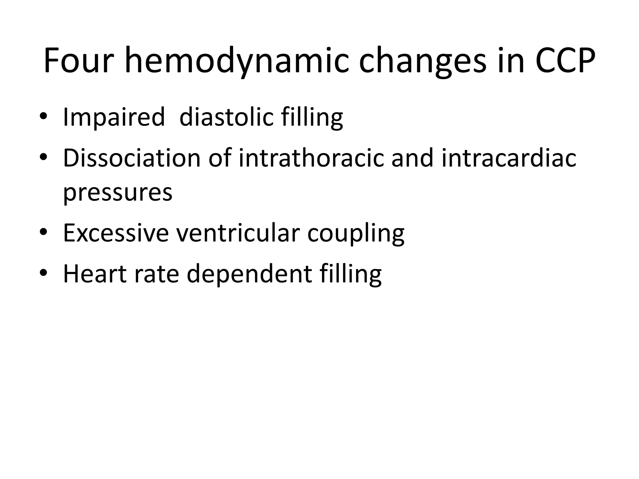
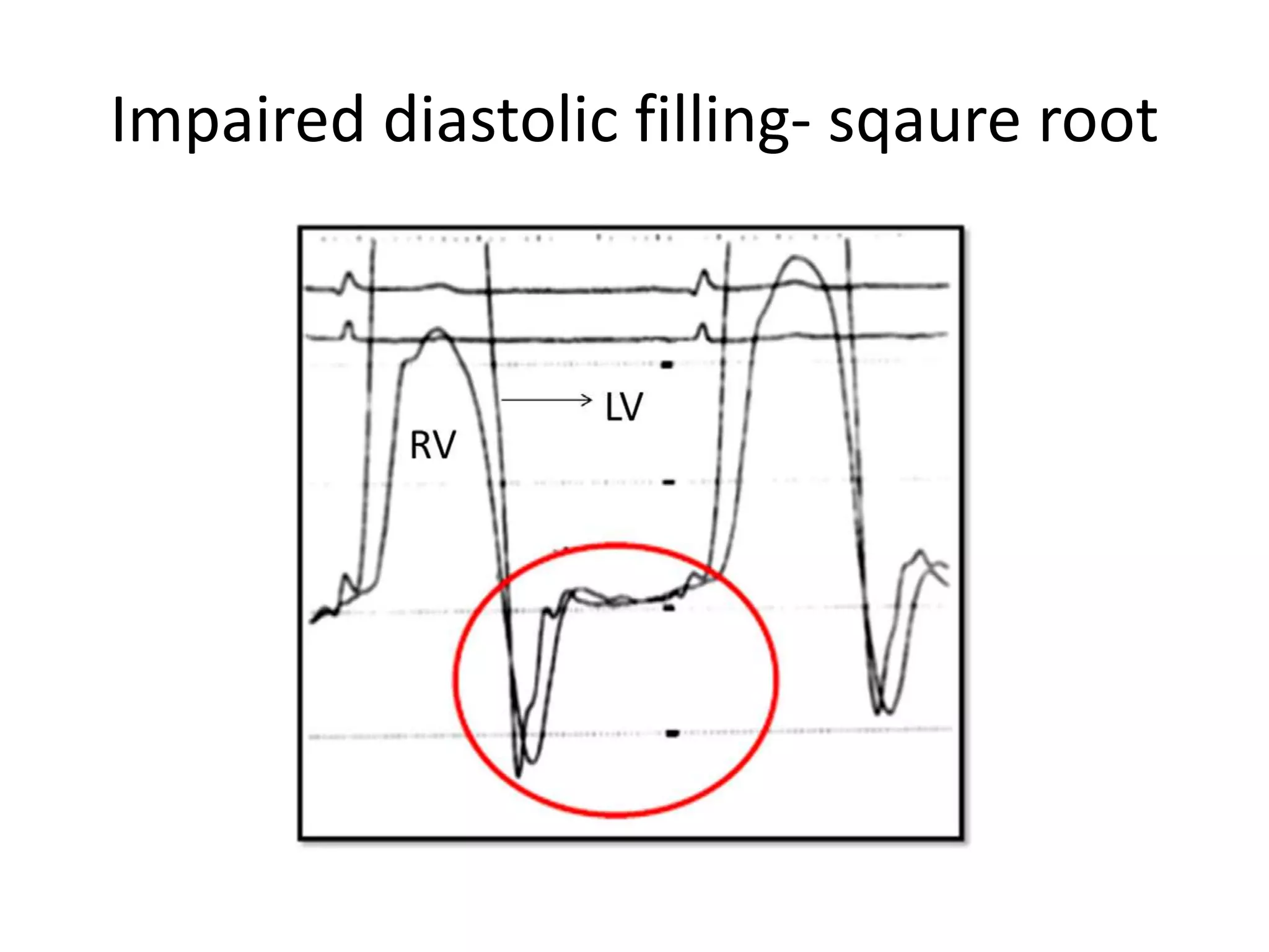
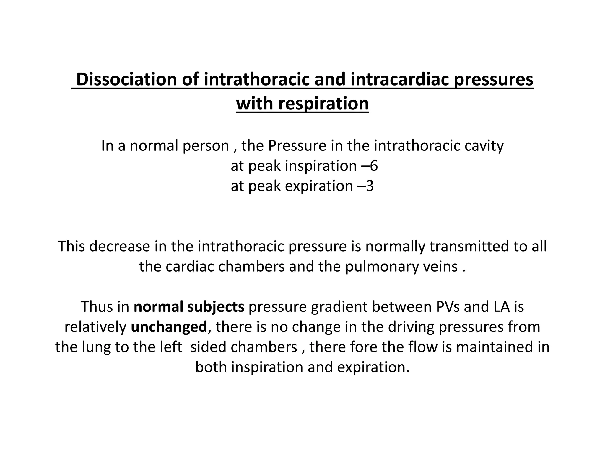
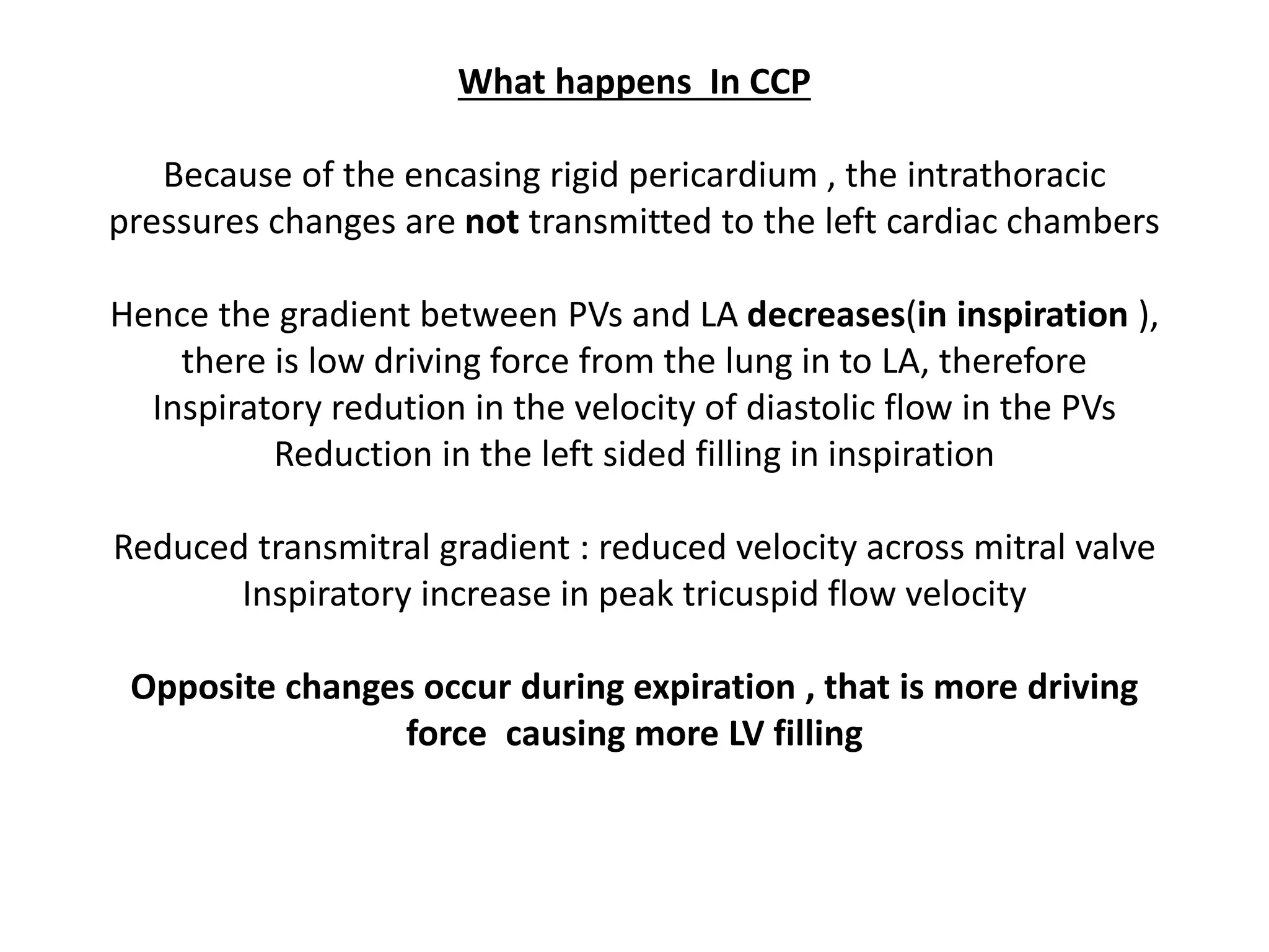
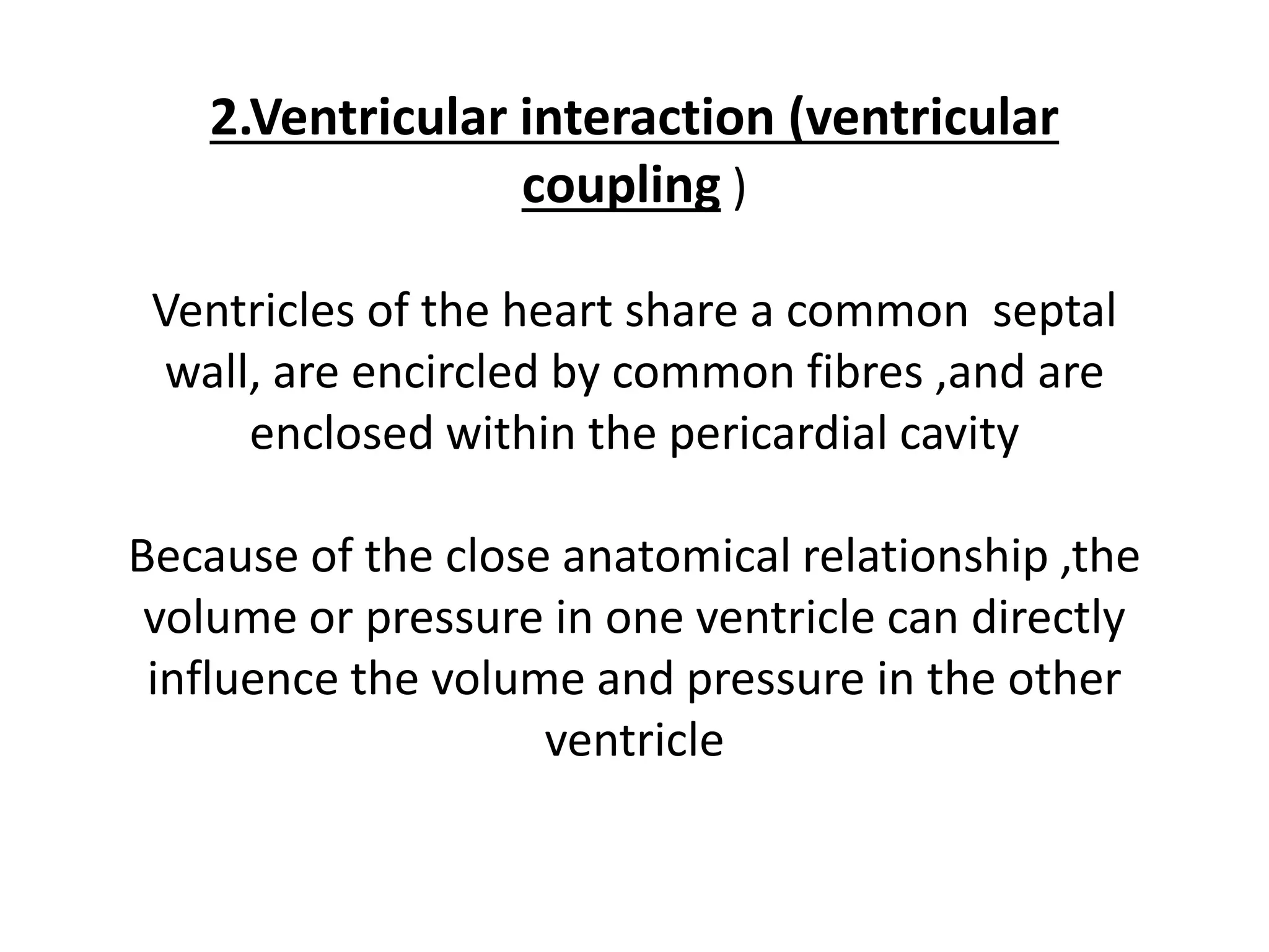
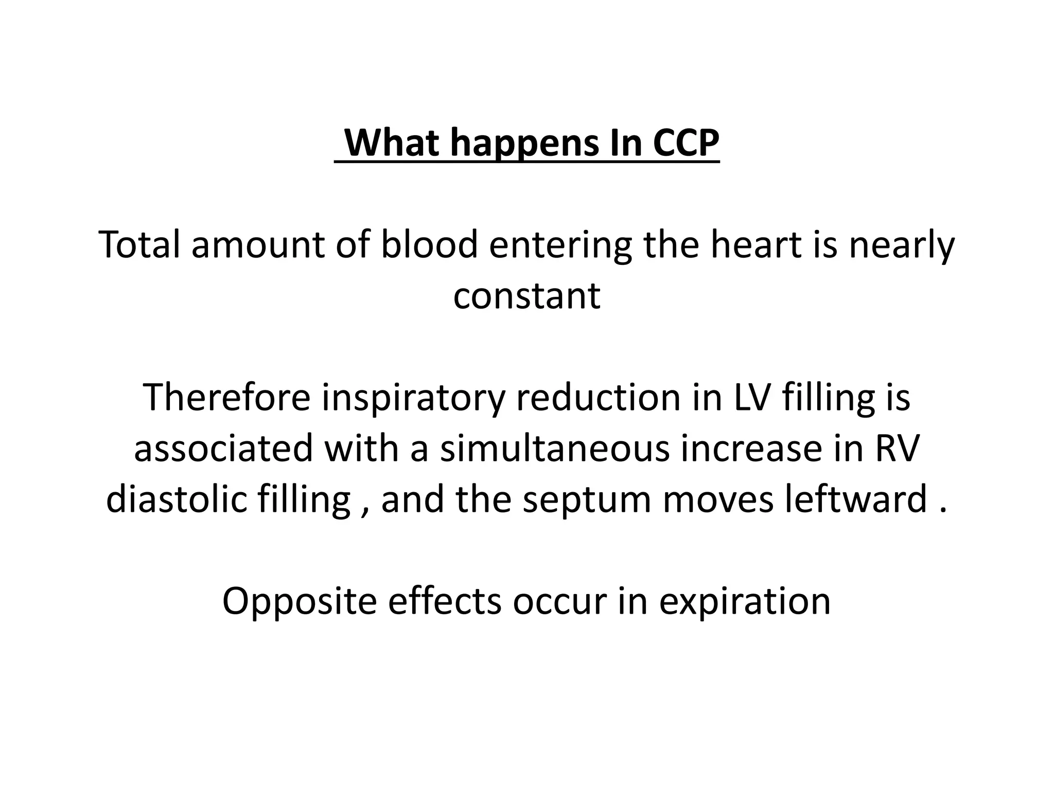
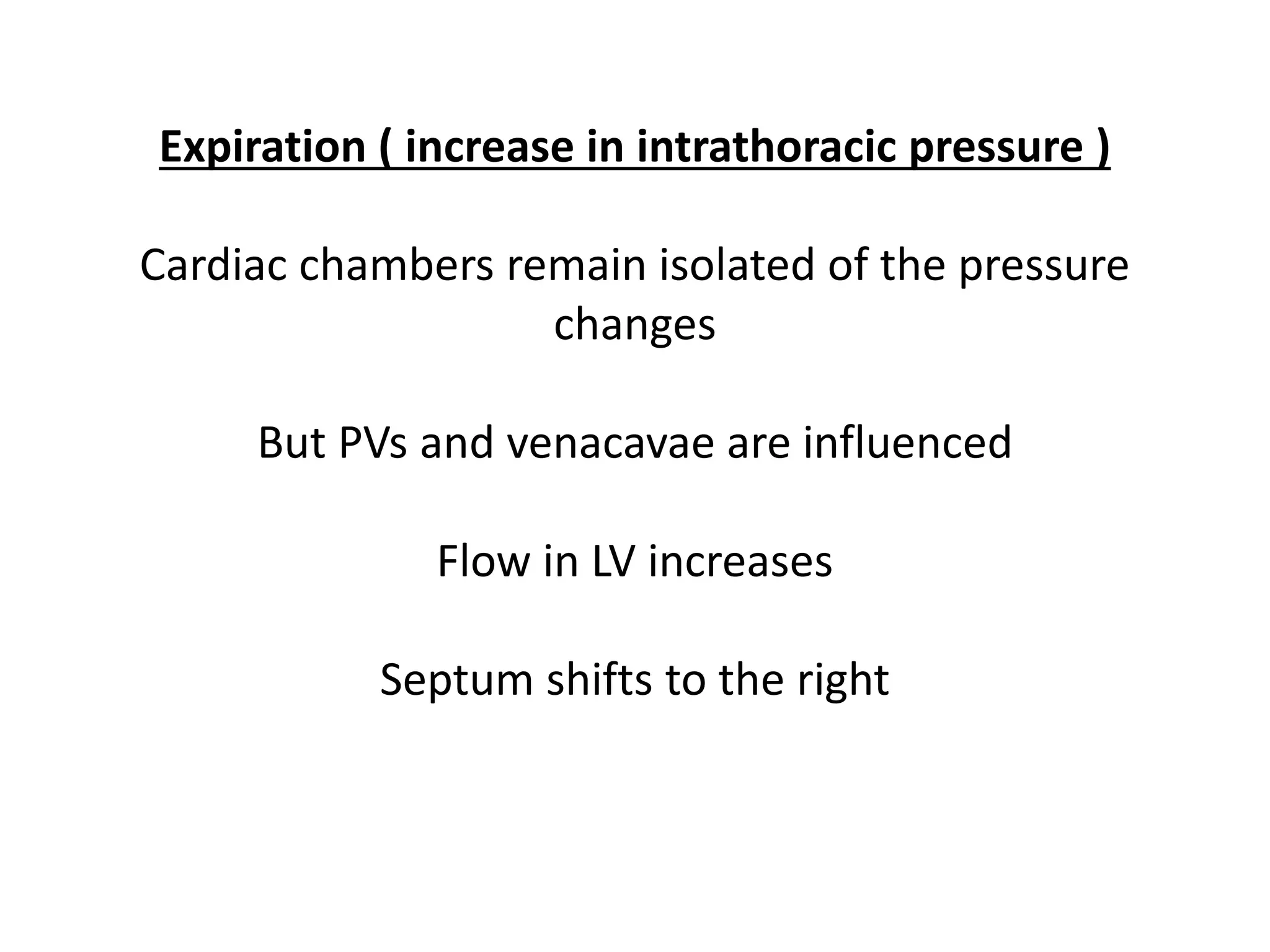
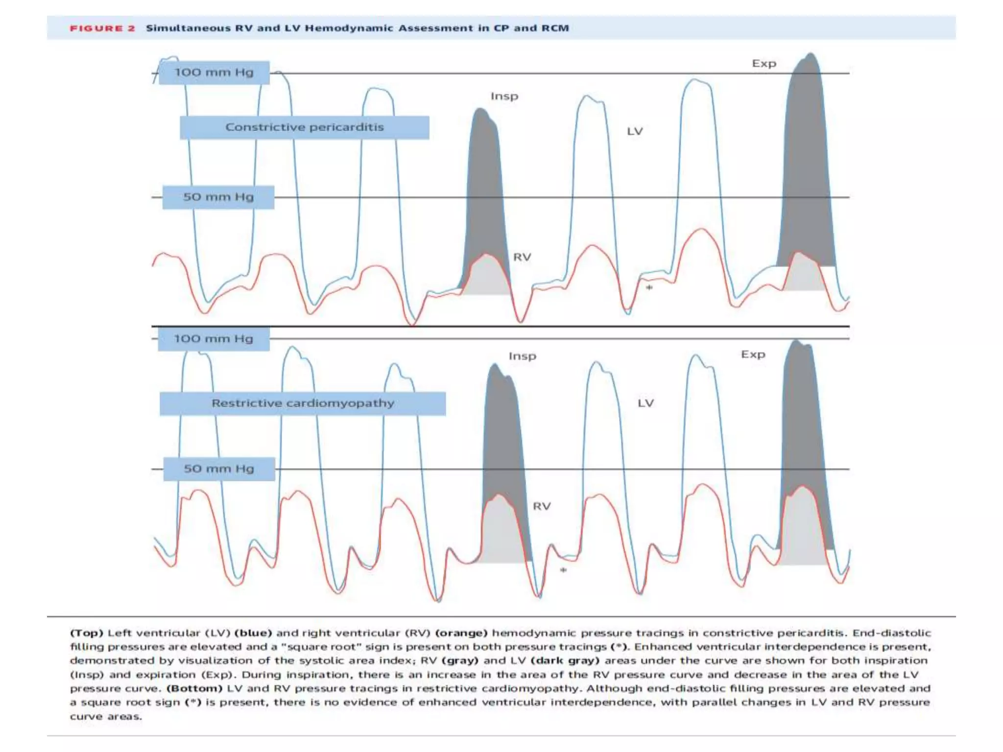
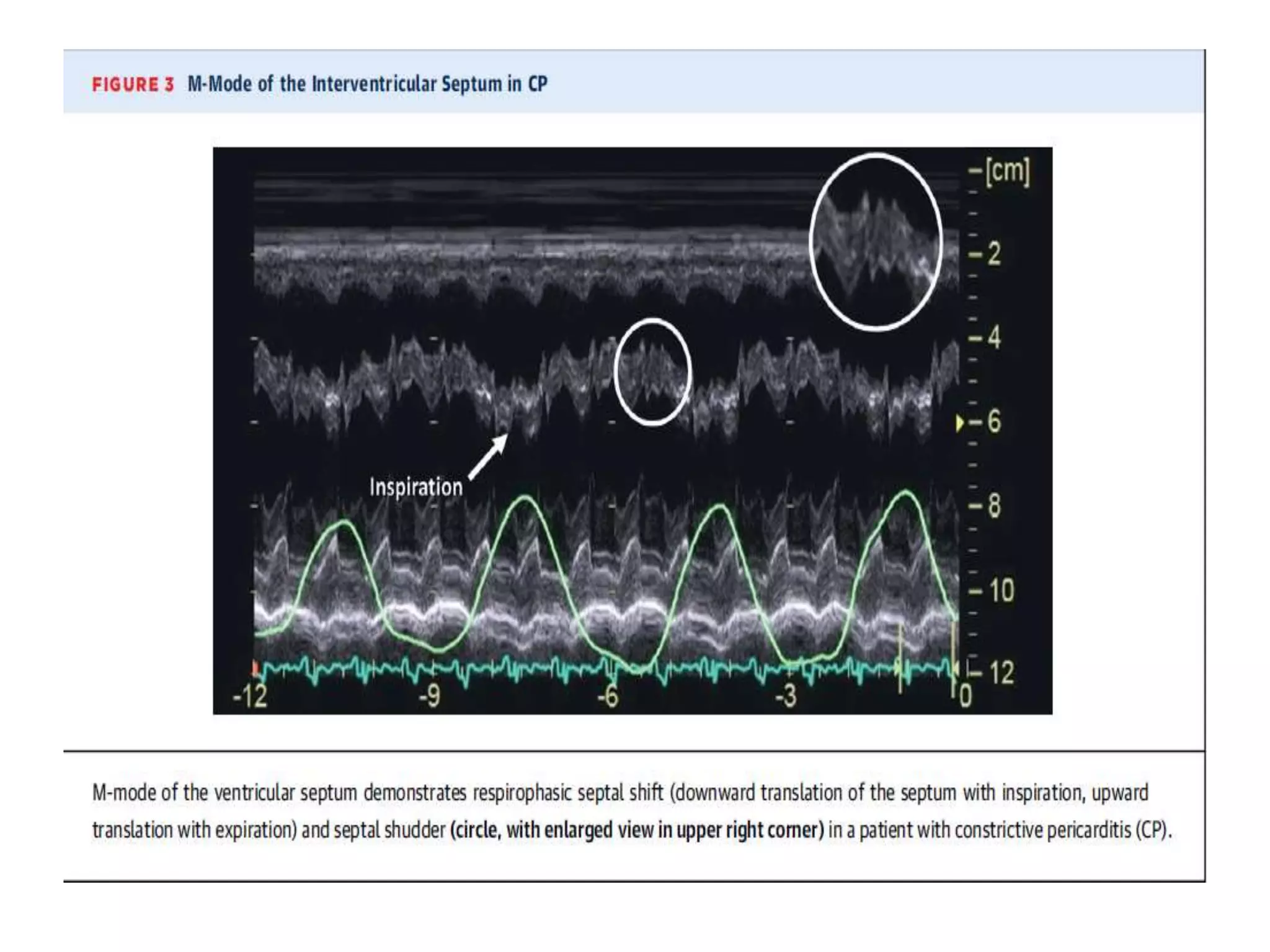
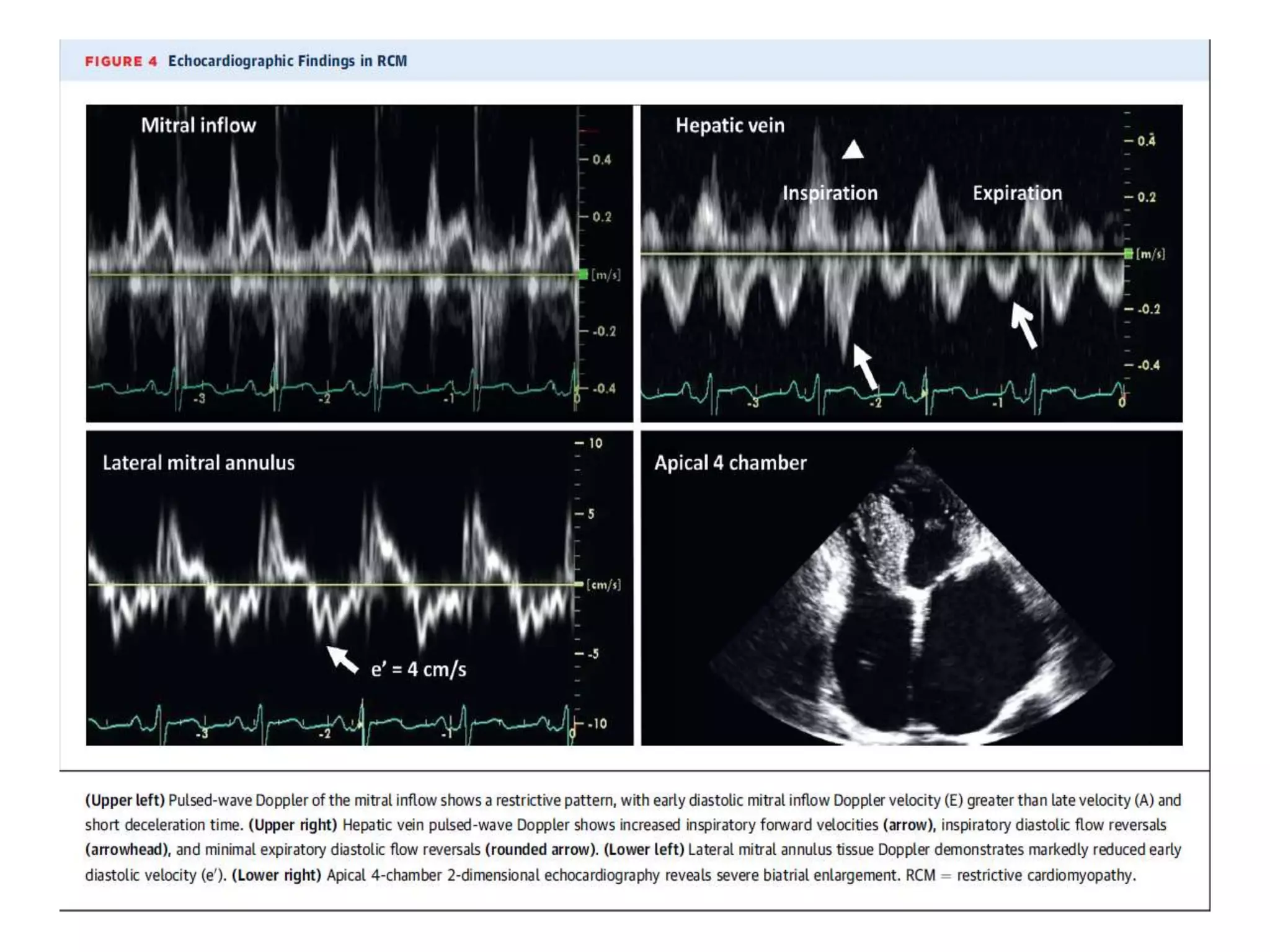
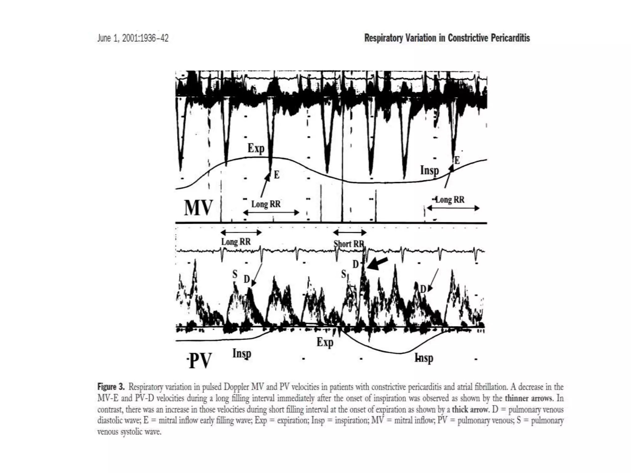
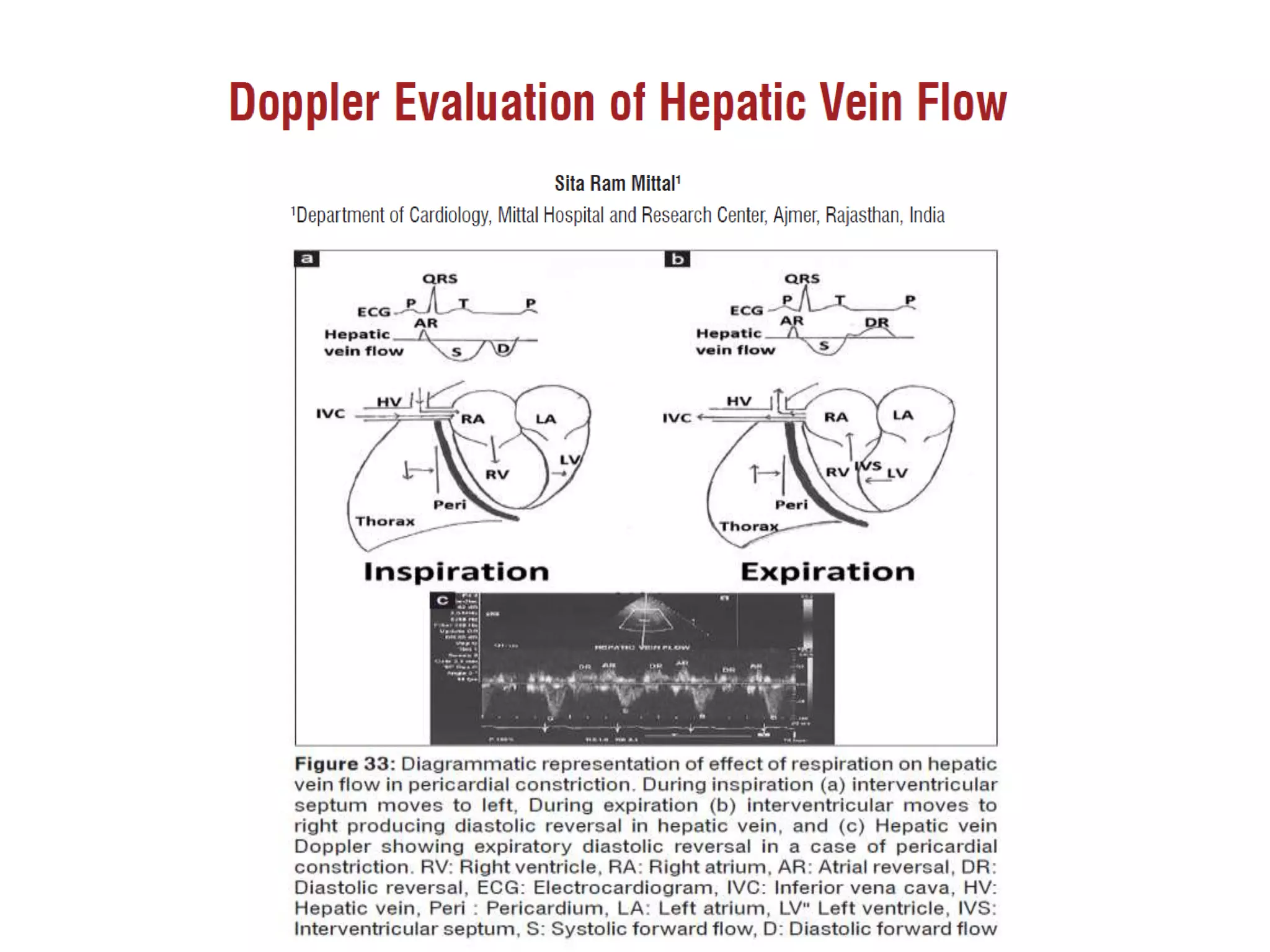
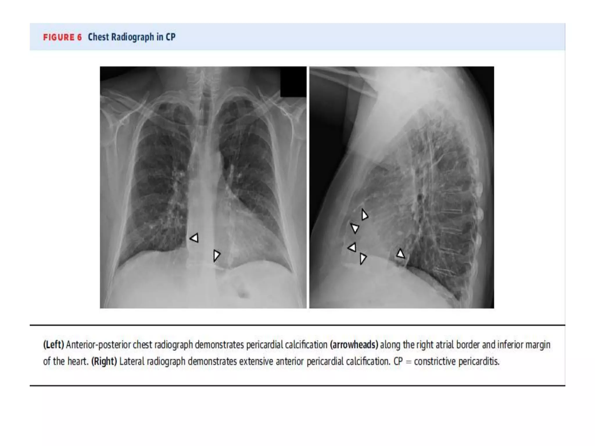
CONSTRICTIVE PERICARDITIS-
PATHOPHYSIOLOGY
Normal pericardium
ETIOLOGY OF CCP
• Idiopathic
• Infective – Tubercular
• Bacterial : staphylococcal , strep, E coli, salmonella, H influenza,
gonococcus, pneumococcal
• Viral : adeno, coxsackie, influenza, lassa
• Fungal:Histoplasma,coccidiomycosis,nocardia,aspergillosis,Blastomy
cosis.
• Others: Guinea worm infestation, hydatid disease , amebiasis,
whipple disease
• Trauma: Blunt: steering wheel injury, CPR
• Penetrating injury: stabs, bullets
• Impalement injury, cardiopericardial surgery
• Intrapericardial electrodes,pacing electrodes and AICD devices
Autoimmune: SLE, rheumatoid arthritis, polyserositis,
scleroderma
Irradiation
Neoplastic: Mesothelioma, undifferentiated carcinoma,
melanoma, metastasis.
Drugs: Methysergide, procainamide, mitomycin C,
Hydralasine
Dialysis related, congenital : mulibreynanism, afibrinogenemia
Asbestosis, amyloidosis, Sarcoidosis, campylobacter,
whipples disease.
Post infarction, post hemopericardium, dresslars.
Peel described four stages in the
development of constriction
Dry stage
Stage of effusion
Stage of absorption
Stage of pericardial constriction
Summarizing Features of variant forms of constrictive pericarditis
Effusive-
constrictive
pericarditis
Pericardial effusion is present, sometimes loculated, with constriction by the
visceral pericardium
Occult constrictive
pericarditis
Haemodynamics are normal at rest, but assume the features of
constriction after an acute volume load
Localised
constrictive
pericarditis
Constriction limited to the right or left ventricle. Ventricular
interdependence reduced or absent
Transient
constrictive
pericarditis
During the resolution of acute pericarditis with effusion, constriction
develops, but then resolves spontaneously over a few weeks
Basic pathology in CCP is the
inability of the ventricles to
dilate due to the thick fibrous
envelope around the heart
Four hemodynamic changes in CCP
• Impaired diastolic filling
• Dissociation of intrathoracic and intracardiac
pressures
• Excessive ventricular coupling
• Heart rate dependent filling
Fibrosed thickened calcified pericardium restricts the
diastolic filling of all the chambers and determines the
diastolic volume of the heart
Symmetrical constricting effect results in elevation and
equalization of diastolic pressures in all four chambers.
Early diastolic filling is unimpeded and hence there is early
rapid filling
It is abruptly halted when the intra cardiac volume reaches
the limit set by a noncompliant pericardium
Avasthe ,Wasir, ML Bhatia, Sujoy B Roy
AIIMS,JIMA 1968
Concluded that haemodynamic effects of CP is due to
mechanical limitation of ventricular filling and possibly also
to emptying of the ventricle if the myocardium is involved
The ventricular function is depressed due to
-Myocardial atrophy
-Myocardial fibrosis
-Obliteration of coronary arteries in the fibrotic pericardium
causing myocardial ischemia
-Coexistent CMP
Grover,Bhatia ,Sujoy ,AIIMS IHJ 1972
Described presence of myocardial factor ---
incriminated it as a cause for incomplete
symptomatic improvement following surgery
Sukumar et al 1966
reported that a delayed early dip and a slow rising
EDP in the ventricular tracing of a patient with CCP
suggests co existing myocardial disease.
Impaired diastolic filling- sqaure root
Dissociation of intrathoracic and intracardiac pressures
with respiration
In a normal person , the Pressure in the intrathoracic cavity
at peak inspiration –6
at peak expiration –3
This decrease in the intrathoracic pressure is normally transmitted to all
the cardiac chambers and the pulmonary veins .
Thus in normal subjects pressure gradient between PVs and LA is
relatively unchanged, there is no change in the driving pressures from
the lung to the left sided chambers , there fore the flow is maintained in
both inspiration and expiration.
What happens In CCP
Because of the encasing rigid pericardium , the intrathoracic
pressures changes are not transmitted to the left cardiac chambers
Hence the gradient between PVs and LA decreases(in inspiration ),
there is low driving force from the lung in to LA, therefore
Inspiratory redution in the velocity of diastolic flow in the PVs
Reduction in the left sided filling in inspiration
Reduced transmitral gradient : reduced velocity across mitral valve
Inspiratory increase in peak tricuspid flow velocity
Opposite changes occur during expiration , that is more driving
force causing more LV filling
2.Ventricular interaction (ventricular
coupling )
Ventricles of the heart share a common septal
wall, are encircled by common fibres ,and are
enclosed within the pericardial cavity
Because of the close anatomical relationship ,the
volume or pressure in one ventricle can directly
influence the volume and pressure in the other
ventricle
Henderson and Prince
Increasing RV volume shifted the IVS
towards the LV and altered the LV filling
characterstics .
The coupling is greatly increased in the
prescense of intact pericardium
What happens In CCP
Total amount of blood entering the heart is nearly
constant
Therefore inspiratory reduction in LV filling is
associated with a simultaneous increase in RV
diastolic filling , and the septum moves leftward .
Opposite effects occur in expiration
Expiration ( increase in intrathoracic pressure )
Cardiac chambers remain isolated of the pressure
changes
But PVs and venacavae are influenced
Flow in LV increases
Septum shifts to the right
-On Expiration-
-There is reduction in flow velocity in the
venacavae
-Increased hepatic venous diastolic flow
reversal
-Decreased transtricuspid flow velocity
Normal
CCP
Which is paradox ?
Kussmaul?
Pulsus paradoxus ?
A DECISION
IS AS IMPORTANT AS
AN INCISION
Thank you

Constrictive pericarditis pathophysiology

  • 1.
  • 3.
  • 4.
    ETIOLOGY OF CCP •Idiopathic • Infective – Tubercular • Bacterial : staphylococcal , strep, E coli, salmonella, H influenza, gonococcus, pneumococcal • Viral : adeno, coxsackie, influenza, lassa • Fungal:Histoplasma,coccidiomycosis,nocardia,aspergillosis,Blastomy cosis. • Others: Guinea worm infestation, hydatid disease , amebiasis, whipple disease • Trauma: Blunt: steering wheel injury, CPR • Penetrating injury: stabs, bullets • Impalement injury, cardiopericardial surgery • Intrapericardial electrodes,pacing electrodes and AICD devices
  • 5.
    Autoimmune: SLE, rheumatoidarthritis, polyserositis, scleroderma Irradiation Neoplastic: Mesothelioma, undifferentiated carcinoma, melanoma, metastasis. Drugs: Methysergide, procainamide, mitomycin C, Hydralasine Dialysis related, congenital : mulibreynanism, afibrinogenemia Asbestosis, amyloidosis, Sarcoidosis, campylobacter, whipples disease. Post infarction, post hemopericardium, dresslars.
  • 6.
    Peel described fourstages in the development of constriction Dry stage Stage of effusion Stage of absorption Stage of pericardial constriction
  • 7.
    Summarizing Features ofvariant forms of constrictive pericarditis Effusive- constrictive pericarditis Pericardial effusion is present, sometimes loculated, with constriction by the visceral pericardium Occult constrictive pericarditis Haemodynamics are normal at rest, but assume the features of constriction after an acute volume load Localised constrictive pericarditis Constriction limited to the right or left ventricle. Ventricular interdependence reduced or absent Transient constrictive pericarditis During the resolution of acute pericarditis with effusion, constriction develops, but then resolves spontaneously over a few weeks
  • 8.
    Basic pathology inCCP is the inability of the ventricles to dilate due to the thick fibrous envelope around the heart
  • 9.
    Four hemodynamic changesin CCP • Impaired diastolic filling • Dissociation of intrathoracic and intracardiac pressures • Excessive ventricular coupling • Heart rate dependent filling
  • 10.
    Fibrosed thickened calcifiedpericardium restricts the diastolic filling of all the chambers and determines the diastolic volume of the heart Symmetrical constricting effect results in elevation and equalization of diastolic pressures in all four chambers. Early diastolic filling is unimpeded and hence there is early rapid filling It is abruptly halted when the intra cardiac volume reaches the limit set by a noncompliant pericardium
  • 11.
    Avasthe ,Wasir, MLBhatia, Sujoy B Roy AIIMS,JIMA 1968 Concluded that haemodynamic effects of CP is due to mechanical limitation of ventricular filling and possibly also to emptying of the ventricle if the myocardium is involved The ventricular function is depressed due to -Myocardial atrophy -Myocardial fibrosis -Obliteration of coronary arteries in the fibrotic pericardium causing myocardial ischemia -Coexistent CMP
  • 12.
    Grover,Bhatia ,Sujoy ,AIIMSIHJ 1972 Described presence of myocardial factor --- incriminated it as a cause for incomplete symptomatic improvement following surgery Sukumar et al 1966 reported that a delayed early dip and a slow rising EDP in the ventricular tracing of a patient with CCP suggests co existing myocardial disease.
  • 13.
  • 14.
    Dissociation of intrathoracicand intracardiac pressures with respiration In a normal person , the Pressure in the intrathoracic cavity at peak inspiration –6 at peak expiration –3 This decrease in the intrathoracic pressure is normally transmitted to all the cardiac chambers and the pulmonary veins . Thus in normal subjects pressure gradient between PVs and LA is relatively unchanged, there is no change in the driving pressures from the lung to the left sided chambers , there fore the flow is maintained in both inspiration and expiration.
  • 15.
    What happens InCCP Because of the encasing rigid pericardium , the intrathoracic pressures changes are not transmitted to the left cardiac chambers Hence the gradient between PVs and LA decreases(in inspiration ), there is low driving force from the lung in to LA, therefore Inspiratory redution in the velocity of diastolic flow in the PVs Reduction in the left sided filling in inspiration Reduced transmitral gradient : reduced velocity across mitral valve Inspiratory increase in peak tricuspid flow velocity Opposite changes occur during expiration , that is more driving force causing more LV filling
  • 16.
    2.Ventricular interaction (ventricular coupling) Ventricles of the heart share a common septal wall, are encircled by common fibres ,and are enclosed within the pericardial cavity Because of the close anatomical relationship ,the volume or pressure in one ventricle can directly influence the volume and pressure in the other ventricle
  • 17.
    Henderson and Prince IncreasingRV volume shifted the IVS towards the LV and altered the LV filling characterstics . The coupling is greatly increased in the prescense of intact pericardium
  • 18.
    What happens InCCP Total amount of blood entering the heart is nearly constant Therefore inspiratory reduction in LV filling is associated with a simultaneous increase in RV diastolic filling , and the septum moves leftward . Opposite effects occur in expiration
  • 19.
    Expiration ( increasein intrathoracic pressure ) Cardiac chambers remain isolated of the pressure changes But PVs and venacavae are influenced Flow in LV increases Septum shifts to the right
  • 20.
    -On Expiration- -There isreduction in flow velocity in the venacavae -Increased hepatic venous diastolic flow reversal -Decreased transtricuspid flow velocity
  • 21.
  • 22.
  • 23.
    Which is paradox? Kussmaul? Pulsus paradoxus ?
  • 54.
    A DECISION IS ASIMPORTANT AS AN INCISION Thank you