This document provides an overview of Streptococcus bacteria, including characteristics, diseases caused, taxonomy, and methods for identification. Key points include:
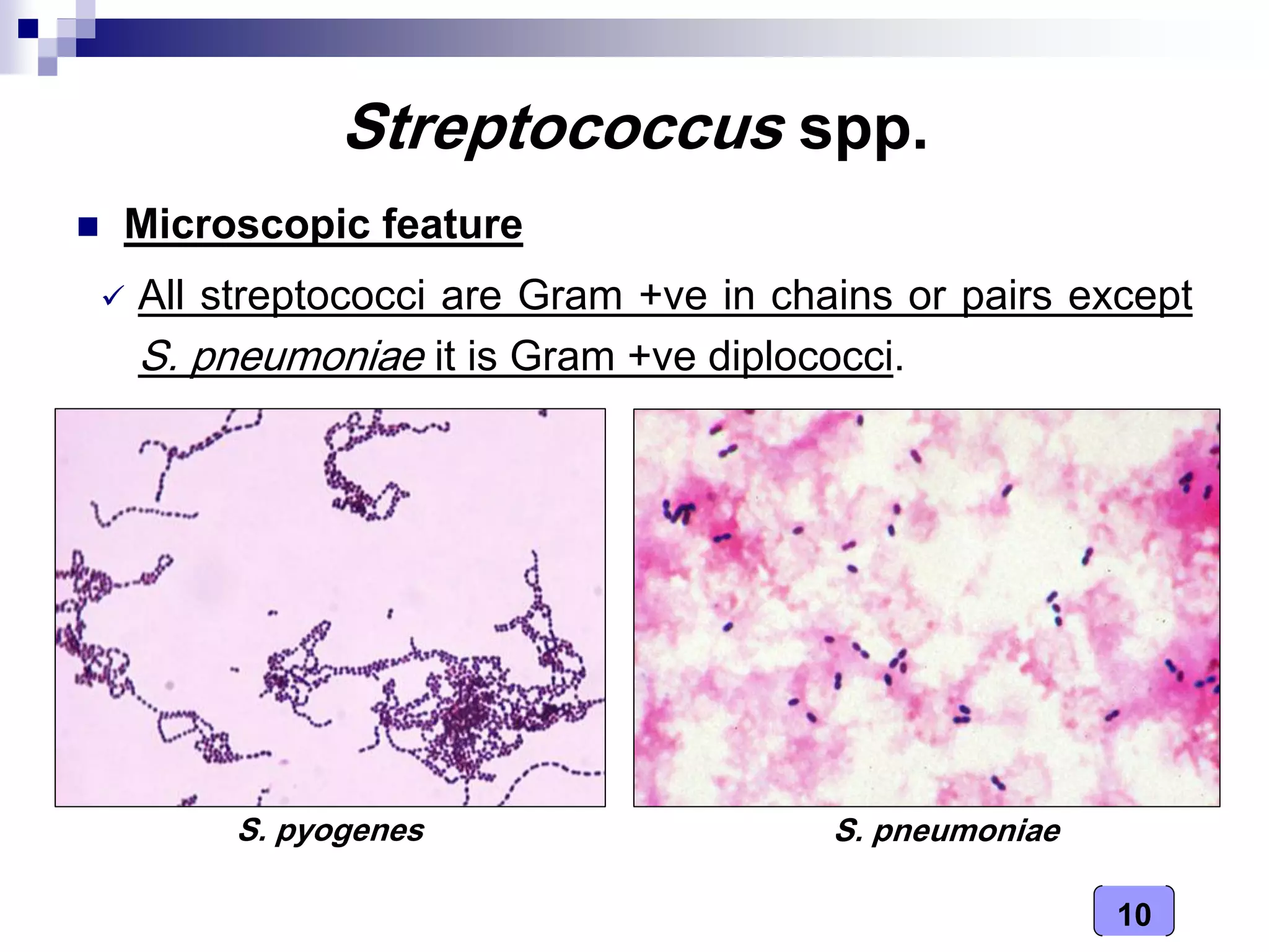
- Streptococcus is a genus of spherical, Gram-positive bacteria that grow in chains. It includes over 50 species that are part of normal oral flora but can also cause diseases.
- Major diseases caused by different Streptococcus species include pharyngitis, pneumonia, toxic shock syndrome, and neonatal infections.
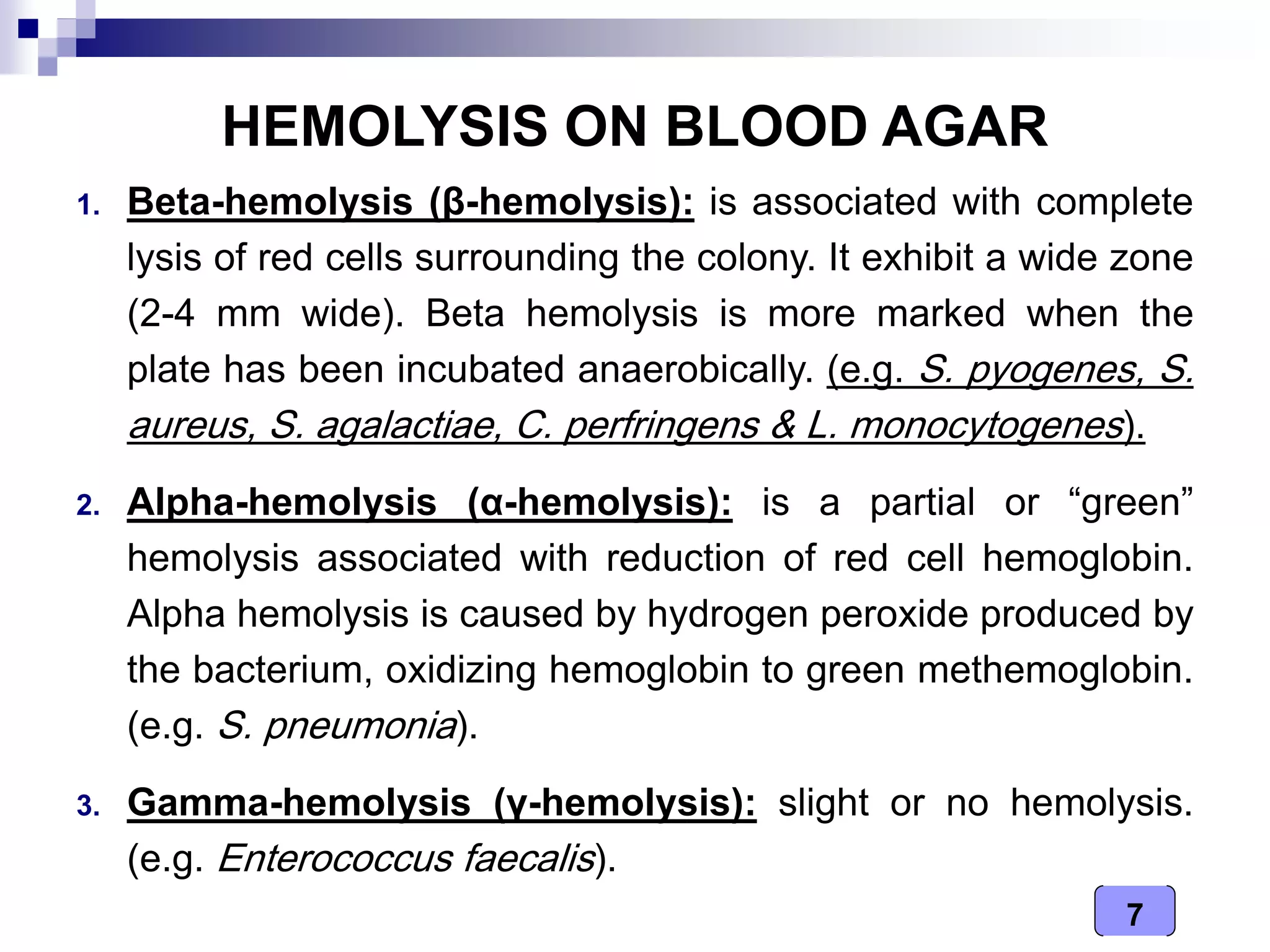
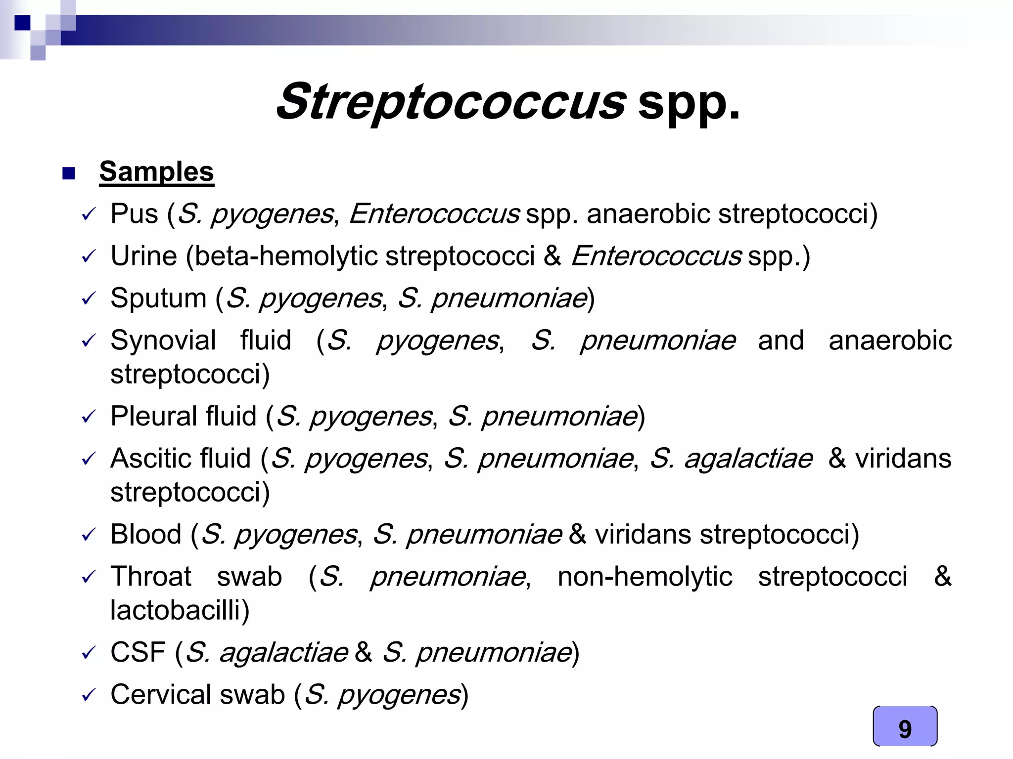
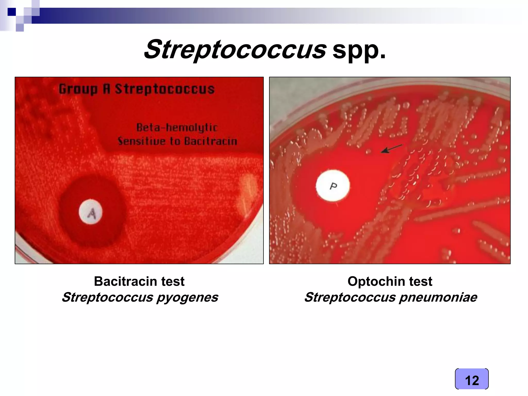
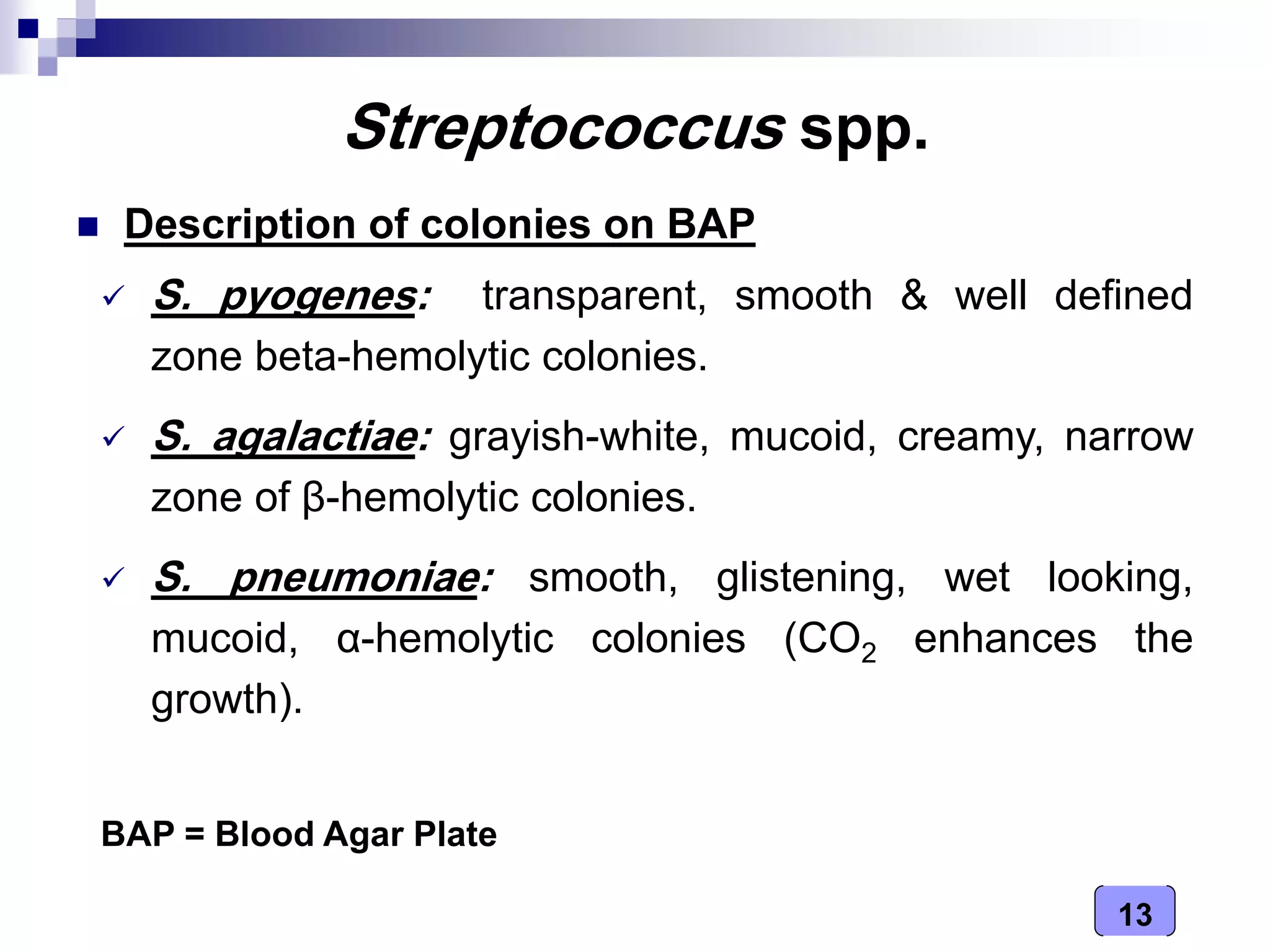
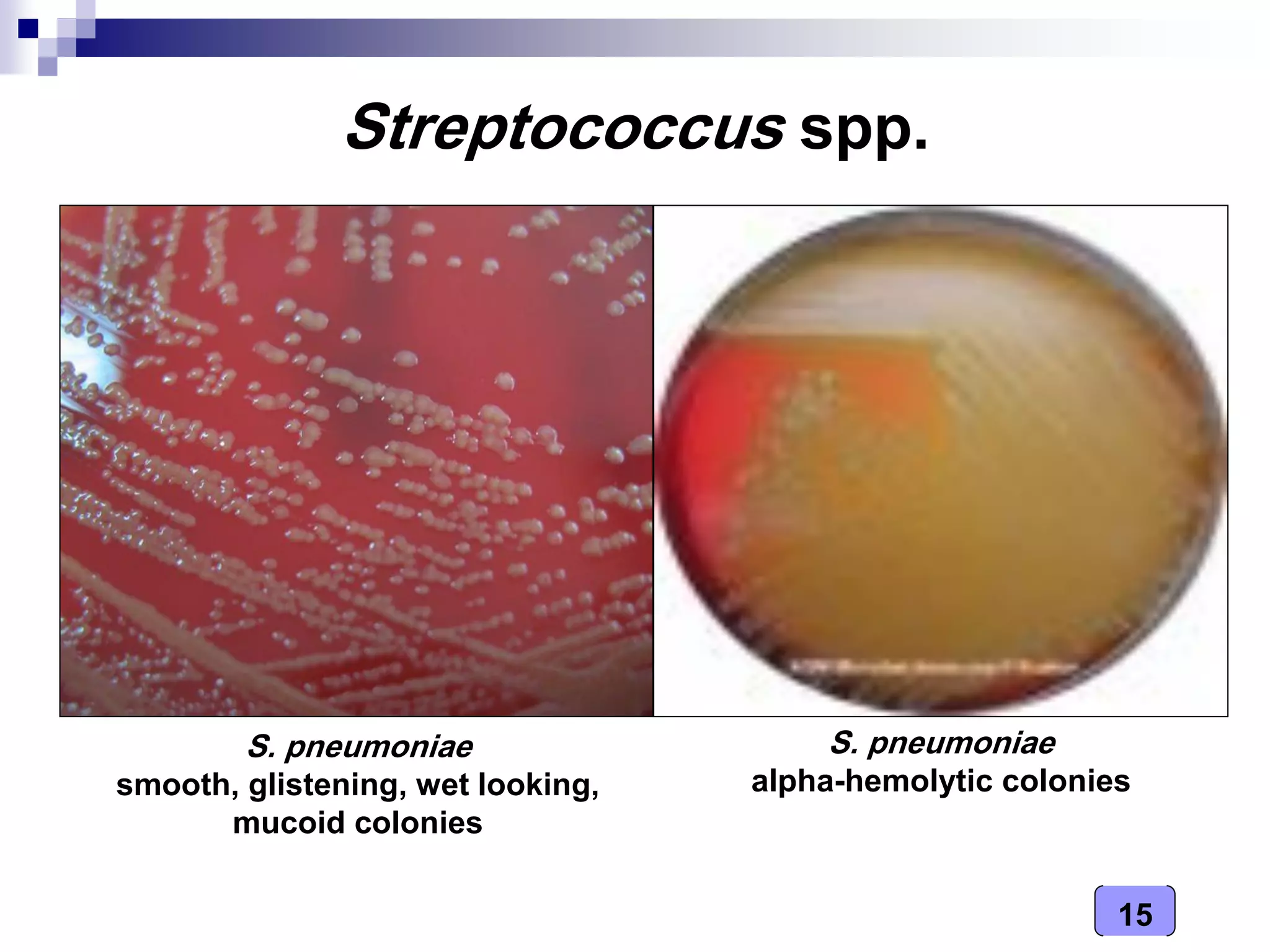
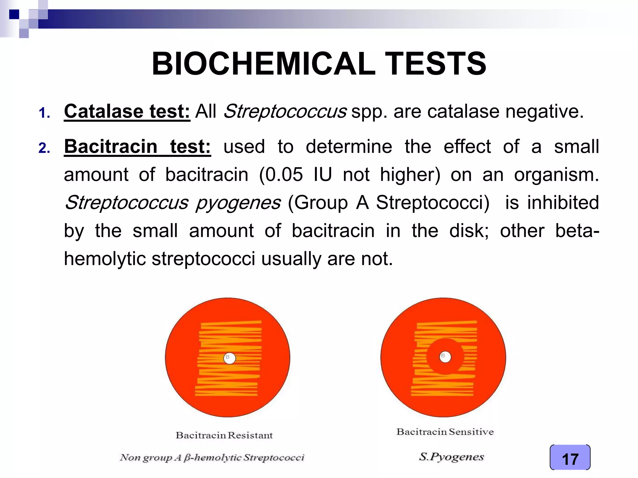
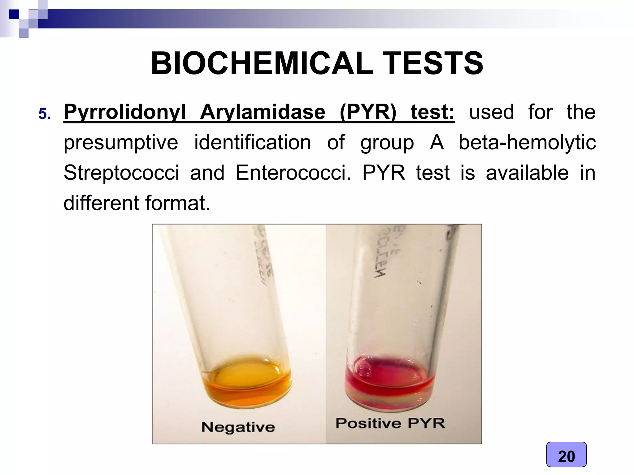
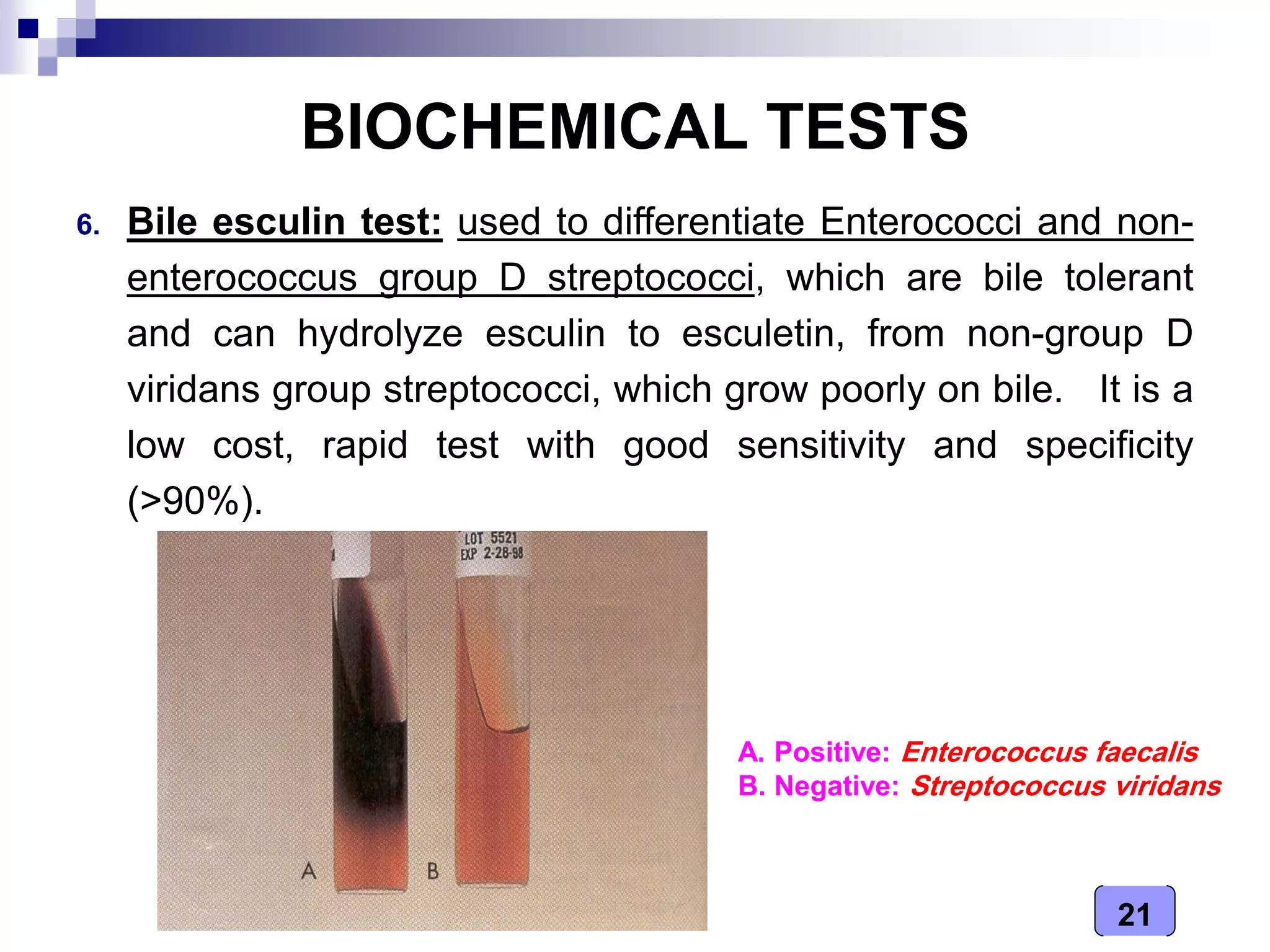
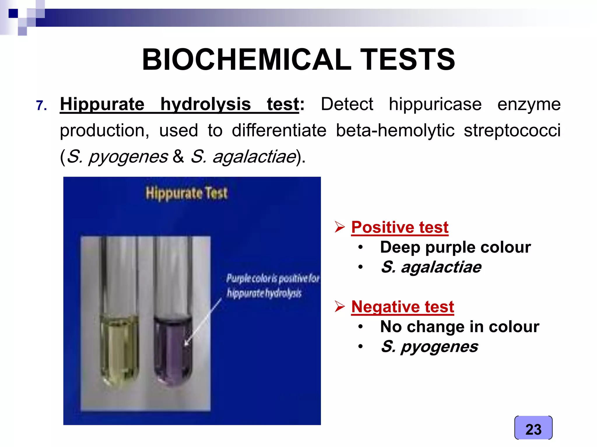
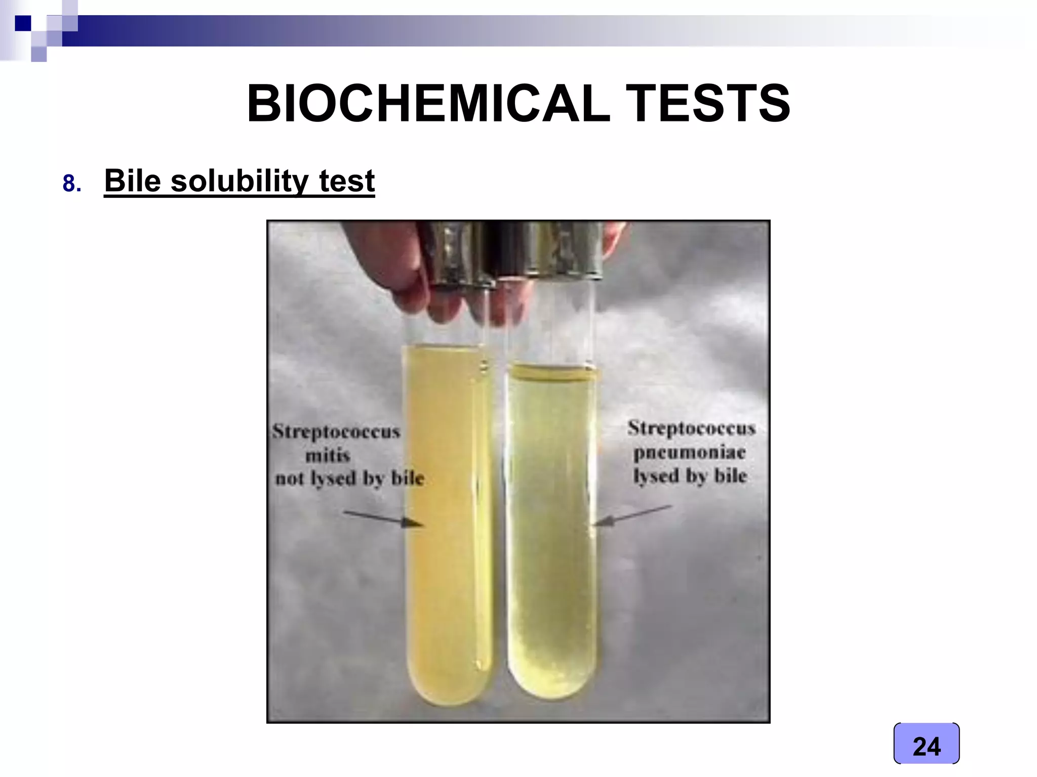
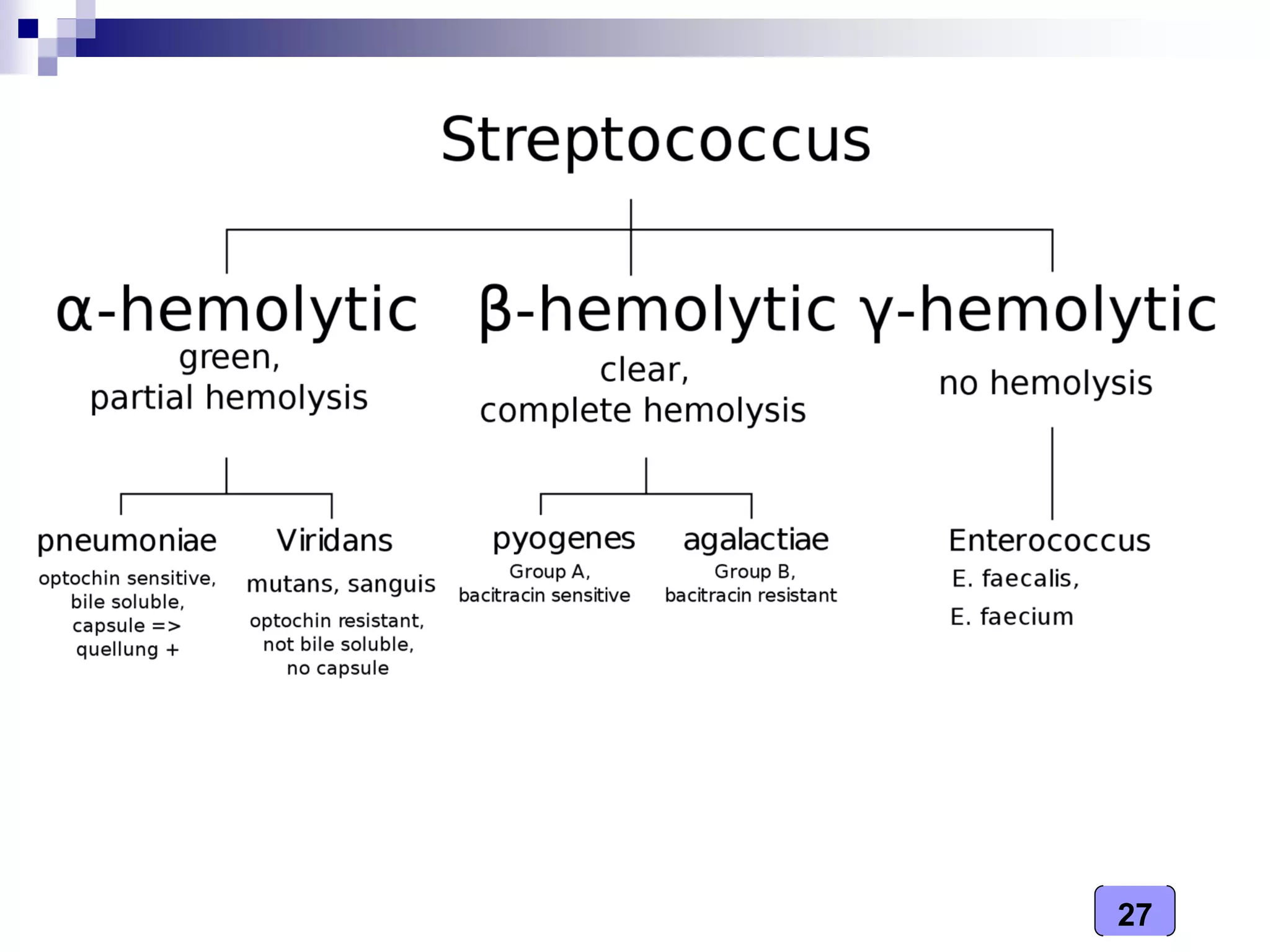
- Identification involves examining colony morphology on blood agar plates, microscopic appearance, and biochemical tests like catalase, optochin, and bile esculin tests.
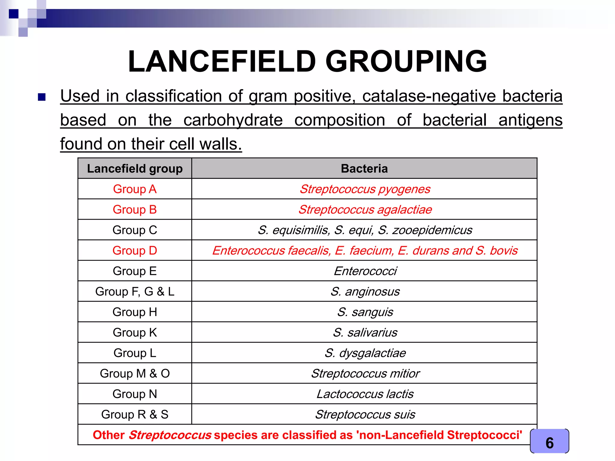
- Classification is based on carbohydrate antigens identified through Lancefield grouping