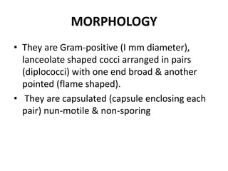
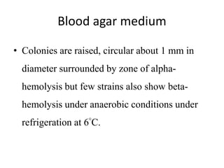
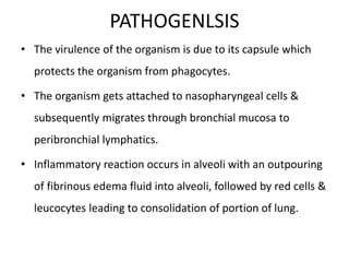
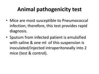
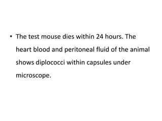
PNEUMOCOCCUS
RATHEESH R L
• Medically important species is
pneumococcus pneumonia
MORPHOLOGY
• They are Gram-positive (I mm diameter),
lanceolate shaped cocci arranged in pairs
(diplococci) with one end broad & another
pointed (flame shaped).
• They are capsulated (capsule enclosing each
pair) nun-motile & non-sporing
CULTURAL CHARACTERISTICS
• They are aerobic/ facultative anaerobic and
grow best at 37 degree C at pH 7.6.
• Growth occurs best in atmosphere of 10-20%
of carbon dioxide.
Serum & glucose broth
• After 24 hours, show uniform turbidity.
• Non-capsulated strains
Blood agar medium
• Colonies are raised, circular about 1 mm in
diameter surrounded by zone of alpha-
hemolysis but few strains also show beta-
hemolysis under anaerobic conditions under
refrigeration at 6°C.
PATHOGENLSIS
• The virulence of the organism is due to its capsule which
protects the organism from phagocytes.
• The organism gets attached to nasopharyngeal cells &
subsequently migrates through bronchial mucosa to
peribronchial lymphatics.
• Inflammatory reaction occurs in alveoli with an outpouring
of fibrinous edema fluid into alveoli, followed by red cells &
leucocytes leading to consolidation of portion of lung.
• The usual lesion in adults is segmental or lobar
in distribution (lobar pneumonia) but in
children & aged, there Is patchy involvement
of lung (bronchopneumonia).
• Microorganisms are found throughout the
exudates & bacteraemia is common.
• Pneumococcal meningitis:
Strep. pneumoniae are the second
most important cause of pyogenic meningitis
after N.meningitides.
It is most serious Pneumococcal
infection more common in children with 40-50%
mortality rate
LABORATORY DIAGNOSIS
• Hematological investigation:
1…..TLC: Shows increase in leucocytes
(> 15000/mm3>.
II…..DLC. Shows increase in
polymorphonuclear cells.
• Bacteriological investigation:
– Specimen: sputum, CSF, pleural fluid, peritoneal
fluid, pericardial fluid & pus discharges.
Microscopy
• Gram staining of the above specimen shows
flame shaped diplococci.
• India ink preparation shows capsule as a clear
halo.
Culture
• Specimen is inoculated on BAM & incubated
at 37 C for 24-48 hours in atmosphere of
5-10% CO 2.
• Colonies develop after 24 hours which appear
as flat, umbonate (depressed at centre)
showing alpha hemolysis (greenish
discoloration).
Biochemical tests
• Sugar fermentation test - Positive with acid
production only.
• Bile solubility test - Positive
• Catalase test - Negative
• Oxidase test - Negative
Serological tests
• Co-agglutination test (COA)
• Latex agglutination test (LA).
• Counter current immuno electrophoresis
(CCIE)
Animal pathogenicity test
• Mice are most susceptible to Pneumococcal
infection; therefore, this test provides rapid
diagnosis.
• Sputum from infected patient is emulsified
with saline & one ml of this suspension is
inoculated/injected intraperitoneally into 2
mice (test & control).
• The test mouse dies within 24 hours. The
heart blood and peritoneal fluid of the animal
shows diplococci within capsules under
microscope.
TREATMENT
• Antibiotics:
– Sulphonamides and penicillin are effective
medication
3. pneumococcus

3. pneumococcus

  • 1.
  • 2.
    • Medically importantspecies is pneumococcus pneumonia
  • 3.
    MORPHOLOGY • They areGram-positive (I mm diameter), lanceolate shaped cocci arranged in pairs (diplococci) with one end broad & another pointed (flame shaped). • They are capsulated (capsule enclosing each pair) nun-motile & non-sporing
  • 5.
    CULTURAL CHARACTERISTICS • Theyare aerobic/ facultative anaerobic and grow best at 37 degree C at pH 7.6. • Growth occurs best in atmosphere of 10-20% of carbon dioxide.
  • 6.
    Serum & glucosebroth • After 24 hours, show uniform turbidity. • Non-capsulated strains
  • 7.
    Blood agar medium •Colonies are raised, circular about 1 mm in diameter surrounded by zone of alpha- hemolysis but few strains also show beta- hemolysis under anaerobic conditions under refrigeration at 6°C.
  • 8.
    PATHOGENLSIS • The virulenceof the organism is due to its capsule which protects the organism from phagocytes. • The organism gets attached to nasopharyngeal cells & subsequently migrates through bronchial mucosa to peribronchial lymphatics. • Inflammatory reaction occurs in alveoli with an outpouring of fibrinous edema fluid into alveoli, followed by red cells & leucocytes leading to consolidation of portion of lung.
  • 9.
    • The usuallesion in adults is segmental or lobar in distribution (lobar pneumonia) but in children & aged, there Is patchy involvement of lung (bronchopneumonia). • Microorganisms are found throughout the exudates & bacteraemia is common.
  • 10.
    • Pneumococcal meningitis: Strep.pneumoniae are the second most important cause of pyogenic meningitis after N.meningitides. It is most serious Pneumococcal infection more common in children with 40-50% mortality rate
  • 11.
    LABORATORY DIAGNOSIS • Hematologicalinvestigation: 1…..TLC: Shows increase in leucocytes (> 15000/mm3>. II…..DLC. Shows increase in polymorphonuclear cells.
  • 12.
    • Bacteriological investigation: –Specimen: sputum, CSF, pleural fluid, peritoneal fluid, pericardial fluid & pus discharges.
  • 13.
    Microscopy • Gram stainingof the above specimen shows flame shaped diplococci. • India ink preparation shows capsule as a clear halo.
  • 14.
    Culture • Specimen isinoculated on BAM & incubated at 37 C for 24-48 hours in atmosphere of 5-10% CO 2. • Colonies develop after 24 hours which appear as flat, umbonate (depressed at centre) showing alpha hemolysis (greenish discoloration).
  • 15.
    Biochemical tests • Sugarfermentation test - Positive with acid production only. • Bile solubility test - Positive • Catalase test - Negative • Oxidase test - Negative
  • 16.
    Serological tests • Co-agglutinationtest (COA) • Latex agglutination test (LA). • Counter current immuno electrophoresis (CCIE)
  • 17.
    Animal pathogenicity test •Mice are most susceptible to Pneumococcal infection; therefore, this test provides rapid diagnosis. • Sputum from infected patient is emulsified with saline & one ml of this suspension is inoculated/injected intraperitoneally into 2 mice (test & control).
  • 18.
    • The testmouse dies within 24 hours. The heart blood and peritoneal fluid of the animal shows diplococci within capsules under microscope.
  • 19.
    TREATMENT • Antibiotics: – Sulphonamidesand penicillin are effective medication