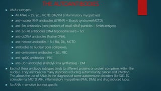
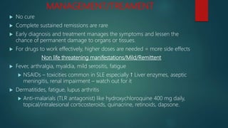
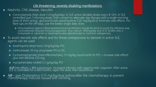
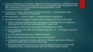
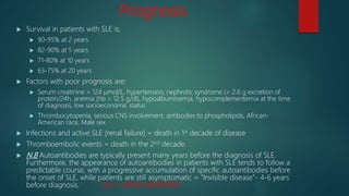
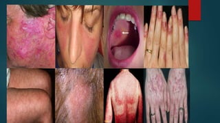
S.L.E.
(SYSTEMIC LUPUS ERYTHEMATOSUS)
By:
Ibiwoye O. O.
 A chronic multisystem autoimmune disease of protean manifestation and
variable behavior
 Lots of organs affected, affect any organ but skin, kidneys, serosal
membrane, joints and heart stand out
 Lots of autoantibodies and variable clinical presentation with so many
overlaps with other autoimmune diseases
 Reason for the development of the criteria with features ≥ 4 = diagnostic
during interval of observations
 F : M = 9:1 Just like all autoimmune diseases
 Affects any age, even early childhood but common in 2nd/3rd decade of life
(15-44 years), 1 in 700 women of childbearing age, 15-50 in 100, 000 of
general population. Blacks > Hispanics > Asians
 Its all about the failure of the body to maintain self tolerance (self-tolerance
means that lymphocytes should not incite an immune response against
human cellular antigens i.e. non-responsiveness of immune system to
substance or tissue with capacity to elicit an immune response)
PATHOPHYSIOLOGY
Inherited susceptibility genes
 Class II MHC HLA-DQ, DR3
 Complement
 Others
Environmental triggers
e.g. UVR, drugs, sex hormones,
infectious agents, stress
Increased burden of
nuclear protein
fragments, other self
antigens
Cellular apoptosis
Activation of T helpers and B cells
specific for self antigens
IgG autoantibody production
Immune complex and autoantibody mediated tissue injury
DIAGNOSTIC CRITERIA/CLINICAL PRESENTATION
 DOPAMIN RASH criteria by ACR has been supplanted by SLICC
 In 2015, revised criteria, both ACR/SLICC = combined. The patients with 4 points out of 16, have definite
diagnosis of SLE. With 3 points highly suggestive SLE, with 2 points probable SLE and with one point
possible SLE are the diagnosis.
 Radiological
 CBC
 Hormonal assay
 HLA
 Renal/Skin biopsy
 Autoantibody screening (serology, IF, ELISA)
 Urinalysis
 LE bodies/cells in 70% patients, now history, not in use anymore. It’s a neutrophil
or macrophage that has engulfed the denatured nucleus of another injured cell by
the immune complex in vitro. Damaged nucleus lose their chromatin pattern =
homogenous = hematoxylin/LE bodies in vivo
 The highly specific markers
 Anti-dsDNA antibodies
 Anti-sm antibodies (99% of people without SLE lack it)
THE AUTOANTIBODIES
 ANAs subtypes
 All ANAs – SS, Scl., MCTD, DM/PM (inflammatory myopathies)
 anti-nuclear RNP antibodies (U1RNP) – Sharp’s syndrome(MCTD)
 anti-Sm antibodies (core proteins of small nRNP particles – Smith antigen),
 anti-Scl-70 antibodies (DNA topoisomerase1) – Scl.
 anti-dsDNA antibodies (Native DNA),
 anti-histone antibodies – Scl. RA, DIL, MCTD
 antibodies to nuclear pore complexes,
 anti-centromere antibodies – Scl., PBC
 anti-sp100 antibodies - PBC
 anti- Jo 1 antibodies (Histidyl-Trna synthetase) - DM
 Each of these antibody subtypes binds to different proteins or protein complexes within the
nucleus. They are found in many disorders including autoimmunity, cancer and infection.
This allows the use of ANAs in the diagnosis of some autoimmune disorders like SLE, SS,
scleroderma, MCTD, AIH, inflammatory myopathies (PMs, DMs) and drug induced lupus.
 So ANA = sensitive but not specific
 Anti cytoplasmic antibodies
 Ribosomes (P proteins)
 Mitochondria
 Lysosomes
 anti-Ro (SS-A) antibodies (RNP) – SS, PBC, Celiac disease, PM, neonatal lupus + congenital heart block
 anti-La (SS-B) antibodies (RNP) – SS, PBC
 Antibodies react with various complexes of phospholipids with proteins = secondary APLS
 Anti-cardiolipin
 Lupus anticoagulant
 Antibodies to beta2-glycoprotein 1
 Others
 Rhematoid factors (antibody reactive with self - IgG)
 C1q
 Heat shock proteins (hsp 70, 90)
 Anti-cellular antibodies;
 RBCs - Anemia
 Platelets - Thrombocytopenia
 WBCs – Leukopenia, lymphopenia
 Thyroid - Hypothyroidism
 Neurons – diffuse CNS lupus
DDX
 Overlapping autoimmune diseases – difficult to differentiate e.g.
MTCD = features of SLE, RA, PMs, Scl. + high titers of anti-RNP
 RA lupus arthritis causes less destruction of the joints than
rheumatoid arthritis.
 Various forms of dermatitis
 Neurologic disorders e.g. epilepsy, MS, psychiatry disorder
 Hematologic diseases e.g. ITP
 Drug-induced Lupus-like syndrome – chronic use and depends on
drug acetylation. ANA/Anti-histone antibodies but no antibodies
to dsDNA and hypocomplementemia which are all present in
idiopathic SLE
 Infections e.g. bacterial endocarditis, histoplasmosis
 Primary/Idiopathic APLS – HLA DR7
MANAGEMENT/TREAMENT
 No cure
 Complete sustained remissions are rare
 Early diagnosis and treatment manages the symptoms and lessen the
chance of permanent damage to organs or tissues.
 For drugs to work effectively, higher doses are needed = more side effects
Non life threatening manifestations/Mild/Remittent
 Fever, arthralgia, myaldia, mild serositis, fatigue
 NSAIDs – toxicities common in SLE especially ↑ Liver enzymes, aseptic
meningitis, renal impairment – watch out for it
 Dermatitides, fatigue, lupus arthritis
 Anti-malarials (TLR antagonist) like hydroxychloroquine 400 mg daily,
topical/intralesional corticosteroids, quinacrine, retinoids, dapsone.
Life threatening, severely disabling manifestations
 Nephritis, CNS disease, Vasculitis
 Corticosteroids (high dose 1-2mg/kg/day). In SLE active (divided doses every 8-12h). In SLE
controlled (just 1 morning dose), then convert to alternate-day therapy with a single morning
dose of short acting glucocorticoids (prednisolone 0.02 mg/kg/d) to minimize side effects. For
flare-ups on the off-days, use the lowest single daily dose.
 Immunizations against influenza/pneumococcal infections should be done to avoid the infections due
corticosteroids-induced immunosuppression. Give calcium 1000mg/day and vit D 50,000 units 3
times/week or calcitonin or bisphosphonates (alendronate) to minimize osteoporosis.
 To avoid steroids side effects and for those unresponsive to steroids in active SLE,
agents can be used
 Azathioprine (least toxic) 3mg/kg/day PO,
 methotrexate 20 mg once/week PO or SC,
 Cyclophosphamide (most effective/toxic) 15 mg/kg once/month IV. PO = increase side effects
give with MESNA 250 mg,
 mycophenolate mofetil 2.5 g/kg/day PO
N.B Side effects = BM suppression, increased infection with opportunistic organism. After active
SLE is controlled for a few months, taper the dose, then discontinue
 NB – give Ondansetron 0.15 mg/kg/dose before/after the chemotherapy to prevent
chemotherapy-induced nausea and vomiting.
 Some manifestations of SLE don’t respond to immunosuppression (clotting disorders,
behavioral abnormalities, end stage GN), Give anticoagulants high dose warfarin with
INR maintained at 2.5-3.0, psychoactive drugs (duloxetine), renal
transplantation/hemodialysis
 Combination therapy high dose IV of glucocorticoids + cyclophosphosmaide +
azathioprine = increase risk of infections
 Plasmapharesis + cytotoxic agents + intravenous Immunoglobulins
 New/experimental therapies in patients with no response to above treatments
 Induce tolerance to DNA by DNA vaccination to modulate immune responses
affecting the Th1, Th2 and, importantly, the T Helpers cell
 Interruption of T/B cell second signals with antibodies to CD40L,
 Immunoablation with high dose cyclophosphamide + or - autologous stem cell
transplantation,
 anti IFN alpha (Rontalizumab , rhuMAb IFNalpha),
 BENLYSTA (belimumab) inhibits B-cell activating factor
 Voclosporin, cyclosporine analog, calcineurin inhibitor, were twice as likely to
achieve complete remission than other drugs,
 Rituximab (treat diseases which are characterized by having too many B cells,
overactive B cells, or dysfunctional B cells with CD20 on their surfaces)
Prognosis
 Survival in patients with SLE is;
 90-95% at 2 years
 82-90% at 5 years
 71-80% at 10 years
 63-75% at 20 years
 Factors with poor prognosis are;
 Serum creatinine > 124 μmol/L, hypertension, nephrotic syndrome (> 2.6 g excretion of
protein/24h, anemia (hb > 12.5 g/dl), hypoalbuminemia, hypocomplementemia at the time
of diagnosis, low socioeconomic status
 Thrombocytopenia, serious CNS involvement, antibodies to phospholipids, African-
American race, Male sex
 Infections and active SLE (renal failure) = death in 1st decade of disease
 Thromboembolic events = death in the 2nd decade.
 N.B Autoantibodies are typically present many years before the diagnosis of SLE.
Furthermore, the appearance of autoantibodies in patients with SLE tends to follow a
predictable course, with a progressive accumulation of specific autoantibodies before
the onset of SLE, while patients are still asymptomatic = “Invisible disease”- 4-6 years
before diagnosis. SLE = GREAT IMITATOR!!!
An ongoing research …….
 How We are Advancing Research on Lupus:
 Funding Promising Research - We fund groundbreaking studies into what
causes lupus, how it progresses, and better to ways to manage lupus.
 Making Clinical Trials More Efficient and Effective - We lead scientific initiatives
to streamline and modernize how clinical trials are designed and conducted
so they can be more efficient and effective.
 Rallying Support to Increase Lupus Research Funding - We conduct
comprehensive efforts to stimulate new federal funding and support for
lupus research.
 - LUPUS FOUNDATION OF AMERICA, INC.
Reference
 Harrison’s principles of internal medicine
 http://www.merckmanuals.com/professional/musculoskeletal-and-connective-
tissue-disorders/autoimmune-rheumatic-disorders/systemic-lupus-
erythematosus-sle.
 http://www.amjmed.com/article/0002-9343(65)90199-3/abstract. American
Journal of Medicine.
 http://www.lupusresearchinstitute.org/lupus-news.
 Dhar JP, Sokol RJ. Lupus and pregnancy: complex yet manageable. Clin Med
Res. 2006 Dec;4(4):310-21.
 Uva L, Miguel D, Pinheiro C, Freitas J, et al. Cutaneous Manifestations of
Systemic Lupus Erythematosus. Autoimmune Diseases. 2012; 2012:15.
 Hochberg MC. Updating the American College of Rheumatology revised
criteria for the classification of systemic lupus erythematosus. Arthritis Rheum
1997; 40: 1725.
SLE?
SLE?

SLE

  • 1.
  • 2.
     A chronicmultisystem autoimmune disease of protean manifestation and variable behavior  Lots of organs affected, affect any organ but skin, kidneys, serosal membrane, joints and heart stand out  Lots of autoantibodies and variable clinical presentation with so many overlaps with other autoimmune diseases  Reason for the development of the criteria with features ≥ 4 = diagnostic during interval of observations  F : M = 9:1 Just like all autoimmune diseases  Affects any age, even early childhood but common in 2nd/3rd decade of life (15-44 years), 1 in 700 women of childbearing age, 15-50 in 100, 000 of general population. Blacks > Hispanics > Asians  Its all about the failure of the body to maintain self tolerance (self-tolerance means that lymphocytes should not incite an immune response against human cellular antigens i.e. non-responsiveness of immune system to substance or tissue with capacity to elicit an immune response)
  • 3.
    PATHOPHYSIOLOGY Inherited susceptibility genes Class II MHC HLA-DQ, DR3  Complement  Others Environmental triggers e.g. UVR, drugs, sex hormones, infectious agents, stress Increased burden of nuclear protein fragments, other self antigens Cellular apoptosis Activation of T helpers and B cells specific for self antigens IgG autoantibody production Immune complex and autoantibody mediated tissue injury
  • 4.
    DIAGNOSTIC CRITERIA/CLINICAL PRESENTATION DOPAMIN RASH criteria by ACR has been supplanted by SLICC  In 2015, revised criteria, both ACR/SLICC = combined. The patients with 4 points out of 16, have definite diagnosis of SLE. With 3 points highly suggestive SLE, with 2 points probable SLE and with one point possible SLE are the diagnosis.
  • 5.
     Radiological  CBC Hormonal assay  HLA  Renal/Skin biopsy  Autoantibody screening (serology, IF, ELISA)  Urinalysis  LE bodies/cells in 70% patients, now history, not in use anymore. It’s a neutrophil or macrophage that has engulfed the denatured nucleus of another injured cell by the immune complex in vitro. Damaged nucleus lose their chromatin pattern = homogenous = hematoxylin/LE bodies in vivo  The highly specific markers  Anti-dsDNA antibodies  Anti-sm antibodies (99% of people without SLE lack it)
  • 7.
    THE AUTOANTIBODIES  ANAssubtypes  All ANAs – SS, Scl., MCTD, DM/PM (inflammatory myopathies)  anti-nuclear RNP antibodies (U1RNP) – Sharp’s syndrome(MCTD)  anti-Sm antibodies (core proteins of small nRNP particles – Smith antigen),  anti-Scl-70 antibodies (DNA topoisomerase1) – Scl.  anti-dsDNA antibodies (Native DNA),  anti-histone antibodies – Scl. RA, DIL, MCTD  antibodies to nuclear pore complexes,  anti-centromere antibodies – Scl., PBC  anti-sp100 antibodies - PBC  anti- Jo 1 antibodies (Histidyl-Trna synthetase) - DM  Each of these antibody subtypes binds to different proteins or protein complexes within the nucleus. They are found in many disorders including autoimmunity, cancer and infection. This allows the use of ANAs in the diagnosis of some autoimmune disorders like SLE, SS, scleroderma, MCTD, AIH, inflammatory myopathies (PMs, DMs) and drug induced lupus.  So ANA = sensitive but not specific
  • 8.
     Anti cytoplasmicantibodies  Ribosomes (P proteins)  Mitochondria  Lysosomes  anti-Ro (SS-A) antibodies (RNP) – SS, PBC, Celiac disease, PM, neonatal lupus + congenital heart block  anti-La (SS-B) antibodies (RNP) – SS, PBC  Antibodies react with various complexes of phospholipids with proteins = secondary APLS  Anti-cardiolipin  Lupus anticoagulant  Antibodies to beta2-glycoprotein 1  Others  Rhematoid factors (antibody reactive with self - IgG)  C1q  Heat shock proteins (hsp 70, 90)  Anti-cellular antibodies;  RBCs - Anemia  Platelets - Thrombocytopenia  WBCs – Leukopenia, lymphopenia  Thyroid - Hypothyroidism  Neurons – diffuse CNS lupus
  • 9.
    DDX  Overlapping autoimmunediseases – difficult to differentiate e.g. MTCD = features of SLE, RA, PMs, Scl. + high titers of anti-RNP  RA lupus arthritis causes less destruction of the joints than rheumatoid arthritis.  Various forms of dermatitis  Neurologic disorders e.g. epilepsy, MS, psychiatry disorder  Hematologic diseases e.g. ITP  Drug-induced Lupus-like syndrome – chronic use and depends on drug acetylation. ANA/Anti-histone antibodies but no antibodies to dsDNA and hypocomplementemia which are all present in idiopathic SLE  Infections e.g. bacterial endocarditis, histoplasmosis  Primary/Idiopathic APLS – HLA DR7
  • 10.
    MANAGEMENT/TREAMENT  No cure Complete sustained remissions are rare  Early diagnosis and treatment manages the symptoms and lessen the chance of permanent damage to organs or tissues.  For drugs to work effectively, higher doses are needed = more side effects Non life threatening manifestations/Mild/Remittent  Fever, arthralgia, myaldia, mild serositis, fatigue  NSAIDs – toxicities common in SLE especially ↑ Liver enzymes, aseptic meningitis, renal impairment – watch out for it  Dermatitides, fatigue, lupus arthritis  Anti-malarials (TLR antagonist) like hydroxychloroquine 400 mg daily, topical/intralesional corticosteroids, quinacrine, retinoids, dapsone.
  • 11.
    Life threatening, severelydisabling manifestations  Nephritis, CNS disease, Vasculitis  Corticosteroids (high dose 1-2mg/kg/day). In SLE active (divided doses every 8-12h). In SLE controlled (just 1 morning dose), then convert to alternate-day therapy with a single morning dose of short acting glucocorticoids (prednisolone 0.02 mg/kg/d) to minimize side effects. For flare-ups on the off-days, use the lowest single daily dose.  Immunizations against influenza/pneumococcal infections should be done to avoid the infections due corticosteroids-induced immunosuppression. Give calcium 1000mg/day and vit D 50,000 units 3 times/week or calcitonin or bisphosphonates (alendronate) to minimize osteoporosis.  To avoid steroids side effects and for those unresponsive to steroids in active SLE, agents can be used  Azathioprine (least toxic) 3mg/kg/day PO,  methotrexate 20 mg once/week PO or SC,  Cyclophosphamide (most effective/toxic) 15 mg/kg once/month IV. PO = increase side effects give with MESNA 250 mg,  mycophenolate mofetil 2.5 g/kg/day PO N.B Side effects = BM suppression, increased infection with opportunistic organism. After active SLE is controlled for a few months, taper the dose, then discontinue  NB – give Ondansetron 0.15 mg/kg/dose before/after the chemotherapy to prevent chemotherapy-induced nausea and vomiting.
  • 12.
     Some manifestationsof SLE don’t respond to immunosuppression (clotting disorders, behavioral abnormalities, end stage GN), Give anticoagulants high dose warfarin with INR maintained at 2.5-3.0, psychoactive drugs (duloxetine), renal transplantation/hemodialysis  Combination therapy high dose IV of glucocorticoids + cyclophosphosmaide + azathioprine = increase risk of infections  Plasmapharesis + cytotoxic agents + intravenous Immunoglobulins  New/experimental therapies in patients with no response to above treatments  Induce tolerance to DNA by DNA vaccination to modulate immune responses affecting the Th1, Th2 and, importantly, the T Helpers cell  Interruption of T/B cell second signals with antibodies to CD40L,  Immunoablation with high dose cyclophosphamide + or - autologous stem cell transplantation,  anti IFN alpha (Rontalizumab , rhuMAb IFNalpha),  BENLYSTA (belimumab) inhibits B-cell activating factor  Voclosporin, cyclosporine analog, calcineurin inhibitor, were twice as likely to achieve complete remission than other drugs,  Rituximab (treat diseases which are characterized by having too many B cells, overactive B cells, or dysfunctional B cells with CD20 on their surfaces)
  • 13.
    Prognosis  Survival inpatients with SLE is;  90-95% at 2 years  82-90% at 5 years  71-80% at 10 years  63-75% at 20 years  Factors with poor prognosis are;  Serum creatinine > 124 μmol/L, hypertension, nephrotic syndrome (> 2.6 g excretion of protein/24h, anemia (hb > 12.5 g/dl), hypoalbuminemia, hypocomplementemia at the time of diagnosis, low socioeconomic status  Thrombocytopenia, serious CNS involvement, antibodies to phospholipids, African- American race, Male sex  Infections and active SLE (renal failure) = death in 1st decade of disease  Thromboembolic events = death in the 2nd decade.  N.B Autoantibodies are typically present many years before the diagnosis of SLE. Furthermore, the appearance of autoantibodies in patients with SLE tends to follow a predictable course, with a progressive accumulation of specific autoantibodies before the onset of SLE, while patients are still asymptomatic = “Invisible disease”- 4-6 years before diagnosis. SLE = GREAT IMITATOR!!!
  • 14.
    An ongoing research…….  How We are Advancing Research on Lupus:  Funding Promising Research - We fund groundbreaking studies into what causes lupus, how it progresses, and better to ways to manage lupus.  Making Clinical Trials More Efficient and Effective - We lead scientific initiatives to streamline and modernize how clinical trials are designed and conducted so they can be more efficient and effective.  Rallying Support to Increase Lupus Research Funding - We conduct comprehensive efforts to stimulate new federal funding and support for lupus research.  - LUPUS FOUNDATION OF AMERICA, INC.
  • 15.
    Reference  Harrison’s principlesof internal medicine  http://www.merckmanuals.com/professional/musculoskeletal-and-connective- tissue-disorders/autoimmune-rheumatic-disorders/systemic-lupus- erythematosus-sle.  http://www.amjmed.com/article/0002-9343(65)90199-3/abstract. American Journal of Medicine.  http://www.lupusresearchinstitute.org/lupus-news.  Dhar JP, Sokol RJ. Lupus and pregnancy: complex yet manageable. Clin Med Res. 2006 Dec;4(4):310-21.  Uva L, Miguel D, Pinheiro C, Freitas J, et al. Cutaneous Manifestations of Systemic Lupus Erythematosus. Autoimmune Diseases. 2012; 2012:15.  Hochberg MC. Updating the American College of Rheumatology revised criteria for the classification of systemic lupus erythematosus. Arthritis Rheum 1997; 40: 1725.
  • 16.