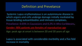
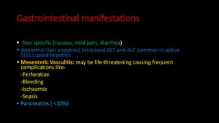
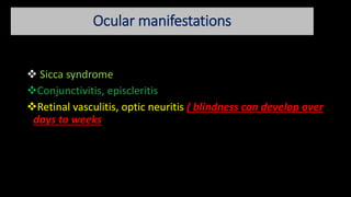
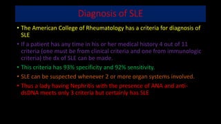
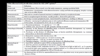
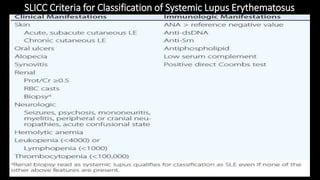
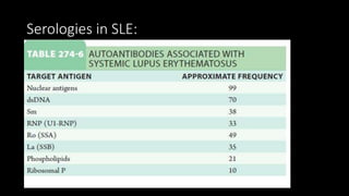
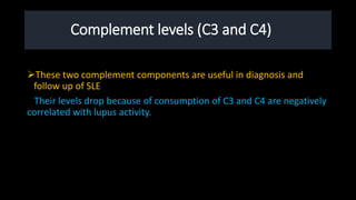
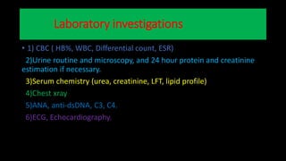
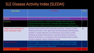
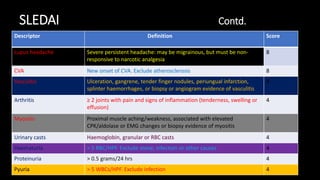
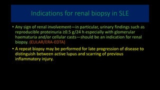
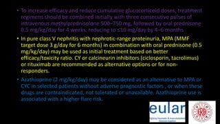
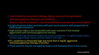
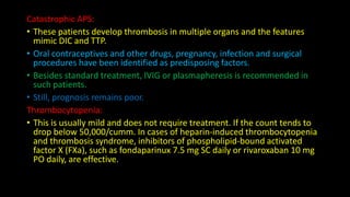
Definition and Prevelance
Systemic Lupus erythematosus is an autoimmune disease in
which organs and cells undergo damage initially mediated by
tissue-binding autoantibodies and immune complexes.
Prevelance: 0.03% in Caucasians and 0.2% in Afro-Caribbeans.
Gender: 90% patients are females of reproductive age
Age: peak age at onset is between 20 and 30 years of age
Lupus is associated with considerable morbidity and a five fold
increase in mortality
Pathophysiology of Lupus
• The cause of SLE is incompletely understood.
• The characteristic feature is Autoantibody production.
• An interplay between genetic susceptibility,
environment, gender and abnormal immune response
leads to autoimmunity.
• SLE is a multigenic disease.
• Defect in the genes encoding for componement eg. C1q,C2,C4A
• Impaired production of these early complement components may
decrease the clearance of apoptotic cells
• Thereby augmenting the pool of available autoantigens, or decrease
the solubility of immune complexes.
• Association with (HLA)-DR2 and HLA-DR3 has been documented.
• Polymorphisms in the Fc receptor genes FCGR2A and FCGR3A have
been associated with SLE nephritis, possibly based on altered
clearance of immune complexes.
• Regulator gene of innate immune system activation and signaling
molecules important in lymphocyte activation and genes that
influence DNA repair (TREX-1) on X chromosome are also associated.
• Common theme: the genes that have been associated with lupus
confer either increased activation or impaired regulation of the innate
or adaptive immune responses, with increased type I interferon often
observed in association with the risk genotype
Environmental Triggers
• Microbial triggers: Epstein Barr virus, cytomegalovirus
• Ultraviolet light exposure is a well-described trigger of lupus flare
• DNA damage by UV and induction of apoptosis of skin cells
concentration of nucleic acids and associated proteins in cell
membrane blebs increasing processing by APC’s
• There is also association between current tobacco use and anti-
double-stranded DNA antibodies and lupus disease activity
Immune responses:
1) Activation of innate immunity( dendritic cells, monocytes/ macrophages) by CpG
DNA, DNA in immune complexes, viral DNA or RNA and RNA in RNA/ protein self
antigens.
2)Abnormal activation pathways in adaptive immunity cells (mature T and B
lymphocytes)
3) Ineffective regulatory CD4+ and CD8+ T cells , B cells and Myeloid derived
suppressor cells.
4)Reduced clearance of immune complexes and apoptic cells.
Consequence immune responses
Self antigens (Nucleosomal DNA/ protein; RNA/Protein in
Sm, Ro and La; phospholipids) are recognized by immune
systems of surface blebs of apoptic cells;
- Thus autoantigens, autoantibodies and immune complexes
persist for prolonged periods ; allowing inflammation and
disease to develop.
Pathogenesis of SLE ( Diagram)
Clinical features of SLE
• Systemic symptoms , particularly fatigue, myalgia/ arthralgia are
always present.
• Fever, prostration, weight loss and anaemia with or without
organ targeted manifestations.
• Severity varies from mild and intermittent to severe and
fulminant.
• Approximately 85% patients may have continuing active disease
( while being treated) or one or more flares of disease activity
annually.
• Permanent complete remissions are rare.
Musculoskeletal Manifestations
 Intermittent Polyarthritis ( commonly Hands, Wrist and Knees)
Joint deformities( Jaccoud’s Arthropathy in hands and feet) develop in only 10% patients due to
Tendon damage.
Joint erosions do not occur.
RHUPUS: Individuals may have rheumatoid like arthritis with erosion and fulfill criteria for both
RA and SLE.
Myositis: Evidenced by
Muscle weakness
Elevated CPK level
Positive MRI scan
Muscle necrosis and inflammation on biopsy
Glucocorticoid therapies (commonly) and Antimalarial therapies (rarely) cause muscle weakness.
Raynaud’s phenomenon
(Secondary)
• A common presentation is Raynaud’s in combination with
Arthralgia or arthritis.
• May antedate other symptoms by months or years
• Features in favour of secondary Raynaud’s include age at onset of over 25
years, absence of a family history of Raynaud’s, and occurrence in a male
Cutaneous manifestations
• 3 distinct types of rashes occur:
-Acute rash ( Classic Butterfly rash ; upto 20% patients):
Raised, erythematous and painful or itchy ;usually invove over cheeks with sparing
nasolabial folds.
- Subacute Cutaneous Lupus Erythematosus (SCLE):
Migratory, non-scarring either annular or psoriaform.
-Chronic Rash( Discoid Lupus Erythematosus):
-Hyperkeratosis
-Follicular plugging and Scarring Alopecia of scalp.
Other skin manifestations:
Periungual Erythema, Vasculitis, Livedo reticularis, Urticaria, Lichen planus like dermatitis, bullae,
and panniculitis( Lupus profundus)
Butterfly(malar)rash
Discood rash
SCLE
Severe secondary Raynaud’s phenomenon Livedo reticularis
DLE
Renal Manifestations:
 Nephritis and infection: leading cause of mortality in first decade of
disease.
 Urine analysis should be checked in any patient of SLE.
 Renal biopsy is recommended for every SLE patients with any clinical
evidence of nephritis.
 Patients with dangerous Proliferative form of Lupus nephritis (ISN III and
IV) usually have Microscopic heamaturia and Protienuria(>500 mg/ 24 hrs)
; approximately one half develop nephrotic syndrome.
 If inadequately treated DPGN will turn into ESRD within 2 years of
diagnosis
 Immunosupression is needed in class III and IV lupus Nephritis and also
class V and nephrotic range Protienuria.
Nervous System Manifestations
• Common manifestations of diffuse CNS Lupus:
 Cognitive dysfunction ( difficulties with memory and reasoning) (17-66%)
Mood disorder
Headache
Seizure(6-51%)
Psychosis (Steroid induced psychosis usually occurs in 1st week of steroid
therapy and at daily dose of >/40 mg of prednisolone or equivalent)
Stroke, TIA(5-18%)
Aseptic Meningitis,
• Myelopathy (Transeverse myelitis frequently associated with
antiphospholipid antibodies)
Vascular Occlusions
• Patients with antiphospholipid antibodies are associated with
hypercoagulability and acute thrombotic events.
Chronic SLE patients may develop accelerated atherosclerosis.
Myocardial Infarction are primary manifestation of accelerated atherosclerosis
Brain ischaemia can be caused by either:
- Focal occlusion ( non inflammatory or associated with vasculitis)
-Embolization from carotid artery plaque or fibrinous vegetations of Libman-
Sacks endocarditis.
Increased risk of atherosclerosis is three to ten fold overall and highest in
women<49 yrs old.
Characteristics associated with increased risk of atherosclerosis
 Older age
 Hypertension
 Dyslipidemia
 Dysfunctional proinflammatory HDL
 Repeated high scores for disease activity
 High cumulative or daily doses of glucocorticoids
 High levels of homocysteine.
Pulmonary manifestations
 Pleurisy with or without pleural effusion(common)
 Lupus pneumonitis.
 Life threatening manifestations:
 Interstitial fibrosis
 Shrinking Lung Syndrome
 Pulmonary HTN, ARDS and intra alveolar hemorrhage
( usually require early aggressive immunosuppressive therapy)
Cardiac manifestations
• Pericarditis is most frequent cardiac manifestation.(Infrequently leads
to tamponade).
Serious cardiac manifestations: Myocarditis and fibrinous
endocarditis of Libman –Sacks.
Endocardial involvement can lead to valvular insufficiencies,
commonly mitral or aortic valves or to embolic events
Myocardial infarction occurs due to accelerated atherosclerosis.
Mortality from atherosclerosis may be upto 10 times.
Hematological manifestations
• Anaemia (50%)
Normocytic normochromic anaemia(anemia of chronic disease)
Hemolytic anaemia
 Leukopenia(<4000/uL)
 Lymphopenia(<1500/uL)
 Thrombocytopenia(<100000/uL)
 Lymhadenopathy
 Splenomegaly
Gastrointestinal manifestations
 Non specific (nausea, mild pain, diarrhea)
 Abnormal liver enzymes( increased AST and ALT common in active
SLE),Lupoid hepatitis
 Mesenteric Vasculitis: may be life threatening causing frequent
complications like-
-Perforation
-Bleeding
-Ischaemia
-Sepsis
• Pancreatitis ( <10%)
Ocular manifestations
 Sicca syndrome
Conjunctivitis, episcleritis
Retinal vasculitis, optic neuritis ( blindness can develop over
days to weeks
Diagnosis of SLE
• The American College of Rheumatology has a criteria for diagnosis of
SLE
• If a patient has any time in his or her medical history 4 out of 11
criteria (one must be from clinical criteria and one from immunologic
criteria) the dx of SLE can be made.
• This criteria has 93% specificity and 92% sensitivity.
• SLE can be suspected whenever 2 or more organ systems involved.
• Thus a lady having Nephritis with the presence of ANA and anti-
dsDNA meets only 3 criteria but certainly has SLE
SLICC Criteria for Classification of Systemic Lupus Erythematosus
Serologies in SLE:
Antinuclear antibody
 ANA is a good screening test. Because 98% of cases show a high titre
(1:80 or more) of this autoantibody.
 A negative test makes the diagnosis highly improbable.
 Specificity of ANA for diagnosis of SLE is quite low(app 40% only)
 ANA may be positive in other rheumatic disorders, such as- Systemic
sclerosis ,Sjogren’s syndrome ,Overlap syndrome ,Antiphospholipid
syndrome,Polymyositis and Rheumatoid arthritis.
Like rheumatoid factor test ANA may also be positive in:
-Chronic infections, Malignancies and in normal individuals.
• Diagnosis strongly suspected at the clinical evaluation before
requesting for ANA.
• A positive test supports dx of SLE.
• A serial titres of ANA in a diagnosed case of SLE is of no clinical value.
• It does not correlate well with disease activity.
• It can remain positive for long periods in the absence of any disease
activity.
Anti-double stranded DNA
• High Specificity for SLE
 Poorly sensitive( only 60%) but when present confirms the diagnosis.
 It often correlates with disease activity.
Antibodies to extractable nuclear antigens (anti-ENA)
 These include anti Sm, anti U1RNP, anti-Ro and anti-La antibodies.
 Anti-ENA are found in about 50% sera which are positive for ANA.
 High titres of anti-UIRNP are associated with Mixed connective tissue
disease(MCTD) which is a subset of SLE with prominent Raynaud’s
phenomenon, Sclerodactyly, proximal myopathy and mild or no renal
involvement.
 Anti-Sm antibody is specifc for SLE and is found in only 10-30% of
patients.
 Anti-Ro is associated with- ANA negative SLE,Congenital heart
block,Neonatal lupus,Sjogren’s syndrome, SCLE
• Anti-La: associated with Sjogren’s syndrome and SLE.
• Anti-histone antibodies : associated with Drug induced SLE
Anticardiolipin antibodies(aCL) and Lupus anticoagulant: is
positive in Antiphospholipid syndrome
Complement levels (C3 and C4)
These two complement components are useful in diagnosis and
follow up of SLE
Their levels drop because of consumption of C3 and C4 are negatively
correlated with lupus activity.
Laboratory investigations
• 1) CBC ( HB%, WBC, Differential count, ESR)
2)Urine routine and microscopy, and 24 hour protein and creatinine
estimation if necessary.
3)Serum chemistry (urea, creatinine, LFT, lipid profile)
4)Chest xray
5)ANA, anti-dsDNA, C3, C4.
6)ECG, Echocardiography.
SLE Disease Activity Index (SLEDAI)
Descriptor Definition Score
Seizure Recent onset, exclude metabolic, infectious or drug causes 8
Psychosis Include hallucinations, incoherence, markedly loose associations,
impoverished thought content, markedly illogical thinking, bizarre,
disorganised or catatonic behaviour. Exclude uraemia and drug causes
8
‘Organic brain syndrome’ or
Acute confusional state
Altered mental function with impaired orientation, memory or other
intellectual function, with rapid onset and fluctuating clinical features,
inability to sustain attention to environment, plus at least 2 of the
following: perceptual disturbance, incoherent speech, insomnia or
daytime drowsiness, or increased or decreased psychomotor activity.
Exclude metabolic, drug or infectious causes
8
Visual disturbance Retinal changes of SLE. Include cytoid bodies, retinal haemorrhages,
serous exudates haemorrhages in choroid or optic neuritis. Exclude
hypertension, infection or drug causes
8
Cranial nerve disorder New onset of sensory or motor neuropathy involving cranial nerves 8
SLEDAI Contd.
Descriptor Definition Score
Lupus headache Severe persistent headache: may be migrainous, but must be non-
responsive to narcotic analgesia.
8
CVA New onset of CVA. Exclude atherosclerosis 8
Vasculitis Ulceration, gangrene, tender finger nodules, periungual infarction,
splinter haemorrhages, or biopsy or angiogram evidence of vasculitis
8
Arthritis ≥ 2 joints with pain and signs of inflammation (tenderness, swelling or
effusion)
4
Myositis Proximal muscle aching/weakness, associated with elevated
CPK/aldolase or EMG changes or biopsy evidence of myositis
4
Urinary casts Haemoglobin, granular or RBC casts 4
Haematuria > 5 RBC/HPF. Exclude stone, infection or other causes 4
Proteinuria > 0.5 grams/24 hrs 4
Pyuria > 5 WBCs/HPF. Exclude infection 4
SLEDAI Contd.
Descriptor Definition Score
Rash Inflammatory type rash 2
Alopecia Abnormal, patchy or diffuse loss of hair 2
Mucosal ulcers Oral or nasal ulcerations 2
Pleurisy Pleuritic chest pain with pleural rub/effusion/pleural thickening 2
Pericarditis Pericardial pain with at least 1 of the following: rub, effusion or ECG or Echo
confirmation
2
Low complement Decrease in CH50, C3 or C4 below the normal limit of Lab 2
Increased DNA binding Increased DNA binding using Farr assay 2
Fever > 38 Deg C. Exclude infection 1
Thrombocytopaenia < 100,000/cu mm, exclude drug causes 1
Leukopaenia < 3000/cu mm, exclude drug causes 1
Management of SLE
Non pharmacological
 Patient education:
1. Written Pamphlets can be very helpful.
2. Opportunity to interact with other previously diagnosed Lupus patients who are
identified by the specialist.
3. The biologic behavior of the disease, in particular the long remitting and relapsing
course of disease must be explained to patient.
4.Patient needs advice regarding marriage, contraception. Pregnancy and breast-feeding.
5.Regarding marriage, physician should openly discuss the risks involved with the patient.
6.Participation of fiancée and other members is crucial.
7. Patient needs to be emphasized that fertility is not impaired inspite of abortions and
frequent foetal wastages.
8. Normal outcome of pregnancy is possible with appropriate care.
9.Contraception is essential during active disease.
 Educating the family.
 Avoidance of Sun-exposure: Photosensitive patients must be advised to
wear protective clothing with long sleeves etc, use sunscreens
(cream/lotions) with sun protector factor (spf) of more than 15 .
 Advice to avoid going outdoors during daytime.
 The ultraviolet rays need to be avoided and hence the unprotected
exposure to low pollution areas such as seashores and hill stations
frequently precipitate relaspse of lupus.
 Computer screen can also be a source of UV rays.
 Photosensitizing drugs (demeclocycline, sparfloxacin, dapsone, amiodarone
etc) are harmful.
Infections Control
1) infection screening particularly necessary when patient is on long
term steroid/ cytotoxic therapy.
2)Renal failure, cardiac vulvular vegetations, and ulcerative
mucocutaneous lesions predispose SLE patients to infections.
3. SLE patient with fever of unknown origin: it may be consequence of
SLE itself or an infection.
4. In such situation elevated CRP in blood favours diagnosis of infection.
5. Patients undergoing splenectomy must receive pneumococcal
vaccine preferably before surgery.
Pharmacological management
Category 1 (mild SLE)
Characterized by: Arthritis, arthralgia, myalgia. Fatigue, mild mucocutaneous
involvement, low grade fever, mild serositis, lupus headache.
 For mild musculoskeletal complaints , NSAIDs and analgesics may suffice.
If symptoms not alleviated by NSAIDs alone, then antimalarials should be
prescribed (chloroquine, Hydroxychloroquine)
These dugs are not only useful for cutaneous manifestations; but also have
immunosuppressive anti-inflammatory effect and sun-blocking properties.
Also have anti-platelet and cholesterol lowering effects.
Dose: Hydroxychloroquine (200 mg BD for 3 months and then 200 mg daily)
The maintenance dose must not exceed 6 mg/kg/day.
• Annual monitoring for vision with perimetry using a red object is
recommended (for chloroquine, 6 monthly monitoring is desirable).
• The drug must be discontinued if a central scotoma is detected at any
stage.
• Use of hydroxychloroquine in pregnancy is controversial.
• When antimalarials are withdrawl after prolonged use, relapse of
lupus activity can occur.
• In refractory cases: Quinacrine can be combined with
Hydroxychloroquine.
• Alternatives include: Dapsone and Thalidomide.
Patients not responding to above measures Low dose
steroid therapy (prednisolone o.3-o.5 mg/kg/day) for 4-
6 weeks followed by slow tapering.
For lupus dermatitis, there is a role of local steroids(
topical cream and ointments) and injection into
unresponsive skin lesions.
Category II (moderate SLE)
-Characterized by : high grade fever, toxaemia, severe
mucocutaneous features, marked photosensitivity,
moderate to severe serositis, lupus pneumonitis, mild to
moderate myocarditis, mesengioproliferative or minimal
change lupus nephritis, haemolytic anaemia and
thrombocytopenia.
• For moderate to severe manifestations:
 Prednisolone 1 mg/kg orally per day.
Antimalarials may be administered concominantly.
High dose of steroids need to be continued till disease activity is well
controlled that usually takes upto 6 weeks when it should be tapered off
slowly over 6 to 12 months.
 In toxic appearing patients: Intravenous pulse Methylprednisolone (15
mg/kg, max 1 gm) over one hour for 3 or 5 consecutive days may achieve
rapid control.
 Dexamethasone 100 mg is a good, cheap and equally effective alternative
steroid for pulse therapy.
 Pulse therapy should be followed by oral prednisolone.
• Calcium supplements (1 gm/day) and vitamin (800units/day) prescribed
along with steroids retard osteoporosis.
• Alendronate 1o mg daily or 70 mg once a week is preferable for prevention
of osteoporosis.
• Majority of patients do not require maintenance of oral prednisolone
beyond 6 months.
• Most enjoy remission with antimalarials and intermittent use of NSAIDs.
• INH prophylaxis: one Indian study has shown: SLE on sterois showed 82%
protection from tuberculosis with INH prophylaxis in one year.
• It may be better to use 2-drug prophylaxis(Rifampicin+ INH or
INH+Ethambutol) for a period of one year.
Category III (Severe SLE)
Characterized by: organ/life threatening features such as focal /diffuse
proliferative glomerulonephritis with or without azotemia/hypertension,
lupus cerebritis with recurrent seizures, acute confusional state, coma,
systemic necrotizing vasculitis such as peripheral gangrene, GI bleeding or
mononeuritis multiplex.
 A combination therapy of high dose daily oral prednisolone (40-
60mg/day) and intravenous cyclophosphamide (0.75 gm/m2; maximum
1gm over 1 hour) is recommended.
Cyclophosphamide pulses are given once a month for 6 months by which
time usually remission is achieved and then a maintenance pulse is
administered every 3 months for a total of 2 years of cytotoxic therapy.
Prednisolone is tapered off or reduced to a very low dose ie 5-7.5
mg/day by 6 months.
Cyclophosphamide can cause haemorrhagic cystitis; but can be
minimized by good hydration and co-prescription of MESNA.
Because of risk of azoospermia and anovulation (which may be
permanent), pre-treatment sperm or ova collection and storage need
to be considered.
Some authorities recommend the above pulse cyclophosphamide
therapy for induction of remission (for first 6 months), then it is
maintained with Azathioprine 2-2.5 mg/kg/day for about 2 years.
• Alternative approach:
-Intravenous pulses of steroids on 3 consecutive days each month,
daily oral administration of cyclophosphamide (2 mg/kg/day) or
azathioprine from the beginning or combination of these 2 agents
along with oral prednisolone.
 Cyclophosphamide based regimens have been shown to achieve renal
preservation.
 Plasmapheresis, methotrexate, cyclosporine and Mycophenolate
mofetil are other options.
Category IV (SLE with miscellaneous features)
- Characterized by antiphospholipid syndrome(recurrent DVT, CVAs, recurrent foetal loss
etc), pure membranous lupus nephritis, chronic sclerosing lupus nephritis, seizures
without other evidence of lupus activity, behavioural disorders without other serious
manifestations, resistant thrombocytopenia or haemolytic anaemia.
- Immunosuppressive therapy does not play any significant role.
- If seizures or psychosis occur as isolated events, only symptomatic treatment. Steroids
are not indicated.
- For Refractory thrombocytopenia, Danazol may be useful. Colchicine and vincristine are
sometimes useful to improve pkatelet count.
- Splenectomy may be indicated when platelet count is less than 50,000/cumm and
maintenance requiremrnt for steroid is high,
- Plasmapheresis, IV immunoglobulin may be required in refractory cases.
- Stem cell transplantation has been reported in some refractory lupus.
Using CYC
Calculate body surface area as shown in the figure(Mosteller Method)
Counsel the potential site effect
To Protect bladder from haemorrhagic cystitis
use vigorous fluid with 1 litre 0.9% NaCl over
Oral Mesna at -2,+2 and +6 as per dose(0.2*CYC dose in mg)
Antiemetic Granietron 1mg +Dexamethasone 10mg PO at -2
Monitoring Of CYC Therapy
• Check WCC weekly for 1st month,every 2 weeks for 2nd & 3rd & monthly
thereafter
• If count <4000/cmm discontinue temporarily.Restart with 25% reduction
When WCC normal with weekly monitoring
• If WCC falling rapidly e.g >2000between tests reduce by 25% pre-emtively
• If WCC <1000 /cmm or <4000/cmm persists for >2 weeks then restart low
dose (e.g. 25-50 mg) after WCC becomes normal
• For IV CYC check WCC the day of the proposed pulse.if <4000 then
postpone until >4000/cmm and reduce dose 25%
• Check WCC 14 days after pulse;if WCC
2000-3000 then reduce dose of next pulse by 20%
1000-2000 then reduce by 40%
Special situations associated with Lupus
LUPUS NEPHRITIS
• Def: Lupus nehritis is defined as the presence of more than +++ or 0.5
gram/24 hr proteinuria or presence of cellular casts of any type.
• So Lupus nephritis occurs in about half of SLE patients.
• Reliable urine examination is the single most important investigation
for early diagnosis of LN.
• Renal tubular acidosis occur more frequently in SLE.
Indications for renal biopsy in SLE
• Any sign of renal involvement—in particular, urinary findings such as
reproducible proteinuria ≥0.5 g/24 h especially with glomerular
haematuria and/or cellular casts—should be an indication for renal
biopsy. (EULAR/ERA-EDTA)
• A repeat biopsy may be performed for late progression of disease to
distinguish between active lupus and scarring of previous
inflammatory injury.
Principles of treatment of LN
• General measures:
- Salt restriction if hypertension present.
-fat restriction if hyperlipidaemia or nephrotic syndrome is present.
-Protein should be restricted if azotaemia is present and calcium
should be supplemented with steroid therapy.
-Meticulous control of hypertension is desirable.
-Pregnancy should be avoided during active lupus nephritis with
suitable contraception.
-NSAIDS should be avoided in the presence of impaired renal function.
Immunosuppressive therapy
Initial treatment:
• For patients with class IIIA or IIIA/C (±V) and class IVA or IVA/C (±V)
LN, MMF target dose: 3 g/day for 6 months, or MPA sodium at
equivalent dose) or low-dose IV CYC total dose 3 g over 3 months) in
combination with glucocorticoids, are recommended as initial
treatment
• In patients with adverse prognostic factors (acute deterioration in
renal function, substantial cellular crescents and/or fibrinoid
necrosis), similar regimens may be used but CYC can also be
prescribed monthly at higher doses (0.75–1 g/m2) for 6 months or
orally (2–2.5 mg/kg/day) for 3 months
• To increase efficacy and reduce cumulative glucocorticoid doses, treatment
regimens should be combined initially with three consecutive pulses of
intravenous methylprednisolone 500–750 mg, followed by oral prednisone
0.5 mg/kg/day for 4 weeks, reducing to ≤10 mg/day by 4–6 months
• In pure class V nephritis with nephrotic-range proteinuria, MPA (MMF
target dose 3 g/day for 6 months) in combination with oral prednisone (0.5
mg/kg/day) may be used as initial treatment based on better
efficacy/toxicity ratio. CY or calcineurin inhibitors (ciclosporin, tacrolimus)
or rituximab are recommended as alternative options or for non-
responders.
• Azathioprine (2 mg/kg/day) may be considered as an alternative to MPA or
CYC in selected patients without adverse prognostic factors , or when these
drugs are contraindicated, not tolerated or unavailable. Azathioprine use is
associated with a higher flare risk.
Subsequent treatment
• In patients improving after initial treatment, subsequent
immunosuppression is recommended with either MPA at lower doses
(initial target MMF dose 2 g/day) or AZA (2 mg/kg/day) for at least 3
years, in combination with low dose prednisone (5–7.5 mg/day).
Gradual drug withdrawal, glucocorticoids first, can then be attempted
• Patients who responded to initial treatment with MPA should remain
on MPA unless pregnancy is contemplated, in which case they should
switch to AZA at least 3 months prior to conception
• Calcineurin inhibitors can be considered in pure class V nephritis
Refractory disease
• For patients who fail treatment with MPA or CY either because of lack
of effect or due to adverse events, we recommendion is that the
treatment is switched from MPA to CYC, or CYC to MPA, or rituximab.
• -Best treated by conservative therapy, dialysis and renal transplantation.
-Immunosuppressive therapy is not beneficial.
-At least 3 months of dialysis is recommended before considering renal transplant.
A high chronicity index correlates with poor renal outcome with progression to
ESRD despite treatment.
High activity index is also associated with poor outcome if not treated
aggressively with immunosuppressive therapy.
Patient with high chronicity index and serum creatinine > 3 mg/dl should not be
treated aggressively unless activity index is also high.
If s. creatinine is chronically high and more than 5 mg/dl, aggressive
immunosuppressive therapy is harmful.
These patients may be managed by dialysis and transplantation in due course.
Indian guideline
LN and pregnancy
• Pregnancy may be planned in stable patients with inactive lupus and
UPCR <50 mg/mmol, for the preceding 6 months, with GFR that
should preferably be >50 ml/min.
• Acceptable medications include hydroxychloroquine, and where
needed, low dose prednisone, azathioprine and/or calcineurin
inhibitors.
• The intensity of treatment should not be reduced in anticipation of
pregnancy.
• During pregnancy, acetylsalicylic acid should be considered to reduce
the risk of pre-eclampsia. Patients should be assessed at least every 4
weeks, preferably by a specialist physician and obstetrician
Antiphospholipid Syndrome
• can present as a subset of SLE
• hallmark of APS is thrombosis (venous and/or arterial).
• The common clinical manifestations of APS comprise recurrent
pregnancy loss, recurrent deep venous thrombosis and
cerebrovascular accidents
Diagnosis (Consensus Classification Criteria)
A. CLINICAL CRITERIA
1. Vascular thrombosis: One or more episodes of
arterial,venous or small-vessel thrombosis, occurring within any tissue
or organ
2. Complications of pregnancy: One or more unexplained
deaths of morphologically normal foetuses at > 10 weeks of gestation,
or One or more premature births of morphologically normal neonates
at < 34 weeks of gestation, or Three or more unexplained consecutive
spontaneous abortions at < 10 weeks of gestation
B. LABORATORY CRITERIA
1. aCL: Anticardiolipin IgG or IgM antibodies present at
moderate or high titres on 2 or more occasions at least 6 weeks apart
2. LAC: Lupus anticoagulant antibodies detected on 2 or more
occasions at least 6 weeks apart
A diagnosis of definite APS requires the presence of one clinical + one
laboratory criterion
Treatment of APS
This can be considered under the following heads:
Deep venous thrombosis:
• The main purpose of treatment here is to prevent pulmonary embolism.
• Standard measures include bed-rest, elevation of the affected limb to allow the
oedema and tenderness to subside
• Anticoagulant therapy: Heparin and warfarin should be started simultaneously so
as to allow an overlap of about 5 days. INR should be adjusted between 3 and 4
on long-term warfarin therapy. The duration of warfarin therapy is life-long in
patients with recurrent venous thrombosis.
• Thrombolytics such as streptokinase, urokinase and tPA can be used but they are
not more effective in preventing pulmonary embolism.
• Thromboendarterectomy and percutaneous insertion of IVC filter may be
considered in special circumstance
Acute arterial thrombosis
• In a patient with APS this usually means a TIA or stroke, with MI and digital
gangrene being less common.
• In some patients with acute stroke (< 3 hours duration), thrombolytics can
be used but the standard of care is usually heparin followed by warfarin.
• Low-dose aspirin is strongly recommended in patients who continue having
thrombotic events despite full anticoagulation.
• APS patients with acute MI can be treated with thrombolytics, angioplasty
or coronary stents.
• Peripheral arterial thrombosis can be treated with thrombolytics or
heparin or angioplasty.
Catastrophic APS:
• These patients develop thrombosis in multiple organs and the features
mimic DIC and TTP.
• Oral contraceptives and other drugs, pregnancy, infection and surgical
procedures have been identified as predisposing factors.
• Besides standard treatment, IVIG or plasmapheresis is recommended in
such patients.
• Still, prognosis remains poor.
Thrombocytopenia:
• This is usually mild and does not require treatment. If the count tends to
drop below 50,000/cumm. In cases of heparin-induced thrombocytopenia
and thrombosis syndrome, inhibitors of phospholipid-bound activated
factor X (FXa), such as fondaparinux 7.5 mg SC daily or rivaroxaban 10 mg
PO daily, are effective.
Pregnancy in SLE
SLE does not impair the women’s fertility except during the periods
of severe disease activity.
There is increased risk of abortions( 2-3 times), Intrauterine growth
retardation and Still birth.
Pregnancy increases the risk of disease flare (40-50% probability).
Risk of flare is double in women who have active disease during
conception.
About 10% develop severe flares; therefore Lupus pregnancy is high
risk pregnancy.
• Laboratory monitoring during pregnancy:
1.Initial evaluation: Hb, WBC, DLC, platelets,Urinalysis with
microscopy, 24 hr urinary estimation of protein and creatinine, blood
urea, glucose, and serum creatinine, serum lipids (if patient is nephrotic
or on steroids), Coomb’s test, aPL( IgG and IgM aCL, LAC), VDRL, anti
ds-DNA, C3. Anti-Ro and Ati- la should be done if there is history of
giving birth to a baby with neonatal lupus.
2.Monthly laboratory estimations: Hb, WBC, DLc, platelets,
urinanalysis (with 24hr analysis if nephritis),anti ds DNA and C3.
3. In case anaemia develops, Peripheral smear should be reviewed and
Coombs test repeated.
General Principles of treatment of lupus
pregnancy
The disease should be in clinical remission for at least 6 months before the
patient plans pregnancy.
At the onset of pregnancy , a complete assessment of disease activity and
severity should be made.
If disease is in remission, pt should be seen once every month in the first
half of pregnancy and more frequentlu later on.
Prime focus is early detection and prompt treatment of lupus flare during
pregnancy and the post partum period.
Drug thrapy:
-Presence of nephritis with or without hypertension is an indication for low
dose daily aspirin from 10 th week till 36 th week for prevention of Pre
eclampsia.
• -pts on long term steroid therapy (>2 yrs) are administered “stress doses”of
steroids during delivery
• Indications of caesarian section:
-Maternal reasons (avascular necrosis of hips with inadequate hip
abduction)
-Foetal reasons( fetal distress, abnormal non stress test, cephalo-pelvic
disproportion and transverse presentation etc.)
 Treatment of Lupus flares in pregnancy:
-Appropriate dose steroid should be given.
-Cyclophosphamide and methotrexate should be avoided during first
trimester.
-Azathioprine and Ciclosporine can be used in pregnancy with active SLE.
• Antiphospholipid syndrome in pregnancy:
 A great risk of thrombosis and foetal loss.
Warfarin must be omitted as early as possible after conception(preferably
the next day her first menses are missed and pregnancy confirmed).
Daily subcutaneous inj of LMW Heparin ( either Enoxaparin 40 mg/day or
Dalteparin 500 units per day) along with low dose aspirin must be
continued until delivery.
The heparin is omitted 12 hours before delivery or caesarian section
whereas low dose aspirin may be continued.
After postpartum bleeding stops ,usually within a week after delivery,
Warfarin is restarted.
Corticosteroids no longer needed, increase maternal morbidity.
• Breast-feeding:
 Majority of drugs are excreted in human milk in variable amounts.
From neonatal perspective: maternal intake of prednisolone 30
mg/day, warfarin, cyclosporine in standard doses and weekly
Chloroquine for malaria prophylaxis are safe.
If the dose of prednisolone os above 30 mg/day, feeding should be
avoided for 4 hours after ingestion of morning steroid.
Breast feeding contraindicated if mother is on azathioprine
cyclophosphamide, hydroxychloroquine for SLE, Salicylates,
indomethacine and sulindac.
Drug Induced Lupus(DIL)
• This is a syndrome of positive ANA associated with symptms, such as fever,
malaise, arthritis or intense arthralgias/myalgias, serositis and/or rash.
• Appears during therapy with certain medication or biologic agents,
• Predominant in whites, has less female predilection than SLE.
• Rarely involves kidney or brain, rarely anti ds-DNA positive.
• Commonly associated with anti histone antibody.
• Usually resolves over several weeks, after discontinuation of medication.
• Most frequent drugs are: anti arrhythmic Procainamide, disopyramide,
Propafenone ; anti hypertensive like Hydralazine, several ACE inhibitor and
Beta blocker, anti tbhyroid drugs like Propylthiouracil, anticonvulsants
Carbamazepine, phenytoin, antipsychotic Chlorpromazine.
• Antibiotics- INH, minocycline, Nitrofurantoin.
• Antirheumatic: sulfasalazine, the diuretic hydrochlorthiazide
• Antihyperlipidemics:Lovastatim and Simvastatin.
• It is appropriate to test for ANA at the first hint of relevant symptoms
and to use test results to help decide whether to withdraw the
suspected agent.
• After withdrawal of the suspected agent, clinical improvement follows
within days to weeks.
• Autoantibodies may take several months to disappear.
• Sometimes, NSAIDs and corticosteroids may be required for a short
period to obtain symptomatic relief
SLE and its Associated Disorders

SLE and its Associated Disorders

  • 3.
    Definition and Prevelance SystemicLupus erythematosus is an autoimmune disease in which organs and cells undergo damage initially mediated by tissue-binding autoantibodies and immune complexes. Prevelance: 0.03% in Caucasians and 0.2% in Afro-Caribbeans. Gender: 90% patients are females of reproductive age Age: peak age at onset is between 20 and 30 years of age Lupus is associated with considerable morbidity and a five fold increase in mortality
  • 4.
    Pathophysiology of Lupus •The cause of SLE is incompletely understood. • The characteristic feature is Autoantibody production. • An interplay between genetic susceptibility, environment, gender and abnormal immune response leads to autoimmunity.
  • 7.
    • SLE isa multigenic disease. • Defect in the genes encoding for componement eg. C1q,C2,C4A • Impaired production of these early complement components may decrease the clearance of apoptotic cells • Thereby augmenting the pool of available autoantigens, or decrease the solubility of immune complexes. • Association with (HLA)-DR2 and HLA-DR3 has been documented. • Polymorphisms in the Fc receptor genes FCGR2A and FCGR3A have been associated with SLE nephritis, possibly based on altered clearance of immune complexes.
  • 8.
    • Regulator geneof innate immune system activation and signaling molecules important in lymphocyte activation and genes that influence DNA repair (TREX-1) on X chromosome are also associated. • Common theme: the genes that have been associated with lupus confer either increased activation or impaired regulation of the innate or adaptive immune responses, with increased type I interferon often observed in association with the risk genotype
  • 9.
    Environmental Triggers • Microbialtriggers: Epstein Barr virus, cytomegalovirus • Ultraviolet light exposure is a well-described trigger of lupus flare • DNA damage by UV and induction of apoptosis of skin cells concentration of nucleic acids and associated proteins in cell membrane blebs increasing processing by APC’s • There is also association between current tobacco use and anti- double-stranded DNA antibodies and lupus disease activity
  • 10.
    Immune responses: 1) Activationof innate immunity( dendritic cells, monocytes/ macrophages) by CpG DNA, DNA in immune complexes, viral DNA or RNA and RNA in RNA/ protein self antigens. 2)Abnormal activation pathways in adaptive immunity cells (mature T and B lymphocytes) 3) Ineffective regulatory CD4+ and CD8+ T cells , B cells and Myeloid derived suppressor cells. 4)Reduced clearance of immune complexes and apoptic cells.
  • 11.
    Consequence immune responses Selfantigens (Nucleosomal DNA/ protein; RNA/Protein in Sm, Ro and La; phospholipids) are recognized by immune systems of surface blebs of apoptic cells; - Thus autoantigens, autoantibodies and immune complexes persist for prolonged periods ; allowing inflammation and disease to develop.
  • 12.
  • 13.
    Clinical features ofSLE • Systemic symptoms , particularly fatigue, myalgia/ arthralgia are always present. • Fever, prostration, weight loss and anaemia with or without organ targeted manifestations. • Severity varies from mild and intermittent to severe and fulminant. • Approximately 85% patients may have continuing active disease ( while being treated) or one or more flares of disease activity annually. • Permanent complete remissions are rare.
  • 14.
    Musculoskeletal Manifestations  IntermittentPolyarthritis ( commonly Hands, Wrist and Knees) Joint deformities( Jaccoud’s Arthropathy in hands and feet) develop in only 10% patients due to Tendon damage. Joint erosions do not occur. RHUPUS: Individuals may have rheumatoid like arthritis with erosion and fulfill criteria for both RA and SLE. Myositis: Evidenced by Muscle weakness Elevated CPK level Positive MRI scan Muscle necrosis and inflammation on biopsy Glucocorticoid therapies (commonly) and Antimalarial therapies (rarely) cause muscle weakness.
  • 15.
    Raynaud’s phenomenon (Secondary) • Acommon presentation is Raynaud’s in combination with Arthralgia or arthritis. • May antedate other symptoms by months or years • Features in favour of secondary Raynaud’s include age at onset of over 25 years, absence of a family history of Raynaud’s, and occurrence in a male
  • 16.
    Cutaneous manifestations • 3distinct types of rashes occur: -Acute rash ( Classic Butterfly rash ; upto 20% patients): Raised, erythematous and painful or itchy ;usually invove over cheeks with sparing nasolabial folds. - Subacute Cutaneous Lupus Erythematosus (SCLE): Migratory, non-scarring either annular or psoriaform. -Chronic Rash( Discoid Lupus Erythematosus): -Hyperkeratosis -Follicular plugging and Scarring Alopecia of scalp. Other skin manifestations: Periungual Erythema, Vasculitis, Livedo reticularis, Urticaria, Lichen planus like dermatitis, bullae, and panniculitis( Lupus profundus)
  • 17.
  • 18.
    Severe secondary Raynaud’sphenomenon Livedo reticularis DLE
  • 19.
    Renal Manifestations:  Nephritisand infection: leading cause of mortality in first decade of disease.  Urine analysis should be checked in any patient of SLE.  Renal biopsy is recommended for every SLE patients with any clinical evidence of nephritis.  Patients with dangerous Proliferative form of Lupus nephritis (ISN III and IV) usually have Microscopic heamaturia and Protienuria(>500 mg/ 24 hrs) ; approximately one half develop nephrotic syndrome.  If inadequately treated DPGN will turn into ESRD within 2 years of diagnosis  Immunosupression is needed in class III and IV lupus Nephritis and also class V and nephrotic range Protienuria.
  • 20.
    Nervous System Manifestations •Common manifestations of diffuse CNS Lupus:  Cognitive dysfunction ( difficulties with memory and reasoning) (17-66%) Mood disorder Headache Seizure(6-51%) Psychosis (Steroid induced psychosis usually occurs in 1st week of steroid therapy and at daily dose of >/40 mg of prednisolone or equivalent) Stroke, TIA(5-18%) Aseptic Meningitis, • Myelopathy (Transeverse myelitis frequently associated with antiphospholipid antibodies)
  • 21.
    Vascular Occlusions • Patientswith antiphospholipid antibodies are associated with hypercoagulability and acute thrombotic events. Chronic SLE patients may develop accelerated atherosclerosis. Myocardial Infarction are primary manifestation of accelerated atherosclerosis Brain ischaemia can be caused by either: - Focal occlusion ( non inflammatory or associated with vasculitis) -Embolization from carotid artery plaque or fibrinous vegetations of Libman- Sacks endocarditis. Increased risk of atherosclerosis is three to ten fold overall and highest in women<49 yrs old.
  • 22.
    Characteristics associated withincreased risk of atherosclerosis  Older age  Hypertension  Dyslipidemia  Dysfunctional proinflammatory HDL  Repeated high scores for disease activity  High cumulative or daily doses of glucocorticoids  High levels of homocysteine.
  • 23.
    Pulmonary manifestations  Pleurisywith or without pleural effusion(common)  Lupus pneumonitis.  Life threatening manifestations:  Interstitial fibrosis  Shrinking Lung Syndrome  Pulmonary HTN, ARDS and intra alveolar hemorrhage ( usually require early aggressive immunosuppressive therapy)
  • 24.
    Cardiac manifestations • Pericarditisis most frequent cardiac manifestation.(Infrequently leads to tamponade). Serious cardiac manifestations: Myocarditis and fibrinous endocarditis of Libman –Sacks. Endocardial involvement can lead to valvular insufficiencies, commonly mitral or aortic valves or to embolic events Myocardial infarction occurs due to accelerated atherosclerosis. Mortality from atherosclerosis may be upto 10 times.
  • 25.
    Hematological manifestations • Anaemia(50%) Normocytic normochromic anaemia(anemia of chronic disease) Hemolytic anaemia  Leukopenia(<4000/uL)  Lymphopenia(<1500/uL)  Thrombocytopenia(<100000/uL)  Lymhadenopathy  Splenomegaly
  • 26.
    Gastrointestinal manifestations  Nonspecific (nausea, mild pain, diarrhea)  Abnormal liver enzymes( increased AST and ALT common in active SLE),Lupoid hepatitis  Mesenteric Vasculitis: may be life threatening causing frequent complications like- -Perforation -Bleeding -Ischaemia -Sepsis • Pancreatitis ( <10%)
  • 27.
    Ocular manifestations  Siccasyndrome Conjunctivitis, episcleritis Retinal vasculitis, optic neuritis ( blindness can develop over days to weeks
  • 28.
    Diagnosis of SLE •The American College of Rheumatology has a criteria for diagnosis of SLE • If a patient has any time in his or her medical history 4 out of 11 criteria (one must be from clinical criteria and one from immunologic criteria) the dx of SLE can be made. • This criteria has 93% specificity and 92% sensitivity. • SLE can be suspected whenever 2 or more organ systems involved. • Thus a lady having Nephritis with the presence of ANA and anti- dsDNA meets only 3 criteria but certainly has SLE
  • 30.
    SLICC Criteria forClassification of Systemic Lupus Erythematosus
  • 31.
  • 33.
    Antinuclear antibody  ANAis a good screening test. Because 98% of cases show a high titre (1:80 or more) of this autoantibody.  A negative test makes the diagnosis highly improbable.  Specificity of ANA for diagnosis of SLE is quite low(app 40% only)  ANA may be positive in other rheumatic disorders, such as- Systemic sclerosis ,Sjogren’s syndrome ,Overlap syndrome ,Antiphospholipid syndrome,Polymyositis and Rheumatoid arthritis. Like rheumatoid factor test ANA may also be positive in: -Chronic infections, Malignancies and in normal individuals.
  • 34.
    • Diagnosis stronglysuspected at the clinical evaluation before requesting for ANA. • A positive test supports dx of SLE. • A serial titres of ANA in a diagnosed case of SLE is of no clinical value. • It does not correlate well with disease activity. • It can remain positive for long periods in the absence of any disease activity.
  • 35.
    Anti-double stranded DNA •High Specificity for SLE  Poorly sensitive( only 60%) but when present confirms the diagnosis.  It often correlates with disease activity.
  • 36.
    Antibodies to extractablenuclear antigens (anti-ENA)  These include anti Sm, anti U1RNP, anti-Ro and anti-La antibodies.  Anti-ENA are found in about 50% sera which are positive for ANA.  High titres of anti-UIRNP are associated with Mixed connective tissue disease(MCTD) which is a subset of SLE with prominent Raynaud’s phenomenon, Sclerodactyly, proximal myopathy and mild or no renal involvement.  Anti-Sm antibody is specifc for SLE and is found in only 10-30% of patients.  Anti-Ro is associated with- ANA negative SLE,Congenital heart block,Neonatal lupus,Sjogren’s syndrome, SCLE
  • 37.
    • Anti-La: associatedwith Sjogren’s syndrome and SLE. • Anti-histone antibodies : associated with Drug induced SLE Anticardiolipin antibodies(aCL) and Lupus anticoagulant: is positive in Antiphospholipid syndrome
  • 38.
    Complement levels (C3and C4) These two complement components are useful in diagnosis and follow up of SLE Their levels drop because of consumption of C3 and C4 are negatively correlated with lupus activity.
  • 39.
    Laboratory investigations • 1)CBC ( HB%, WBC, Differential count, ESR) 2)Urine routine and microscopy, and 24 hour protein and creatinine estimation if necessary. 3)Serum chemistry (urea, creatinine, LFT, lipid profile) 4)Chest xray 5)ANA, anti-dsDNA, C3, C4. 6)ECG, Echocardiography.
  • 40.
    SLE Disease ActivityIndex (SLEDAI) Descriptor Definition Score Seizure Recent onset, exclude metabolic, infectious or drug causes 8 Psychosis Include hallucinations, incoherence, markedly loose associations, impoverished thought content, markedly illogical thinking, bizarre, disorganised or catatonic behaviour. Exclude uraemia and drug causes 8 ‘Organic brain syndrome’ or Acute confusional state Altered mental function with impaired orientation, memory or other intellectual function, with rapid onset and fluctuating clinical features, inability to sustain attention to environment, plus at least 2 of the following: perceptual disturbance, incoherent speech, insomnia or daytime drowsiness, or increased or decreased psychomotor activity. Exclude metabolic, drug or infectious causes 8 Visual disturbance Retinal changes of SLE. Include cytoid bodies, retinal haemorrhages, serous exudates haemorrhages in choroid or optic neuritis. Exclude hypertension, infection or drug causes 8 Cranial nerve disorder New onset of sensory or motor neuropathy involving cranial nerves 8
  • 41.
    SLEDAI Contd. Descriptor DefinitionScore Lupus headache Severe persistent headache: may be migrainous, but must be non- responsive to narcotic analgesia. 8 CVA New onset of CVA. Exclude atherosclerosis 8 Vasculitis Ulceration, gangrene, tender finger nodules, periungual infarction, splinter haemorrhages, or biopsy or angiogram evidence of vasculitis 8 Arthritis ≥ 2 joints with pain and signs of inflammation (tenderness, swelling or effusion) 4 Myositis Proximal muscle aching/weakness, associated with elevated CPK/aldolase or EMG changes or biopsy evidence of myositis 4 Urinary casts Haemoglobin, granular or RBC casts 4 Haematuria > 5 RBC/HPF. Exclude stone, infection or other causes 4 Proteinuria > 0.5 grams/24 hrs 4 Pyuria > 5 WBCs/HPF. Exclude infection 4
  • 42.
    SLEDAI Contd. Descriptor DefinitionScore Rash Inflammatory type rash 2 Alopecia Abnormal, patchy or diffuse loss of hair 2 Mucosal ulcers Oral or nasal ulcerations 2 Pleurisy Pleuritic chest pain with pleural rub/effusion/pleural thickening 2 Pericarditis Pericardial pain with at least 1 of the following: rub, effusion or ECG or Echo confirmation 2 Low complement Decrease in CH50, C3 or C4 below the normal limit of Lab 2 Increased DNA binding Increased DNA binding using Farr assay 2 Fever > 38 Deg C. Exclude infection 1 Thrombocytopaenia < 100,000/cu mm, exclude drug causes 1 Leukopaenia < 3000/cu mm, exclude drug causes 1
  • 43.
  • 44.
    Non pharmacological  Patienteducation: 1. Written Pamphlets can be very helpful. 2. Opportunity to interact with other previously diagnosed Lupus patients who are identified by the specialist. 3. The biologic behavior of the disease, in particular the long remitting and relapsing course of disease must be explained to patient. 4.Patient needs advice regarding marriage, contraception. Pregnancy and breast-feeding. 5.Regarding marriage, physician should openly discuss the risks involved with the patient. 6.Participation of fiancée and other members is crucial. 7. Patient needs to be emphasized that fertility is not impaired inspite of abortions and frequent foetal wastages. 8. Normal outcome of pregnancy is possible with appropriate care. 9.Contraception is essential during active disease.
  • 45.
     Educating thefamily.  Avoidance of Sun-exposure: Photosensitive patients must be advised to wear protective clothing with long sleeves etc, use sunscreens (cream/lotions) with sun protector factor (spf) of more than 15 .  Advice to avoid going outdoors during daytime.  The ultraviolet rays need to be avoided and hence the unprotected exposure to low pollution areas such as seashores and hill stations frequently precipitate relaspse of lupus.  Computer screen can also be a source of UV rays.  Photosensitizing drugs (demeclocycline, sparfloxacin, dapsone, amiodarone etc) are harmful.
  • 46.
    Infections Control 1) infectionscreening particularly necessary when patient is on long term steroid/ cytotoxic therapy. 2)Renal failure, cardiac vulvular vegetations, and ulcerative mucocutaneous lesions predispose SLE patients to infections. 3. SLE patient with fever of unknown origin: it may be consequence of SLE itself or an infection. 4. In such situation elevated CRP in blood favours diagnosis of infection. 5. Patients undergoing splenectomy must receive pneumococcal vaccine preferably before surgery.
  • 47.
    Pharmacological management Category 1(mild SLE) Characterized by: Arthritis, arthralgia, myalgia. Fatigue, mild mucocutaneous involvement, low grade fever, mild serositis, lupus headache.  For mild musculoskeletal complaints , NSAIDs and analgesics may suffice. If symptoms not alleviated by NSAIDs alone, then antimalarials should be prescribed (chloroquine, Hydroxychloroquine) These dugs are not only useful for cutaneous manifestations; but also have immunosuppressive anti-inflammatory effect and sun-blocking properties. Also have anti-platelet and cholesterol lowering effects. Dose: Hydroxychloroquine (200 mg BD for 3 months and then 200 mg daily) The maintenance dose must not exceed 6 mg/kg/day.
  • 48.
    • Annual monitoringfor vision with perimetry using a red object is recommended (for chloroquine, 6 monthly monitoring is desirable). • The drug must be discontinued if a central scotoma is detected at any stage. • Use of hydroxychloroquine in pregnancy is controversial. • When antimalarials are withdrawl after prolonged use, relapse of lupus activity can occur. • In refractory cases: Quinacrine can be combined with Hydroxychloroquine. • Alternatives include: Dapsone and Thalidomide.
  • 49.
    Patients not respondingto above measures Low dose steroid therapy (prednisolone o.3-o.5 mg/kg/day) for 4- 6 weeks followed by slow tapering. For lupus dermatitis, there is a role of local steroids( topical cream and ointments) and injection into unresponsive skin lesions.
  • 50.
    Category II (moderateSLE) -Characterized by : high grade fever, toxaemia, severe mucocutaneous features, marked photosensitivity, moderate to severe serositis, lupus pneumonitis, mild to moderate myocarditis, mesengioproliferative or minimal change lupus nephritis, haemolytic anaemia and thrombocytopenia.
  • 51.
    • For moderateto severe manifestations:  Prednisolone 1 mg/kg orally per day. Antimalarials may be administered concominantly. High dose of steroids need to be continued till disease activity is well controlled that usually takes upto 6 weeks when it should be tapered off slowly over 6 to 12 months.  In toxic appearing patients: Intravenous pulse Methylprednisolone (15 mg/kg, max 1 gm) over one hour for 3 or 5 consecutive days may achieve rapid control.  Dexamethasone 100 mg is a good, cheap and equally effective alternative steroid for pulse therapy.  Pulse therapy should be followed by oral prednisolone.
  • 52.
    • Calcium supplements(1 gm/day) and vitamin (800units/day) prescribed along with steroids retard osteoporosis. • Alendronate 1o mg daily or 70 mg once a week is preferable for prevention of osteoporosis. • Majority of patients do not require maintenance of oral prednisolone beyond 6 months. • Most enjoy remission with antimalarials and intermittent use of NSAIDs. • INH prophylaxis: one Indian study has shown: SLE on sterois showed 82% protection from tuberculosis with INH prophylaxis in one year. • It may be better to use 2-drug prophylaxis(Rifampicin+ INH or INH+Ethambutol) for a period of one year.
  • 53.
    Category III (SevereSLE) Characterized by: organ/life threatening features such as focal /diffuse proliferative glomerulonephritis with or without azotemia/hypertension, lupus cerebritis with recurrent seizures, acute confusional state, coma, systemic necrotizing vasculitis such as peripheral gangrene, GI bleeding or mononeuritis multiplex.  A combination therapy of high dose daily oral prednisolone (40- 60mg/day) and intravenous cyclophosphamide (0.75 gm/m2; maximum 1gm over 1 hour) is recommended. Cyclophosphamide pulses are given once a month for 6 months by which time usually remission is achieved and then a maintenance pulse is administered every 3 months for a total of 2 years of cytotoxic therapy.
  • 54.
    Prednisolone is taperedoff or reduced to a very low dose ie 5-7.5 mg/day by 6 months. Cyclophosphamide can cause haemorrhagic cystitis; but can be minimized by good hydration and co-prescription of MESNA. Because of risk of azoospermia and anovulation (which may be permanent), pre-treatment sperm or ova collection and storage need to be considered. Some authorities recommend the above pulse cyclophosphamide therapy for induction of remission (for first 6 months), then it is maintained with Azathioprine 2-2.5 mg/kg/day for about 2 years.
  • 55.
    • Alternative approach: -Intravenouspulses of steroids on 3 consecutive days each month, daily oral administration of cyclophosphamide (2 mg/kg/day) or azathioprine from the beginning or combination of these 2 agents along with oral prednisolone.  Cyclophosphamide based regimens have been shown to achieve renal preservation.  Plasmapheresis, methotrexate, cyclosporine and Mycophenolate mofetil are other options.
  • 56.
    Category IV (SLEwith miscellaneous features) - Characterized by antiphospholipid syndrome(recurrent DVT, CVAs, recurrent foetal loss etc), pure membranous lupus nephritis, chronic sclerosing lupus nephritis, seizures without other evidence of lupus activity, behavioural disorders without other serious manifestations, resistant thrombocytopenia or haemolytic anaemia. - Immunosuppressive therapy does not play any significant role. - If seizures or psychosis occur as isolated events, only symptomatic treatment. Steroids are not indicated. - For Refractory thrombocytopenia, Danazol may be useful. Colchicine and vincristine are sometimes useful to improve pkatelet count. - Splenectomy may be indicated when platelet count is less than 50,000/cumm and maintenance requiremrnt for steroid is high, - Plasmapheresis, IV immunoglobulin may be required in refractory cases. - Stem cell transplantation has been reported in some refractory lupus.
  • 57.
    Using CYC Calculate bodysurface area as shown in the figure(Mosteller Method) Counsel the potential site effect To Protect bladder from haemorrhagic cystitis use vigorous fluid with 1 litre 0.9% NaCl over Oral Mesna at -2,+2 and +6 as per dose(0.2*CYC dose in mg) Antiemetic Granietron 1mg +Dexamethasone 10mg PO at -2
  • 58.
    Monitoring Of CYCTherapy • Check WCC weekly for 1st month,every 2 weeks for 2nd & 3rd & monthly thereafter • If count <4000/cmm discontinue temporarily.Restart with 25% reduction When WCC normal with weekly monitoring • If WCC falling rapidly e.g >2000between tests reduce by 25% pre-emtively • If WCC <1000 /cmm or <4000/cmm persists for >2 weeks then restart low dose (e.g. 25-50 mg) after WCC becomes normal • For IV CYC check WCC the day of the proposed pulse.if <4000 then postpone until >4000/cmm and reduce dose 25% • Check WCC 14 days after pulse;if WCC 2000-3000 then reduce dose of next pulse by 20% 1000-2000 then reduce by 40%
  • 59.
    Special situations associatedwith Lupus LUPUS NEPHRITIS • Def: Lupus nehritis is defined as the presence of more than +++ or 0.5 gram/24 hr proteinuria or presence of cellular casts of any type. • So Lupus nephritis occurs in about half of SLE patients. • Reliable urine examination is the single most important investigation for early diagnosis of LN. • Renal tubular acidosis occur more frequently in SLE.
  • 61.
    Indications for renalbiopsy in SLE • Any sign of renal involvement—in particular, urinary findings such as reproducible proteinuria ≥0.5 g/24 h especially with glomerular haematuria and/or cellular casts—should be an indication for renal biopsy. (EULAR/ERA-EDTA) • A repeat biopsy may be performed for late progression of disease to distinguish between active lupus and scarring of previous inflammatory injury.
  • 62.
    Principles of treatmentof LN • General measures: - Salt restriction if hypertension present. -fat restriction if hyperlipidaemia or nephrotic syndrome is present. -Protein should be restricted if azotaemia is present and calcium should be supplemented with steroid therapy. -Meticulous control of hypertension is desirable. -Pregnancy should be avoided during active lupus nephritis with suitable contraception. -NSAIDS should be avoided in the presence of impaired renal function.
  • 63.
    Immunosuppressive therapy Initial treatment: •For patients with class IIIA or IIIA/C (±V) and class IVA or IVA/C (±V) LN, MMF target dose: 3 g/day for 6 months, or MPA sodium at equivalent dose) or low-dose IV CYC total dose 3 g over 3 months) in combination with glucocorticoids, are recommended as initial treatment • In patients with adverse prognostic factors (acute deterioration in renal function, substantial cellular crescents and/or fibrinoid necrosis), similar regimens may be used but CYC can also be prescribed monthly at higher doses (0.75–1 g/m2) for 6 months or orally (2–2.5 mg/kg/day) for 3 months
  • 64.
    • To increaseefficacy and reduce cumulative glucocorticoid doses, treatment regimens should be combined initially with three consecutive pulses of intravenous methylprednisolone 500–750 mg, followed by oral prednisone 0.5 mg/kg/day for 4 weeks, reducing to ≤10 mg/day by 4–6 months • In pure class V nephritis with nephrotic-range proteinuria, MPA (MMF target dose 3 g/day for 6 months) in combination with oral prednisone (0.5 mg/kg/day) may be used as initial treatment based on better efficacy/toxicity ratio. CY or calcineurin inhibitors (ciclosporin, tacrolimus) or rituximab are recommended as alternative options or for non- responders. • Azathioprine (2 mg/kg/day) may be considered as an alternative to MPA or CYC in selected patients without adverse prognostic factors , or when these drugs are contraindicated, not tolerated or unavailable. Azathioprine use is associated with a higher flare risk.
  • 65.
    Subsequent treatment • Inpatients improving after initial treatment, subsequent immunosuppression is recommended with either MPA at lower doses (initial target MMF dose 2 g/day) or AZA (2 mg/kg/day) for at least 3 years, in combination with low dose prednisone (5–7.5 mg/day). Gradual drug withdrawal, glucocorticoids first, can then be attempted • Patients who responded to initial treatment with MPA should remain on MPA unless pregnancy is contemplated, in which case they should switch to AZA at least 3 months prior to conception • Calcineurin inhibitors can be considered in pure class V nephritis
  • 66.
    Refractory disease • Forpatients who fail treatment with MPA or CY either because of lack of effect or due to adverse events, we recommendion is that the treatment is switched from MPA to CYC, or CYC to MPA, or rituximab.
  • 67.
    • -Best treatedby conservative therapy, dialysis and renal transplantation. -Immunosuppressive therapy is not beneficial. -At least 3 months of dialysis is recommended before considering renal transplant. A high chronicity index correlates with poor renal outcome with progression to ESRD despite treatment. High activity index is also associated with poor outcome if not treated aggressively with immunosuppressive therapy. Patient with high chronicity index and serum creatinine > 3 mg/dl should not be treated aggressively unless activity index is also high. If s. creatinine is chronically high and more than 5 mg/dl, aggressive immunosuppressive therapy is harmful. These patients may be managed by dialysis and transplantation in due course. Indian guideline
  • 68.
    LN and pregnancy •Pregnancy may be planned in stable patients with inactive lupus and UPCR <50 mg/mmol, for the preceding 6 months, with GFR that should preferably be >50 ml/min. • Acceptable medications include hydroxychloroquine, and where needed, low dose prednisone, azathioprine and/or calcineurin inhibitors. • The intensity of treatment should not be reduced in anticipation of pregnancy. • During pregnancy, acetylsalicylic acid should be considered to reduce the risk of pre-eclampsia. Patients should be assessed at least every 4 weeks, preferably by a specialist physician and obstetrician
  • 69.
    Antiphospholipid Syndrome • canpresent as a subset of SLE • hallmark of APS is thrombosis (venous and/or arterial). • The common clinical manifestations of APS comprise recurrent pregnancy loss, recurrent deep venous thrombosis and cerebrovascular accidents
  • 70.
    Diagnosis (Consensus ClassificationCriteria) A. CLINICAL CRITERIA 1. Vascular thrombosis: One or more episodes of arterial,venous or small-vessel thrombosis, occurring within any tissue or organ 2. Complications of pregnancy: One or more unexplained deaths of morphologically normal foetuses at > 10 weeks of gestation, or One or more premature births of morphologically normal neonates at < 34 weeks of gestation, or Three or more unexplained consecutive spontaneous abortions at < 10 weeks of gestation
  • 71.
    B. LABORATORY CRITERIA 1.aCL: Anticardiolipin IgG or IgM antibodies present at moderate or high titres on 2 or more occasions at least 6 weeks apart 2. LAC: Lupus anticoagulant antibodies detected on 2 or more occasions at least 6 weeks apart A diagnosis of definite APS requires the presence of one clinical + one laboratory criterion
  • 72.
    Treatment of APS Thiscan be considered under the following heads: Deep venous thrombosis: • The main purpose of treatment here is to prevent pulmonary embolism. • Standard measures include bed-rest, elevation of the affected limb to allow the oedema and tenderness to subside • Anticoagulant therapy: Heparin and warfarin should be started simultaneously so as to allow an overlap of about 5 days. INR should be adjusted between 3 and 4 on long-term warfarin therapy. The duration of warfarin therapy is life-long in patients with recurrent venous thrombosis. • Thrombolytics such as streptokinase, urokinase and tPA can be used but they are not more effective in preventing pulmonary embolism. • Thromboendarterectomy and percutaneous insertion of IVC filter may be considered in special circumstance
  • 73.
    Acute arterial thrombosis •In a patient with APS this usually means a TIA or stroke, with MI and digital gangrene being less common. • In some patients with acute stroke (< 3 hours duration), thrombolytics can be used but the standard of care is usually heparin followed by warfarin. • Low-dose aspirin is strongly recommended in patients who continue having thrombotic events despite full anticoagulation. • APS patients with acute MI can be treated with thrombolytics, angioplasty or coronary stents. • Peripheral arterial thrombosis can be treated with thrombolytics or heparin or angioplasty.
  • 74.
    Catastrophic APS: • Thesepatients develop thrombosis in multiple organs and the features mimic DIC and TTP. • Oral contraceptives and other drugs, pregnancy, infection and surgical procedures have been identified as predisposing factors. • Besides standard treatment, IVIG or plasmapheresis is recommended in such patients. • Still, prognosis remains poor. Thrombocytopenia: • This is usually mild and does not require treatment. If the count tends to drop below 50,000/cumm. In cases of heparin-induced thrombocytopenia and thrombosis syndrome, inhibitors of phospholipid-bound activated factor X (FXa), such as fondaparinux 7.5 mg SC daily or rivaroxaban 10 mg PO daily, are effective.
  • 75.
    Pregnancy in SLE SLEdoes not impair the women’s fertility except during the periods of severe disease activity. There is increased risk of abortions( 2-3 times), Intrauterine growth retardation and Still birth. Pregnancy increases the risk of disease flare (40-50% probability). Risk of flare is double in women who have active disease during conception. About 10% develop severe flares; therefore Lupus pregnancy is high risk pregnancy.
  • 76.
    • Laboratory monitoringduring pregnancy: 1.Initial evaluation: Hb, WBC, DLC, platelets,Urinalysis with microscopy, 24 hr urinary estimation of protein and creatinine, blood urea, glucose, and serum creatinine, serum lipids (if patient is nephrotic or on steroids), Coomb’s test, aPL( IgG and IgM aCL, LAC), VDRL, anti ds-DNA, C3. Anti-Ro and Ati- la should be done if there is history of giving birth to a baby with neonatal lupus. 2.Monthly laboratory estimations: Hb, WBC, DLc, platelets, urinanalysis (with 24hr analysis if nephritis),anti ds DNA and C3. 3. In case anaemia develops, Peripheral smear should be reviewed and Coombs test repeated.
  • 77.
    General Principles oftreatment of lupus pregnancy The disease should be in clinical remission for at least 6 months before the patient plans pregnancy. At the onset of pregnancy , a complete assessment of disease activity and severity should be made. If disease is in remission, pt should be seen once every month in the first half of pregnancy and more frequentlu later on. Prime focus is early detection and prompt treatment of lupus flare during pregnancy and the post partum period. Drug thrapy: -Presence of nephritis with or without hypertension is an indication for low dose daily aspirin from 10 th week till 36 th week for prevention of Pre eclampsia.
  • 78.
    • -pts onlong term steroid therapy (>2 yrs) are administered “stress doses”of steroids during delivery • Indications of caesarian section: -Maternal reasons (avascular necrosis of hips with inadequate hip abduction) -Foetal reasons( fetal distress, abnormal non stress test, cephalo-pelvic disproportion and transverse presentation etc.)  Treatment of Lupus flares in pregnancy: -Appropriate dose steroid should be given. -Cyclophosphamide and methotrexate should be avoided during first trimester. -Azathioprine and Ciclosporine can be used in pregnancy with active SLE.
  • 79.
    • Antiphospholipid syndromein pregnancy:  A great risk of thrombosis and foetal loss. Warfarin must be omitted as early as possible after conception(preferably the next day her first menses are missed and pregnancy confirmed). Daily subcutaneous inj of LMW Heparin ( either Enoxaparin 40 mg/day or Dalteparin 500 units per day) along with low dose aspirin must be continued until delivery. The heparin is omitted 12 hours before delivery or caesarian section whereas low dose aspirin may be continued. After postpartum bleeding stops ,usually within a week after delivery, Warfarin is restarted. Corticosteroids no longer needed, increase maternal morbidity.
  • 80.
    • Breast-feeding:  Majorityof drugs are excreted in human milk in variable amounts. From neonatal perspective: maternal intake of prednisolone 30 mg/day, warfarin, cyclosporine in standard doses and weekly Chloroquine for malaria prophylaxis are safe. If the dose of prednisolone os above 30 mg/day, feeding should be avoided for 4 hours after ingestion of morning steroid. Breast feeding contraindicated if mother is on azathioprine cyclophosphamide, hydroxychloroquine for SLE, Salicylates, indomethacine and sulindac.
  • 81.
    Drug Induced Lupus(DIL) •This is a syndrome of positive ANA associated with symptms, such as fever, malaise, arthritis or intense arthralgias/myalgias, serositis and/or rash. • Appears during therapy with certain medication or biologic agents, • Predominant in whites, has less female predilection than SLE. • Rarely involves kidney or brain, rarely anti ds-DNA positive. • Commonly associated with anti histone antibody. • Usually resolves over several weeks, after discontinuation of medication. • Most frequent drugs are: anti arrhythmic Procainamide, disopyramide, Propafenone ; anti hypertensive like Hydralazine, several ACE inhibitor and Beta blocker, anti tbhyroid drugs like Propylthiouracil, anticonvulsants Carbamazepine, phenytoin, antipsychotic Chlorpromazine.
  • 82.
    • Antibiotics- INH,minocycline, Nitrofurantoin. • Antirheumatic: sulfasalazine, the diuretic hydrochlorthiazide • Antihyperlipidemics:Lovastatim and Simvastatin. • It is appropriate to test for ANA at the first hint of relevant symptoms and to use test results to help decide whether to withdraw the suspected agent. • After withdrawal of the suspected agent, clinical improvement follows within days to weeks. • Autoantibodies may take several months to disappear. • Sometimes, NSAIDs and corticosteroids may be required for a short period to obtain symptomatic relief