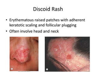
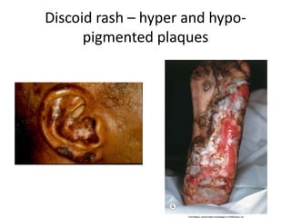
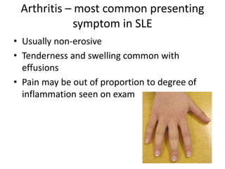
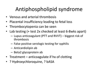
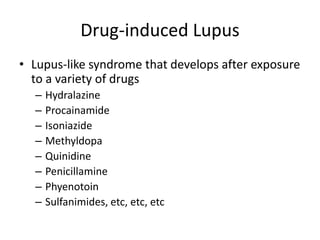
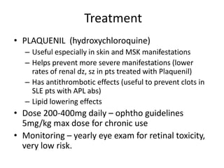
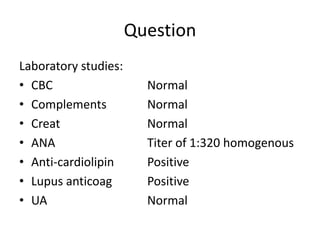
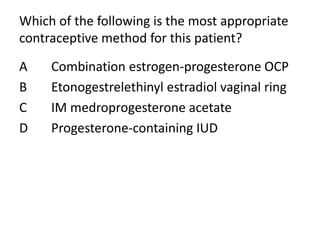
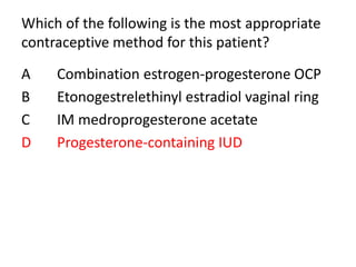
Systemic Lupus Erythematosus
Vivian Stone, MD
Virginia Mason Medical Center
Lupus
– Multisystem, inflammatory autoimmune disease
with diverse manifestations
– Chronic, remitting and relapsing clinical course
– Associated with autoantibody production against
components of the cell nucleus
– Development of immune-complexes that cause
injury to skin, joints and serosal membranes
Forms of Lupus
• Systemic Lupus Erythematosus (SLE)
• Chronic Cutaneous Lupus Erythematosus
– Discoid Lupus Erythematosus (DLE)
– Subacute Cutaneous Lupus Erythematosus (SCLE)
• Drug-induced Lupus
Epidemiology
• Primarily a disease of woman
– Female:male ratio of 9:1 during childbearing age,
but 2:1 during childhood or after 65
• Prevalence ~1:2000 people in the US
– Up to 1:700 in women of childbearing age
• More common in AA
– 1:1000 white women
– 1:250 AA women
Genetics
• Higher frequency in first degree relatives
• Occurs concordantly in 25%-50% monozygotic
twins and in 5% of dizygotic twins
• Polygenetic
– More common in pts with complement deficiency
– HLA association (DR and DG loci)
Lupus pathogenesis
•Multiple immunologic abnormalities and loss of self-tolerance
•Dysregulated activation of T and B cell lymphocytes
•Autoantibody production and immune complex formation resulting in tissue damage
Systemic Lupus Erythematosus
Basic, Applied and Clinical Aspects
2016, Pages 273-279
Genes associated with SLE
Tsokos, NEJM 2011
IFNs and SLE
•  IFNa levels seen in serum of SLE patients
• Treatment with Type 1 IFNs in humans has led
to lupus-like illness
• “Interferon signature” is seen in many SLE
patients in microarray analysis
• IFN response genes are  in SLE patients
Autoantibodies in Autoimmune Disease
• ANA – best screening test for SLE
– + in significant titer (1:160 or higher) in almost all SLE pts
– Titers of 1:40 seen in 20% of healthy controls
– Titers of 1:320 seen in 3% of healthy controls
• dsDNA → SLE
• Smith ab → SLE
• Anti-histone → Drug-induced SLE
• Anti-RNP → MCTD (high titer),
SLE
• Anti-Ro, La → SLE or Sjogrens
• Scl-70 (topoisomerase) → Systemic Sclerosis
• Anticentromere → CREST
4/11
criteria
needed to
dx SLE
Clinical manifestations – initial/ever
• Constitutional 50% / 70-100%
• Arthritis/Arthralgia 65% / 85-95%
• Skin 73% / 80-90%
• Renal 16-38% / 34-73%
• Pleurisy 17% / 30-45%
• Pericarditis 8% / up to 48%
• CNS 12-21% / 25-75%
Clinical manifestations
• Photosensitivity
– Skin rash secondary to UV light
– Feeling “flu-like” in the sun
– Counsel sun protection!
• Oral ulcers
– Oral or nasopharyngeal
– Usually PAINLESS
Malar Rash
Fixed erythema, flat or raised, over the malar eminences,
tending to spare the nasolabial folds
Differential Diagnosis?
Discoid Rash
• Erythematous raised patches with adherent
keratotic scaling and follicular plugging
• Often involve head and neck
Discoid rash – hyper and hypo-
pigmented plaques
Arthritis – most common presenting
symptom in SLE
• Usually non-erosive
• Tenderness and swelling common with
effusions
• Pain may be out of proportion to degree of
inflammation seen on exam
Jacoud’s arthropathy
• Deforming, but non-erosive form of
arthropathy seen in SLE patients
CNS lupus
• Only 2 manifestations are included in the ACR SLE
criteria, more in the newer classification criteria
– Seizures
– Psychosis
• More common in patients with APLS
• CSF findings often nonspecific
– Mild pleocytosis and protein elevation in seen in up to 60%
• Most common MRI finding – small white matter
lesions
• Acute vasculitis is RARELY seen on angiography
Serositis
• Pleuritis
– Convincing hx of pleuritic pain
– Rub heard by a physician
– Evidence of a pleural effusion
• Pericarditis
– EKG
– Rub
– Pericardial effusion
Hematologic
• Leukopenia or lymphopenia
• Hemolytic anemia
– Warm hemolytic anemia
– Coomb’s positive
• Thrombocytopenia, ITP, TTP
• Elevated ESR/CRP
• Lupus anticoagulant (prolonged PTT, RVVT)
Libman Sacks Endocarditis
Antiphospholipid syndrome
• Venous and arterial thrombosis
• Placental insufficiency leading to fetal loss
• Thrombocytopenia can be seen
• Lab testing (+ test 2x checked at least 6-8wks apart)
– Lupus anticoagulant (PTT and RVVT) – biggest risk of
thrombosis
– False positive serologic testing for syphilis
– Anticardiolipin ab
– Beta2 glycoprotein ab
• Treatment – anticoagulate if hx of clotting
• ? Hydroxychloroquine, ? bASA
Question
• A 23 y/o man is evaluated during a f/u visit.
One month ago was dx with SLE, treatment
with pred 20mg was initiated with partial
improvement in joint pain and swelling. He
now report oral ulcers, ankle swelling, fatigue,
nausea and low grade fever.
• On physical, temp is 38.1, BP 146/92, pulse
102, RR 18. Raised malar erythema present.
He has an erythematous ulceration on the
hard palate. MSK exam reveals synovitis of
PIP and knees, bil pitting edema bil ankles
Question
Laboratory studies:
• Hemoglobin 9.1 g/dL
• Leukocyte count 3900/µL
• ESR 102 mm/h
• C3 Decreased
• C4 Decreased
• ANA Titer of 1:640 homogenous
• antidsDNA Positive
• UA 3+ protein, 4-6 RBC, 4-6 WBC,
no casts
• 24 hr urine 1.1 g prot/24 hrs
Which of the following is the most
appropriate next step in management?
A IV cyclophosphomide
B Kidney Biopsy
C MR angiography of the renal arteries
D Mycophenolate mofetil
Which of the following is the most
appropriate next step in management?
A IV cyclophosphomide
B Kidney Biopsy
C MR angiography of the renal arteries
D Mycophenolate mofetil
Lupus nephritis
• Develops during the first year in 50% of lupus
patients
• Occurs in 50% of white and 75% of AA pts
• Clinical features at diagnosis
– PROTEINURIA in almost all cases (nephrotic
syndrome rare)
– Hematuria
– Renal insufficiency
– +ANA, +dsDNA, decreased complements
– Usually ASYMPTOMATIC
Lupus Nephritis
Lupus Nephritis
Diffuse Proliferative Nephritis
• characterized by endocapillary proliferation caused by
immune complex deposits involving > 50% of glomeruli in a
biopsy specimen.
Membranous Nephritis
• chronic immune complex deposition results in mesangial
proliferation and glomerular basement membrane thickening
Lupus nephritis
• Worst prognosis in Class III or IV disease
• Requires aggressive immunosuppression to
prevent renal damage
– High dose steroids
– Cyclophosphamide or mycophenolate
– Tacrolimus add-on if not a complete response
• Class III or IV associated with HTN
– treat with ACE-I
Drug-induced Lupus
• Lupus-like syndrome that develops after exposure
to a variety of drugs
– Hydralazine
– Procainamide
– Isoniazide
– Methyldopa
– Quinidine
– Penicillamine
– Phyenotoin
– Sulfanimides, etc, etc, etc
Drug-induced Lupus
• +ANA, +++histone, neg dsDNA
• Constitutional sx common
– Malaise, low grade fever, myalgia
• Arthralgia
• Usually <4/11 ACR criteria
• Dermatologic, renal and CNS manifestations
RARELY occur
• Disease rapidly remits after w/d of drug, but ab
can persist for 6mo-1 yr
TNF-Induced SLE
• + ANA in ~15% of patient treated with anti-
TNF agents
• +dsDNA, and +anti-histone ab can be seen
• Skin involvement most common sx
• Can also have renal dz, serositis and CNS dz
• Symptoms usually remit with d/c of drug
• Occasionally additional immunosuppression is
needed for severe cases
Treatment
• PLAQUENIL (hydroxychloroquine)
– Useful especially in skin and MSK manifestations
– Helps prevent more severe manifestations (lower
rates of renal dz, sz in pts treated with Plaquenil)
– Has antithrombotic effects (useful to prevent clots in
SLE pts with APL abs)
– Lipid lowering effects
• Dose 200-400mg daily – ophtho guidelines
5mg/kg max dose for chronic use
• Monitoring – yearly eye exam for retinal toxicity,
very low risk.
Treatment of SLE – other agents
• Prednisone
• Cyclophosphomide – for severe manifestations
• Mycophenolate – first line for renal disease in
most cases
• Azathioprine
• Methotrexate – esp for arthritis
• Belimumab –> effect modest. Benefit may not
outweight cost – other than prednisone and HCQ,
the only FDA approved treatment of lupus.
• Rituximab
• Tacrolimus
Vitamin D and lupus
• Low vit D associated with higher disease
activity in lupus
• Higher vit D levels associated with improved
proteinuria.
• Treat to target to 40 ng/ml.
Question
• A 45-year-old woman is admitted to the hospital for
evaluation of a 6-week history of progressive, dull chest
pressure associated with mild dyspnea and nausea. At
onset, the chest pain occurred during physical exertion
(housework) and was relieved by rest within 5 minutes. For
the past several days, the patient has had similar episodes
that occurred with minimal activity, such as walking, and
also at rest, including an episode this morning, which she
described as 8/10 in severity of the pain and lasted for 10
to 15 minutes. The chest discomfort is not pleuritic or
positional and is not related to eating. She has
hypertension, treated for the past 6 years, and systemic
lupus erythematosus for 24 years, with a history of
pericarditis, arthritis, and a photosensitive facial rash. Her
medications include prednisone, hydroxychloroquine,
aspirin, and enalapril.
Question
• On physical examination, she is afebrile, blood
pressure is 132/78 mm Hg, pulse is 86/min,
and respiration rate is 18/min. Oxygen
saturation on ambient air is 98%. BMI is 25.
Her lungs are clear to auscultation. Estimated
central venous pressure is normal; there is no
Kussmaul sign or hepatojugular reflux. Cardiac
auscultation reveals regular rhythm with
normal S1 and S2 and no murmur, rub, or
gallop.
Question
Laboratory studies
• Erythrocyte sedimentation rate 39 mm/h
• Creatine kinase 65 U/L
• Creatine kinase MB fraction 3%
• Troponin normal
• Electrocardiogram demonstrates sinus rhythm, with a
rate of 92/min. There are symmetric T-wave inversions
in leads V1 through V4; there is no ST-segment
depression or elevation. Chest radiograph shows a
normal cardiac silhouette with no infiltrate or edema.
Which of the following diagnostic tests is most
appropriate at this time?
A Coronary angiography
B Exercise stress echocardiogram
C High-sensitivity C-reactive protein level
D Transthoracic echocardiography
Which of the following diagnostic tests is most
appropriate at this time?
A Coronary angiography
B Exercise stress echocardiogram
C High-sensitivity C-reactive protein level
D Transthoracic echocardiography
Complications from SLE
• Major cause of death in SLE patients is
accelerated atherosclerosis (mortality from MI
10x greater in SLE patients)
• Autopsy studies show severe atherosclerosis
in 40% of SLE patients compares with 2% of
age and sex matched controls.
• Monitor weight, HTN, HL, tobacco, and DM
Atherosclerosis in SLE, RA and controls
Roman et al Circulation 2007
Question
• A 22 y/o woman seeks preconception counseling and
treatment of recently diagnosed SLE. She reports
fatigue and hand pain with morning stiffness 15 min.
• On Exam, VS normal. + malar erythema. TTP bil PIP,
no other synovitis.
• Labs
– WBC 3.3
– C3 normal
– C4 decreased
– Creat normal
– UA normal
– ANA 1:160, homogenous.
– Anti-dsDNA positive
– Anti-cardiolipin ab positive
Which of the following is the most appropriate
treatment?
A Azathioprine
B Hydroxychloroquine
C Mycophenolate Mophetil
D Prednisone
E No treatment at this time
Which of the following is the most appropriate
treatment?
A Azathioprine
B Hydroxychloroquine
C Mycophenolate Mophetil
D Prednisone
E No treatment at this time
SLE and pregnancy
• Timing of pregnancy
– Healthy mother, healthy pregnancy, healthy baby
• Lupus should be under control prior to attempting pregnancy
• Some patients should be advised against pregnancy
– Hx of severe SLE with now renal insuff, proteinuria.
• Testing prior to pregnancy
– APL ab
– SSA, SSB ab
• Fetal heart block in 1%
– UA, complement levels
• High risk OB
• Treatments
– Hydroxychloroquine – safe throughout pregnancy
– Steroids
– For more serious manifestation – AZA, tac or cyclosporine ok
Question
• A 38 y/o woman is evaluated during routine f/u
for a 20 yr hx of SLE. In recent years, disease
acitivity has been quiescent. She currently
reports fatigue, alopecia, and occ hand arthralgia.
She seeks advice about contraception options
other than barrier methods
• The patient was also recently diagnosed with
osteoporosis. She had a 22-week fetal loss during
her first pregnancy, followed by 3 successful
pregnancies while taking ASA and enoxaparin.
Current meds: HCQ, low dose prednisone,
alendronate, calcium/D
• On PE, VS normal. Tenderness MCPs and PIPs
bilaterally
Question
Laboratory studies:
• CBC Normal
• Complements Normal
• Creat Normal
• ANA Titer of 1:320 homogenous
• Anti-cardiolipin Positive
• Lupus anticoag Positive
• UA Normal
Which of the following is the most appropriate
contraceptive method for this patient?
A Combination estrogen-progesterone OCP
B Etonogestrelethinyl estradiol vaginal ring
C IM medroprogesterone acetate
D Progesterone-containing IUD
Which of the following is the most appropriate
contraceptive method for this patient?
A Combination estrogen-progesterone OCP
B Etonogestrelethinyl estradiol vaginal ring
C IM medroprogesterone acetate
D Progesterone-containing IUD
Take home points
• Lupus gives a pattern of symptoms – recognize
pattern to make the diagnosis, do not rely on
labs alone.
• Prevent problems in SLE
– Monitor kidneys for proteinuria!
– Use HCQ in all SLE patients
– Screen for vit D deficiency
– Screen and manage for CV risk factors early!
Questions?

Systemic lupus erythematosus2019

  • 1.
    Systemic Lupus Erythematosus VivianStone, MD Virginia Mason Medical Center
  • 2.
    Lupus – Multisystem, inflammatoryautoimmune disease with diverse manifestations – Chronic, remitting and relapsing clinical course – Associated with autoantibody production against components of the cell nucleus – Development of immune-complexes that cause injury to skin, joints and serosal membranes
  • 3.
    Forms of Lupus •Systemic Lupus Erythematosus (SLE) • Chronic Cutaneous Lupus Erythematosus – Discoid Lupus Erythematosus (DLE) – Subacute Cutaneous Lupus Erythematosus (SCLE) • Drug-induced Lupus
  • 4.
    Epidemiology • Primarily adisease of woman – Female:male ratio of 9:1 during childbearing age, but 2:1 during childhood or after 65 • Prevalence ~1:2000 people in the US – Up to 1:700 in women of childbearing age • More common in AA – 1:1000 white women – 1:250 AA women
  • 5.
    Genetics • Higher frequencyin first degree relatives • Occurs concordantly in 25%-50% monozygotic twins and in 5% of dizygotic twins • Polygenetic – More common in pts with complement deficiency – HLA association (DR and DG loci)
  • 6.
    Lupus pathogenesis •Multiple immunologicabnormalities and loss of self-tolerance •Dysregulated activation of T and B cell lymphocytes •Autoantibody production and immune complex formation resulting in tissue damage Systemic Lupus Erythematosus Basic, Applied and Clinical Aspects 2016, Pages 273-279
  • 7.
    Genes associated withSLE Tsokos, NEJM 2011
  • 8.
    IFNs and SLE • IFNa levels seen in serum of SLE patients • Treatment with Type 1 IFNs in humans has led to lupus-like illness • “Interferon signature” is seen in many SLE patients in microarray analysis • IFN response genes are  in SLE patients
  • 10.
    Autoantibodies in AutoimmuneDisease • ANA – best screening test for SLE – + in significant titer (1:160 or higher) in almost all SLE pts – Titers of 1:40 seen in 20% of healthy controls – Titers of 1:320 seen in 3% of healthy controls • dsDNA → SLE • Smith ab → SLE • Anti-histone → Drug-induced SLE • Anti-RNP → MCTD (high titer), SLE • Anti-Ro, La → SLE or Sjogrens • Scl-70 (topoisomerase) → Systemic Sclerosis • Anticentromere → CREST
  • 11.
  • 15.
    Clinical manifestations –initial/ever • Constitutional 50% / 70-100% • Arthritis/Arthralgia 65% / 85-95% • Skin 73% / 80-90% • Renal 16-38% / 34-73% • Pleurisy 17% / 30-45% • Pericarditis 8% / up to 48% • CNS 12-21% / 25-75%
  • 16.
    Clinical manifestations • Photosensitivity –Skin rash secondary to UV light – Feeling “flu-like” in the sun – Counsel sun protection! • Oral ulcers – Oral or nasopharyngeal – Usually PAINLESS
  • 17.
    Malar Rash Fixed erythema,flat or raised, over the malar eminences, tending to spare the nasolabial folds
  • 18.
  • 19.
    Discoid Rash • Erythematousraised patches with adherent keratotic scaling and follicular plugging • Often involve head and neck
  • 20.
    Discoid rash –hyper and hypo- pigmented plaques
  • 21.
    Arthritis – mostcommon presenting symptom in SLE • Usually non-erosive • Tenderness and swelling common with effusions • Pain may be out of proportion to degree of inflammation seen on exam
  • 22.
    Jacoud’s arthropathy • Deforming,but non-erosive form of arthropathy seen in SLE patients
  • 23.
    CNS lupus • Only2 manifestations are included in the ACR SLE criteria, more in the newer classification criteria – Seizures – Psychosis • More common in patients with APLS • CSF findings often nonspecific – Mild pleocytosis and protein elevation in seen in up to 60% • Most common MRI finding – small white matter lesions • Acute vasculitis is RARELY seen on angiography
  • 25.
    Serositis • Pleuritis – Convincinghx of pleuritic pain – Rub heard by a physician – Evidence of a pleural effusion • Pericarditis – EKG – Rub – Pericardial effusion
  • 26.
    Hematologic • Leukopenia orlymphopenia • Hemolytic anemia – Warm hemolytic anemia – Coomb’s positive • Thrombocytopenia, ITP, TTP • Elevated ESR/CRP • Lupus anticoagulant (prolonged PTT, RVVT)
  • 27.
  • 28.
    Antiphospholipid syndrome • Venousand arterial thrombosis • Placental insufficiency leading to fetal loss • Thrombocytopenia can be seen • Lab testing (+ test 2x checked at least 6-8wks apart) – Lupus anticoagulant (PTT and RVVT) – biggest risk of thrombosis – False positive serologic testing for syphilis – Anticardiolipin ab – Beta2 glycoprotein ab • Treatment – anticoagulate if hx of clotting • ? Hydroxychloroquine, ? bASA
  • 29.
    Question • A 23y/o man is evaluated during a f/u visit. One month ago was dx with SLE, treatment with pred 20mg was initiated with partial improvement in joint pain and swelling. He now report oral ulcers, ankle swelling, fatigue, nausea and low grade fever. • On physical, temp is 38.1, BP 146/92, pulse 102, RR 18. Raised malar erythema present. He has an erythematous ulceration on the hard palate. MSK exam reveals synovitis of PIP and knees, bil pitting edema bil ankles
  • 30.
    Question Laboratory studies: • Hemoglobin9.1 g/dL • Leukocyte count 3900/µL • ESR 102 mm/h • C3 Decreased • C4 Decreased • ANA Titer of 1:640 homogenous • antidsDNA Positive • UA 3+ protein, 4-6 RBC, 4-6 WBC, no casts • 24 hr urine 1.1 g prot/24 hrs
  • 31.
    Which of thefollowing is the most appropriate next step in management? A IV cyclophosphomide B Kidney Biopsy C MR angiography of the renal arteries D Mycophenolate mofetil
  • 32.
    Which of thefollowing is the most appropriate next step in management? A IV cyclophosphomide B Kidney Biopsy C MR angiography of the renal arteries D Mycophenolate mofetil
  • 33.
    Lupus nephritis • Developsduring the first year in 50% of lupus patients • Occurs in 50% of white and 75% of AA pts • Clinical features at diagnosis – PROTEINURIA in almost all cases (nephrotic syndrome rare) – Hematuria – Renal insufficiency – +ANA, +dsDNA, decreased complements – Usually ASYMPTOMATIC
  • 34.
  • 35.
  • 36.
    Diffuse Proliferative Nephritis •characterized by endocapillary proliferation caused by immune complex deposits involving > 50% of glomeruli in a biopsy specimen.
  • 37.
    Membranous Nephritis • chronicimmune complex deposition results in mesangial proliferation and glomerular basement membrane thickening
  • 38.
    Lupus nephritis • Worstprognosis in Class III or IV disease • Requires aggressive immunosuppression to prevent renal damage – High dose steroids – Cyclophosphamide or mycophenolate – Tacrolimus add-on if not a complete response • Class III or IV associated with HTN – treat with ACE-I
  • 39.
    Drug-induced Lupus • Lupus-likesyndrome that develops after exposure to a variety of drugs – Hydralazine – Procainamide – Isoniazide – Methyldopa – Quinidine – Penicillamine – Phyenotoin – Sulfanimides, etc, etc, etc
  • 40.
    Drug-induced Lupus • +ANA,+++histone, neg dsDNA • Constitutional sx common – Malaise, low grade fever, myalgia • Arthralgia • Usually <4/11 ACR criteria • Dermatologic, renal and CNS manifestations RARELY occur • Disease rapidly remits after w/d of drug, but ab can persist for 6mo-1 yr
  • 41.
    TNF-Induced SLE • +ANA in ~15% of patient treated with anti- TNF agents • +dsDNA, and +anti-histone ab can be seen • Skin involvement most common sx • Can also have renal dz, serositis and CNS dz • Symptoms usually remit with d/c of drug • Occasionally additional immunosuppression is needed for severe cases
  • 42.
    Treatment • PLAQUENIL (hydroxychloroquine) –Useful especially in skin and MSK manifestations – Helps prevent more severe manifestations (lower rates of renal dz, sz in pts treated with Plaquenil) – Has antithrombotic effects (useful to prevent clots in SLE pts with APL abs) – Lipid lowering effects • Dose 200-400mg daily – ophtho guidelines 5mg/kg max dose for chronic use • Monitoring – yearly eye exam for retinal toxicity, very low risk.
  • 43.
    Treatment of SLE– other agents • Prednisone • Cyclophosphomide – for severe manifestations • Mycophenolate – first line for renal disease in most cases • Azathioprine • Methotrexate – esp for arthritis • Belimumab –> effect modest. Benefit may not outweight cost – other than prednisone and HCQ, the only FDA approved treatment of lupus. • Rituximab • Tacrolimus
  • 44.
    Vitamin D andlupus • Low vit D associated with higher disease activity in lupus • Higher vit D levels associated with improved proteinuria. • Treat to target to 40 ng/ml.
  • 45.
    Question • A 45-year-oldwoman is admitted to the hospital for evaluation of a 6-week history of progressive, dull chest pressure associated with mild dyspnea and nausea. At onset, the chest pain occurred during physical exertion (housework) and was relieved by rest within 5 minutes. For the past several days, the patient has had similar episodes that occurred with minimal activity, such as walking, and also at rest, including an episode this morning, which she described as 8/10 in severity of the pain and lasted for 10 to 15 minutes. The chest discomfort is not pleuritic or positional and is not related to eating. She has hypertension, treated for the past 6 years, and systemic lupus erythematosus for 24 years, with a history of pericarditis, arthritis, and a photosensitive facial rash. Her medications include prednisone, hydroxychloroquine, aspirin, and enalapril.
  • 46.
    Question • On physicalexamination, she is afebrile, blood pressure is 132/78 mm Hg, pulse is 86/min, and respiration rate is 18/min. Oxygen saturation on ambient air is 98%. BMI is 25. Her lungs are clear to auscultation. Estimated central venous pressure is normal; there is no Kussmaul sign or hepatojugular reflux. Cardiac auscultation reveals regular rhythm with normal S1 and S2 and no murmur, rub, or gallop.
  • 47.
    Question Laboratory studies • Erythrocytesedimentation rate 39 mm/h • Creatine kinase 65 U/L • Creatine kinase MB fraction 3% • Troponin normal • Electrocardiogram demonstrates sinus rhythm, with a rate of 92/min. There are symmetric T-wave inversions in leads V1 through V4; there is no ST-segment depression or elevation. Chest radiograph shows a normal cardiac silhouette with no infiltrate or edema.
  • 48.
    Which of thefollowing diagnostic tests is most appropriate at this time? A Coronary angiography B Exercise stress echocardiogram C High-sensitivity C-reactive protein level D Transthoracic echocardiography
  • 49.
    Which of thefollowing diagnostic tests is most appropriate at this time? A Coronary angiography B Exercise stress echocardiogram C High-sensitivity C-reactive protein level D Transthoracic echocardiography
  • 50.
    Complications from SLE •Major cause of death in SLE patients is accelerated atherosclerosis (mortality from MI 10x greater in SLE patients) • Autopsy studies show severe atherosclerosis in 40% of SLE patients compares with 2% of age and sex matched controls. • Monitor weight, HTN, HL, tobacco, and DM
  • 51.
    Atherosclerosis in SLE,RA and controls Roman et al Circulation 2007
  • 52.
    Question • A 22y/o woman seeks preconception counseling and treatment of recently diagnosed SLE. She reports fatigue and hand pain with morning stiffness 15 min. • On Exam, VS normal. + malar erythema. TTP bil PIP, no other synovitis. • Labs – WBC 3.3 – C3 normal – C4 decreased – Creat normal – UA normal – ANA 1:160, homogenous. – Anti-dsDNA positive – Anti-cardiolipin ab positive
  • 53.
    Which of thefollowing is the most appropriate treatment? A Azathioprine B Hydroxychloroquine C Mycophenolate Mophetil D Prednisone E No treatment at this time
  • 54.
    Which of thefollowing is the most appropriate treatment? A Azathioprine B Hydroxychloroquine C Mycophenolate Mophetil D Prednisone E No treatment at this time
  • 55.
    SLE and pregnancy •Timing of pregnancy – Healthy mother, healthy pregnancy, healthy baby • Lupus should be under control prior to attempting pregnancy • Some patients should be advised against pregnancy – Hx of severe SLE with now renal insuff, proteinuria. • Testing prior to pregnancy – APL ab – SSA, SSB ab • Fetal heart block in 1% – UA, complement levels • High risk OB • Treatments – Hydroxychloroquine – safe throughout pregnancy – Steroids – For more serious manifestation – AZA, tac or cyclosporine ok
  • 56.
    Question • A 38y/o woman is evaluated during routine f/u for a 20 yr hx of SLE. In recent years, disease acitivity has been quiescent. She currently reports fatigue, alopecia, and occ hand arthralgia. She seeks advice about contraception options other than barrier methods • The patient was also recently diagnosed with osteoporosis. She had a 22-week fetal loss during her first pregnancy, followed by 3 successful pregnancies while taking ASA and enoxaparin. Current meds: HCQ, low dose prednisone, alendronate, calcium/D • On PE, VS normal. Tenderness MCPs and PIPs bilaterally
  • 57.
    Question Laboratory studies: • CBCNormal • Complements Normal • Creat Normal • ANA Titer of 1:320 homogenous • Anti-cardiolipin Positive • Lupus anticoag Positive • UA Normal
  • 58.
    Which of thefollowing is the most appropriate contraceptive method for this patient? A Combination estrogen-progesterone OCP B Etonogestrelethinyl estradiol vaginal ring C IM medroprogesterone acetate D Progesterone-containing IUD
  • 59.
    Which of thefollowing is the most appropriate contraceptive method for this patient? A Combination estrogen-progesterone OCP B Etonogestrelethinyl estradiol vaginal ring C IM medroprogesterone acetate D Progesterone-containing IUD
  • 60.
    Take home points •Lupus gives a pattern of symptoms – recognize pattern to make the diagnosis, do not rely on labs alone. • Prevent problems in SLE – Monitor kidneys for proteinuria! – Use HCQ in all SLE patients – Screen for vit D deficiency – Screen and manage for CV risk factors early!
  • 61.