Downloaded 129 times
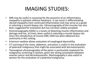
![Infectious agents have been suggested as possible triggers of dermatomyositis. These include the
following:
• Viruses (coxsackie , parvovirus, [HTLV-1], HIV)
• Toxoplasma species
• Borrelia species
Cases of drug-induced dermatomyositis have been reported. Dermatomyositis-like skin changes
have been reported with hydroxyurea in patients with chronic, myelogenous leukemia or
thrombocytosis. Other agents that may trigger the disease include the following:
• Statins
• Penicillamine
• Anti–tumor necrosis factor drugs
• Interferon
• Cyclophosphamide
• Bacillus Calmette-Guérin (BCG) vaccine
• Quinidine
• Phenylbutazone](https://image.slidesharecdn.com/dermatomyositis-161213091044/85/Dermatomyositis-5-320.jpg)
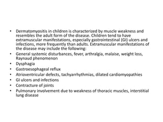
![PATHOPHYSIOLOGY:
Dermatomyositis occurs as a result of a humoral attack against the muscle capillaries and
small arterioles (endothelium of the endomysial blood vessels). The disease starts when
putative antibodies or other factors activate C3, forming C3b and C4b fragments that lead to
formation of C3bNEO and membrane attack complex (MAC), which are deposited in the
endomysial vasculature. Complement C5b-9 MAC is deposited and is needed in preparing the
cell for destruction in antibody-mediated disease. B cells and CD4 (helper) cells are also
present in abundance in the inflammatory reaction associated with the blood vessels.
As the disease progresses, the capillaries are destroyed, and the muscles undergo
microinfarction. Perifascicular atrophy occurs in the beginning; however, as the disease
advances, necrotic and degenerative fibers are present throughout the muscle. The
pathogenesis of the cutaneous component of dermatomyositis is poorly understood, but is
thought to be similar to that of muscle involvement. Studies on the pathogenesis of the
muscle component have been controversial. Some suggest that the myopathy in
dermatomyositis is pathogenically different from that in polymyositis. The former is probably
caused by complement-mediated (terminal attack complex) vascular inflammation, the latter
by the direct cytotoxic effect of CD8+ lymphocytes on muscle. However, other cytokine
studies suggest that some of the inflammatory processes may be similar. One report has
linked tumor necrosis factor (TNF) abnormalities with dermatomyositis. [21]](https://image.slidesharecdn.com/dermatomyositis-161213091044/85/Dermatomyositis-6-320.jpg)
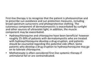
![LABORATORY DIAGNOSIS
• Muscle enzyme levels are often abnormal
during the course of dermatomyositis, except
in patients with amyopathic dermatomyositis
(ADM). The most sensitive/specific enzyme
abnormality is elevated creatine kinase (CK),
but aldolase studies and other tests (eg, for
aspartate aminotransferase [AST] or lactic
dehydrogenase [LDH]) may also yield
abnormal results.](https://image.slidesharecdn.com/dermatomyositis-161213091044/85/Dermatomyositis-9-320.jpg)

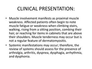
Dermatomyositis is an inflammatory myopathy that affects the skin and muscles. It is characterized by progressive proximal muscle weakness, elevated muscle enzymes, abnormal electromyography and muscle biopsy findings. The cause is unknown but genetic, immunological, infectious and environmental factors may play a role. Treatment involves sun protection, immunosuppressants like hydroxychloroquine and methotrexate, and corticosteroids like prednisone to control muscle and skin symptoms.


