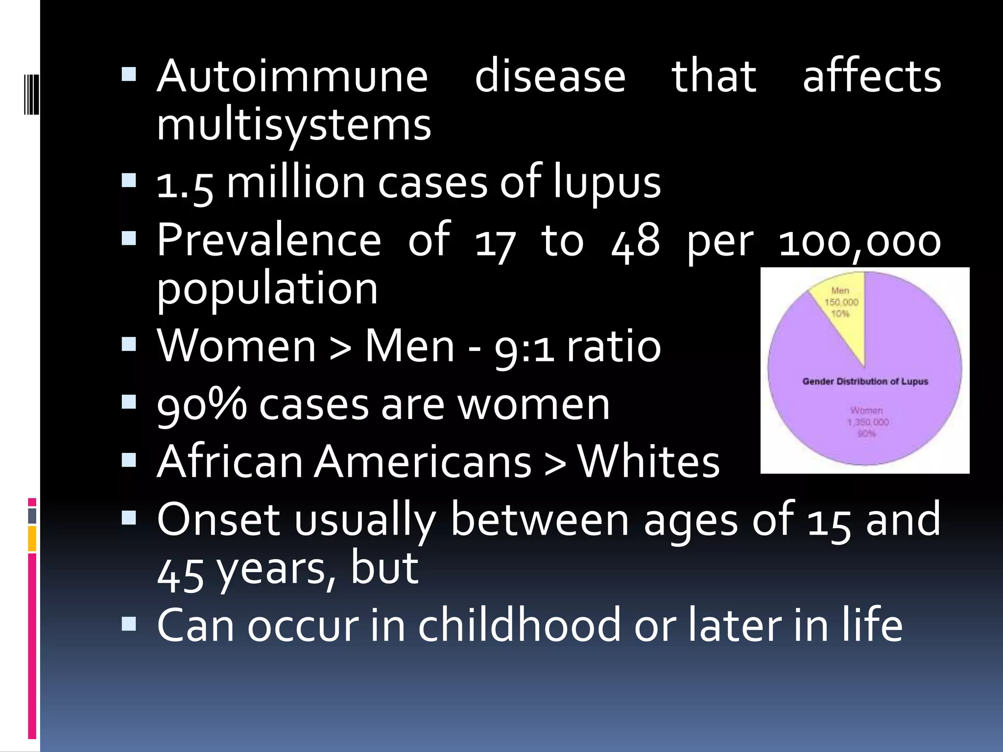
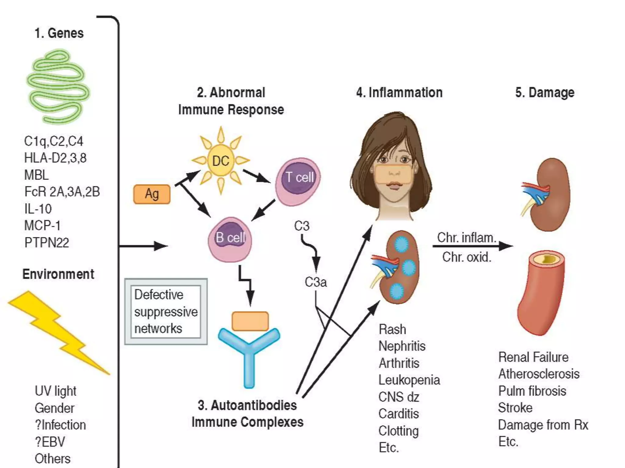
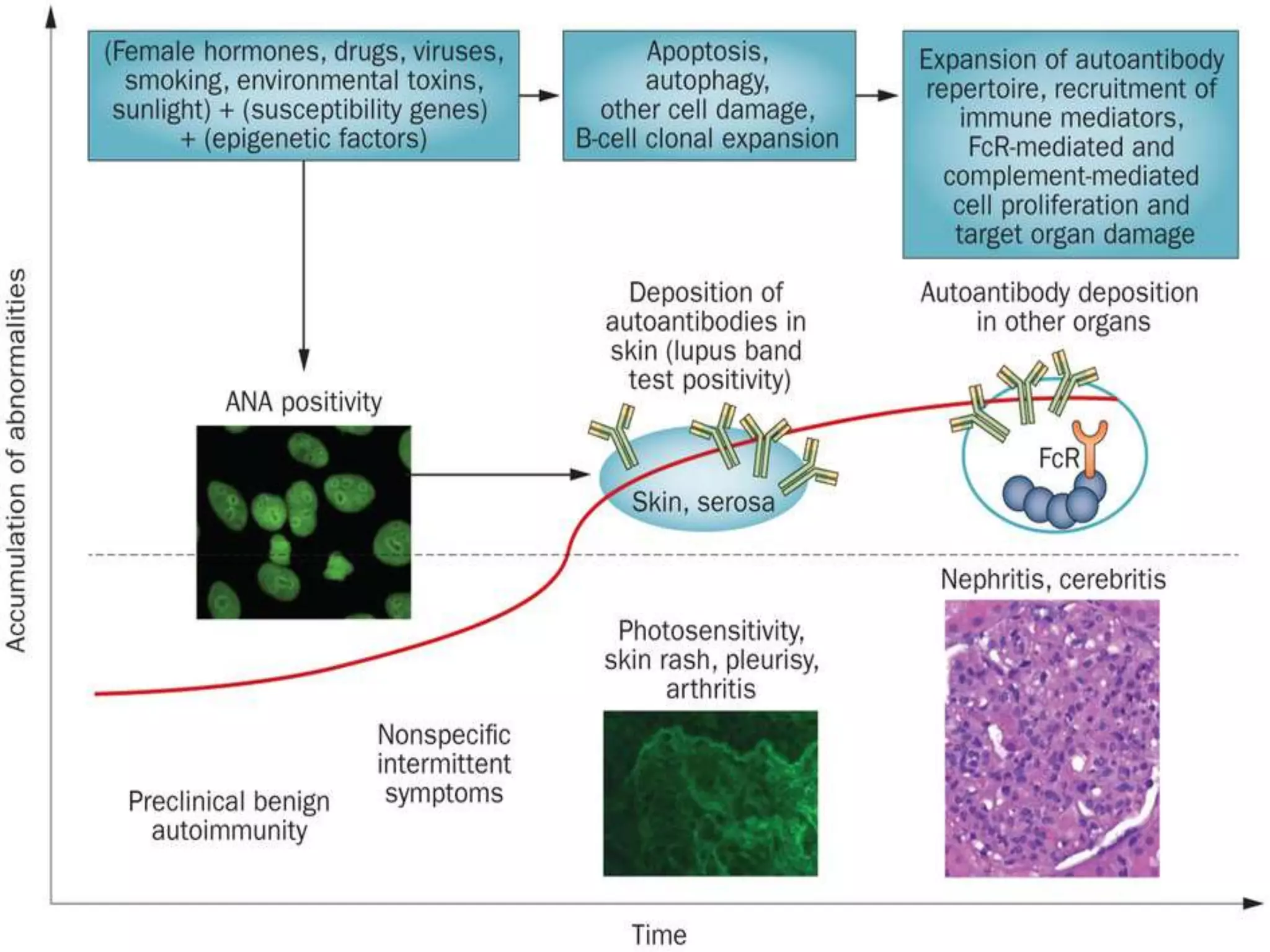
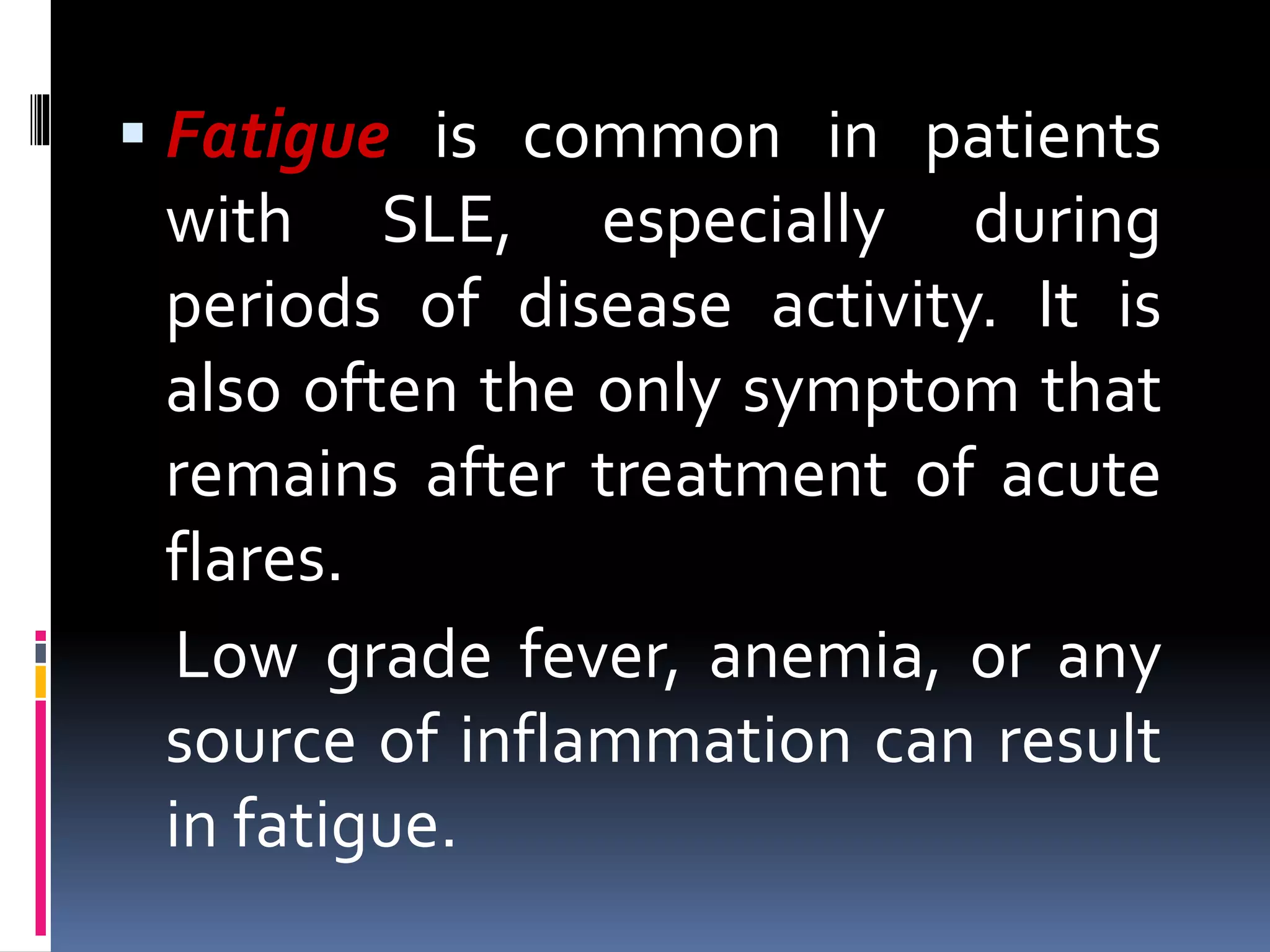
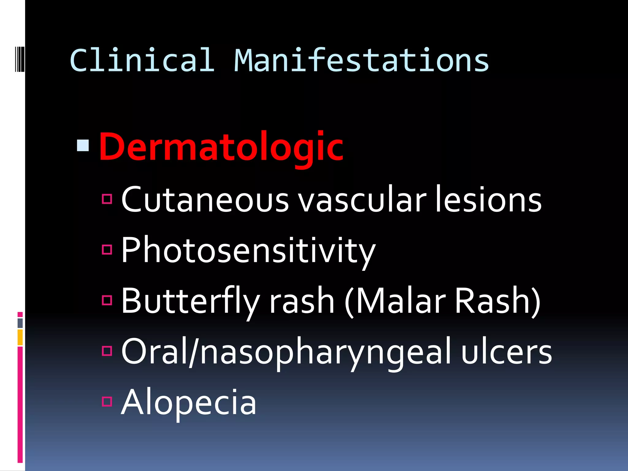
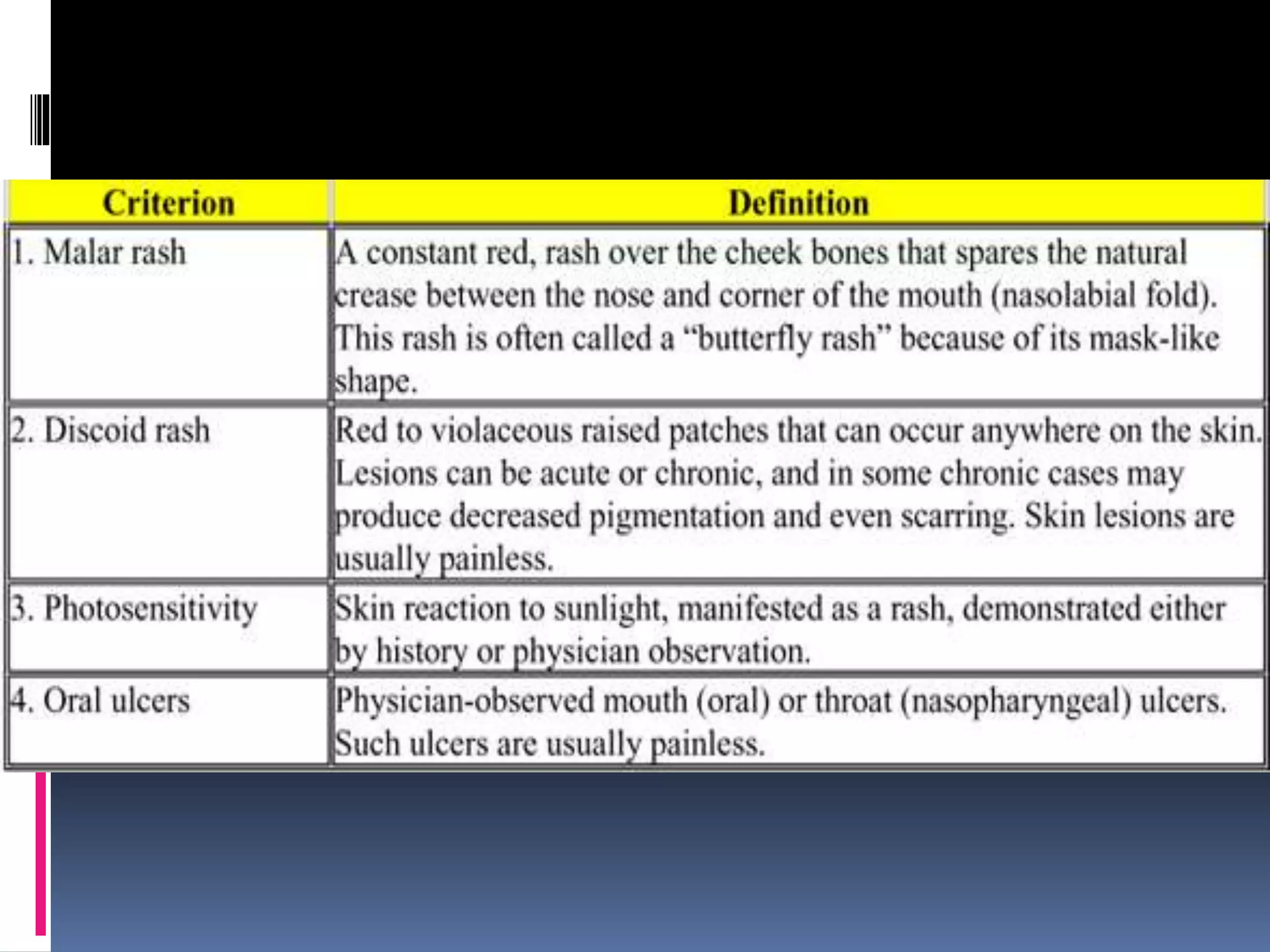
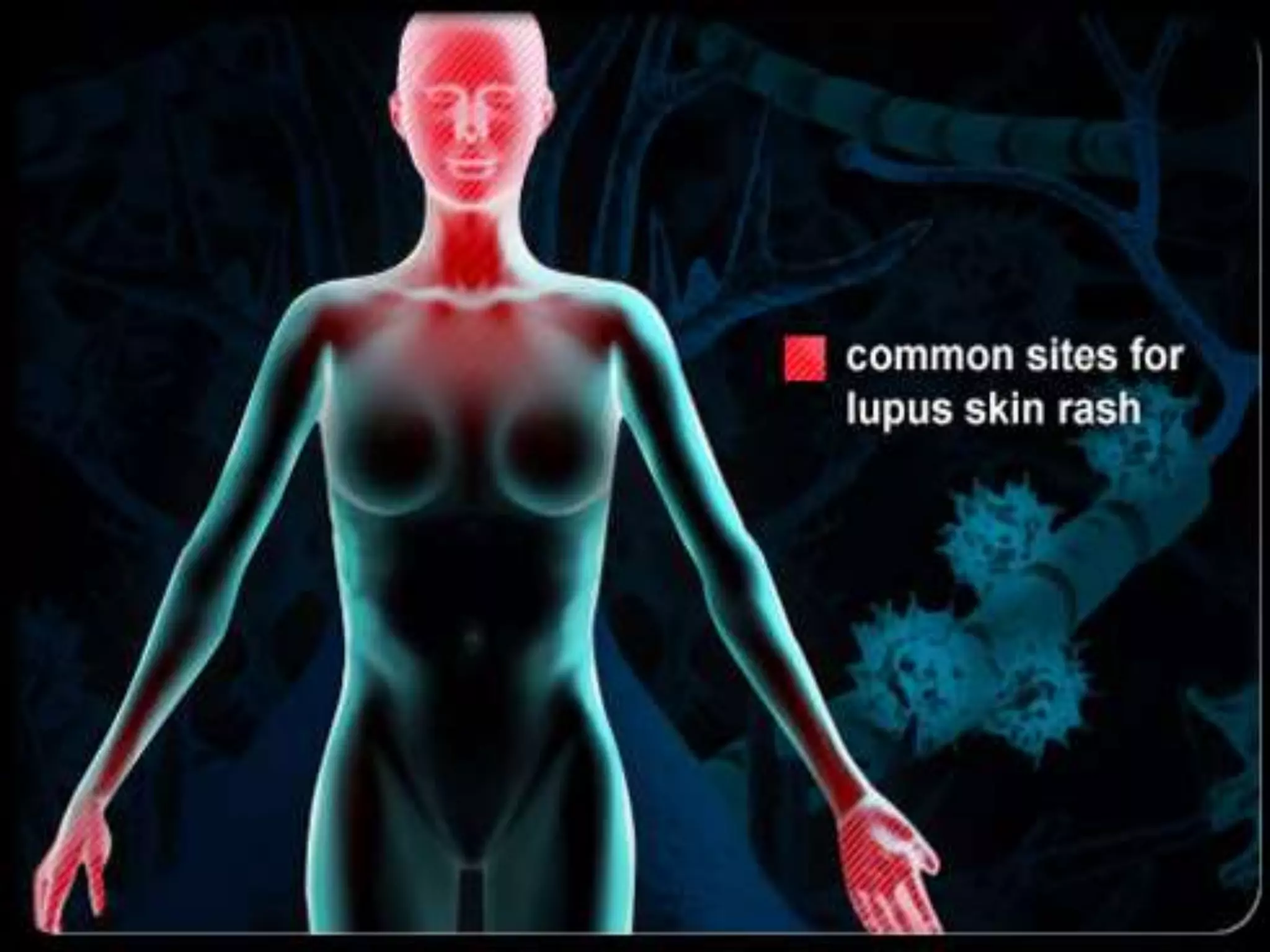
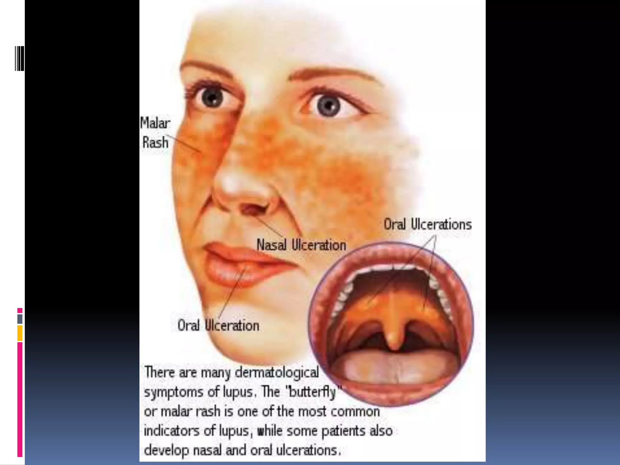
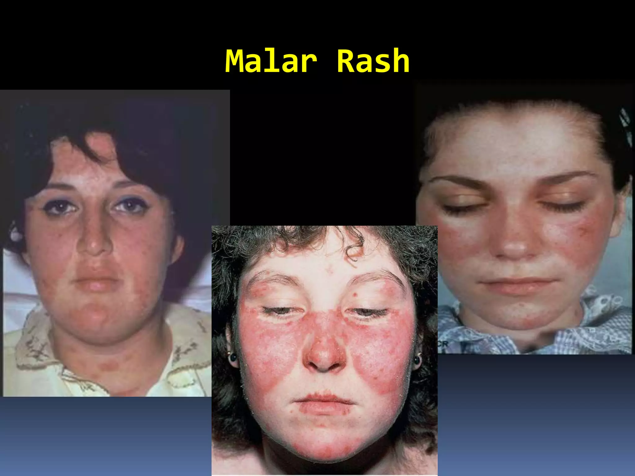
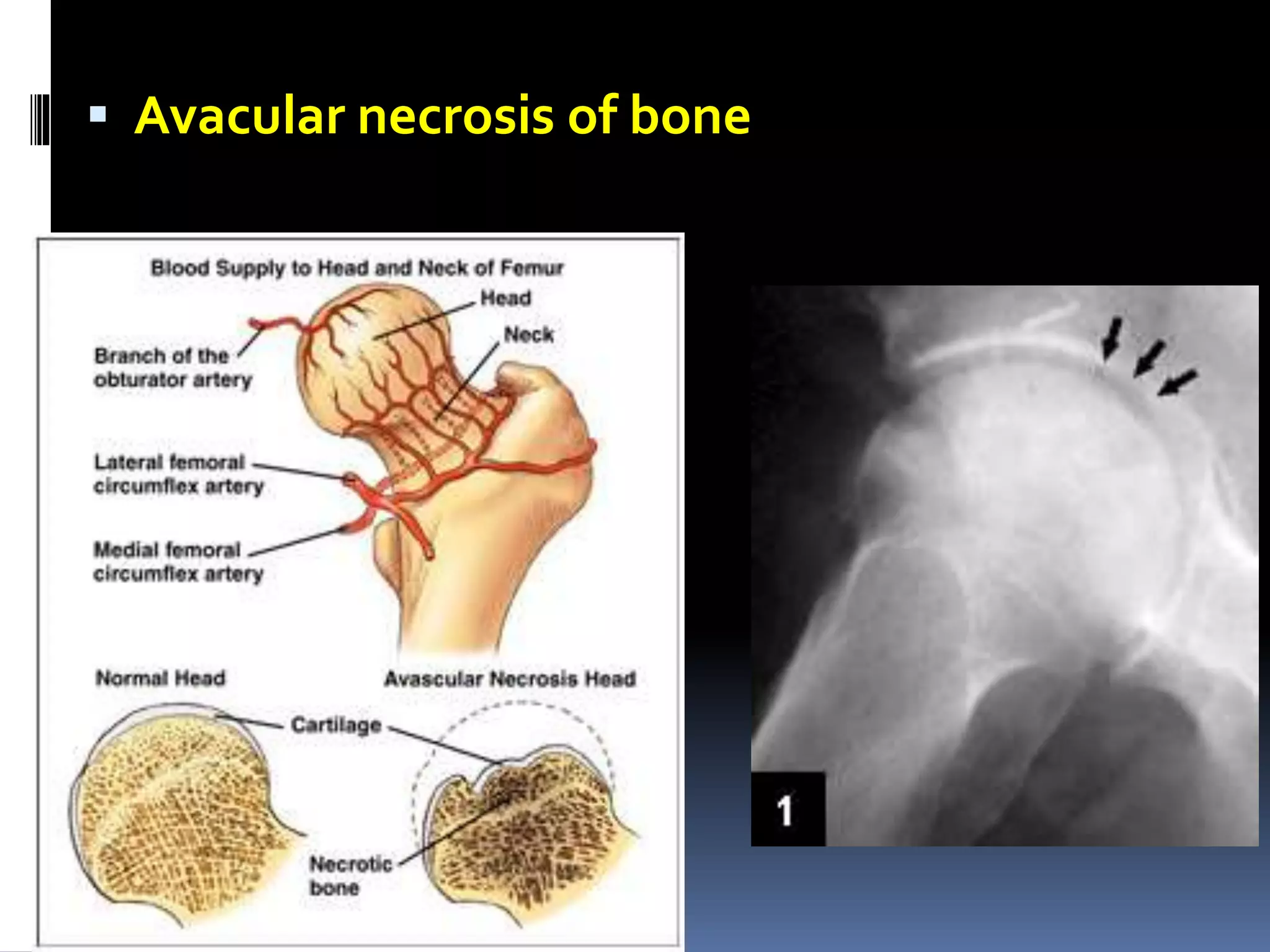
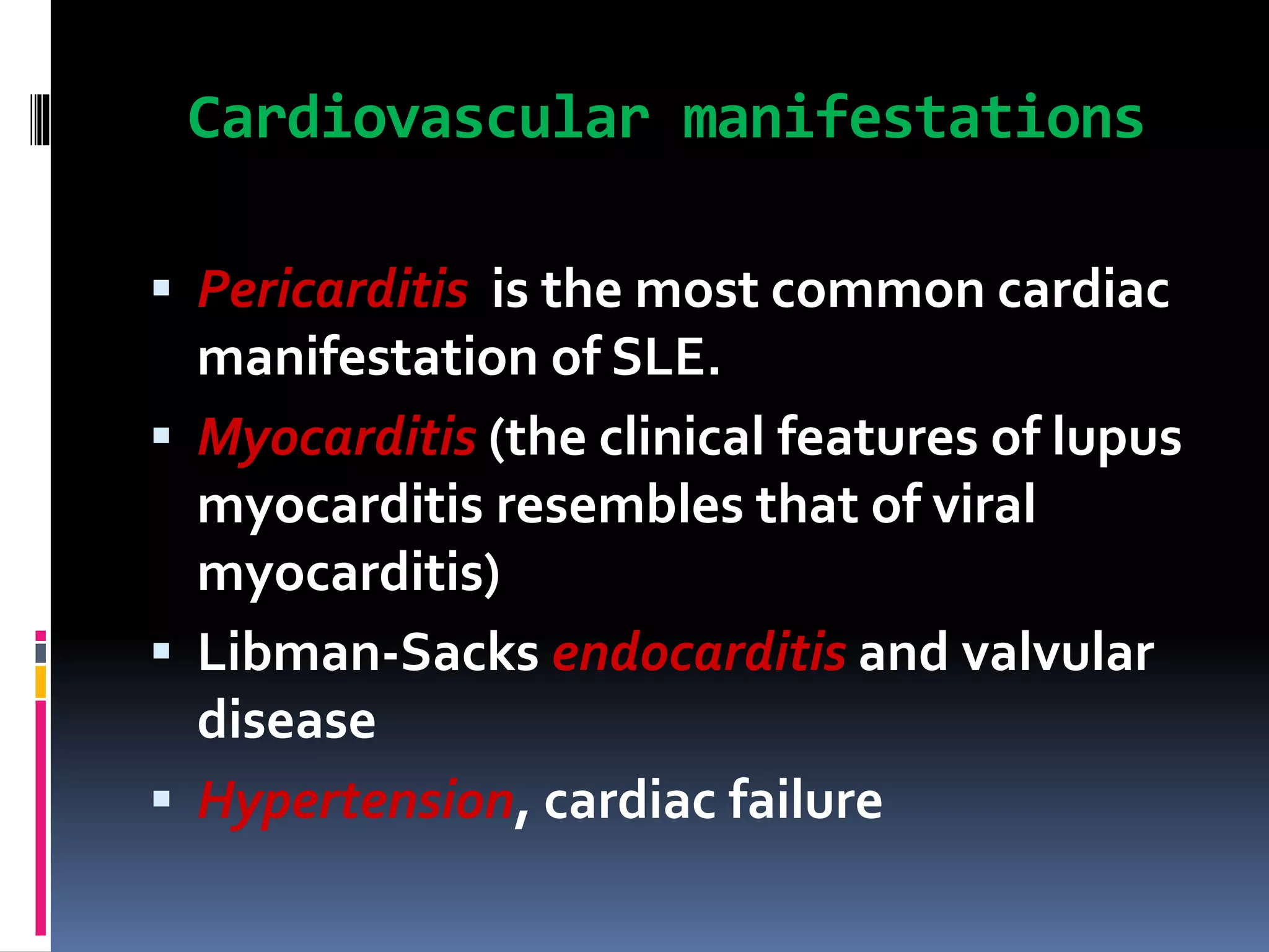
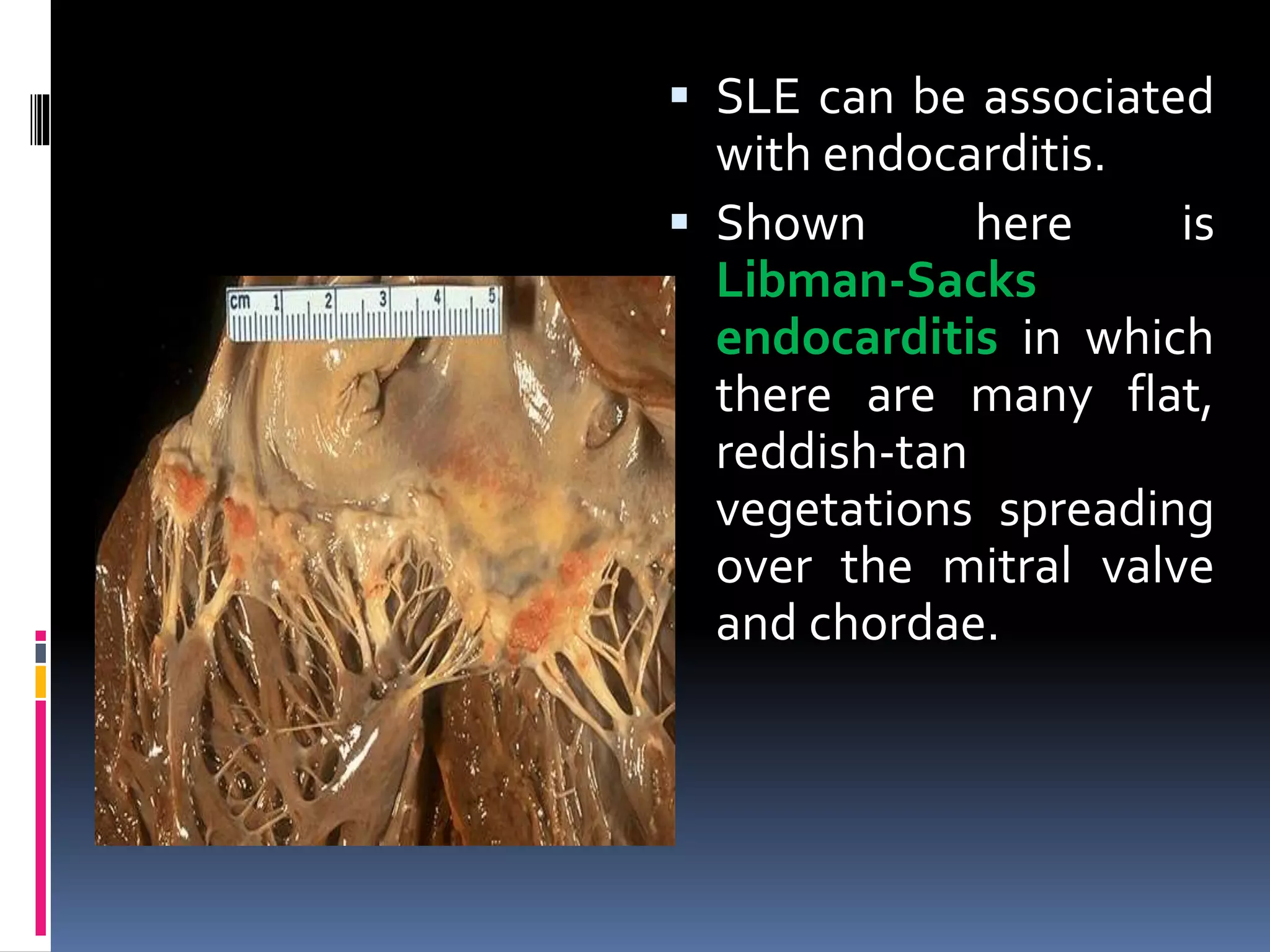
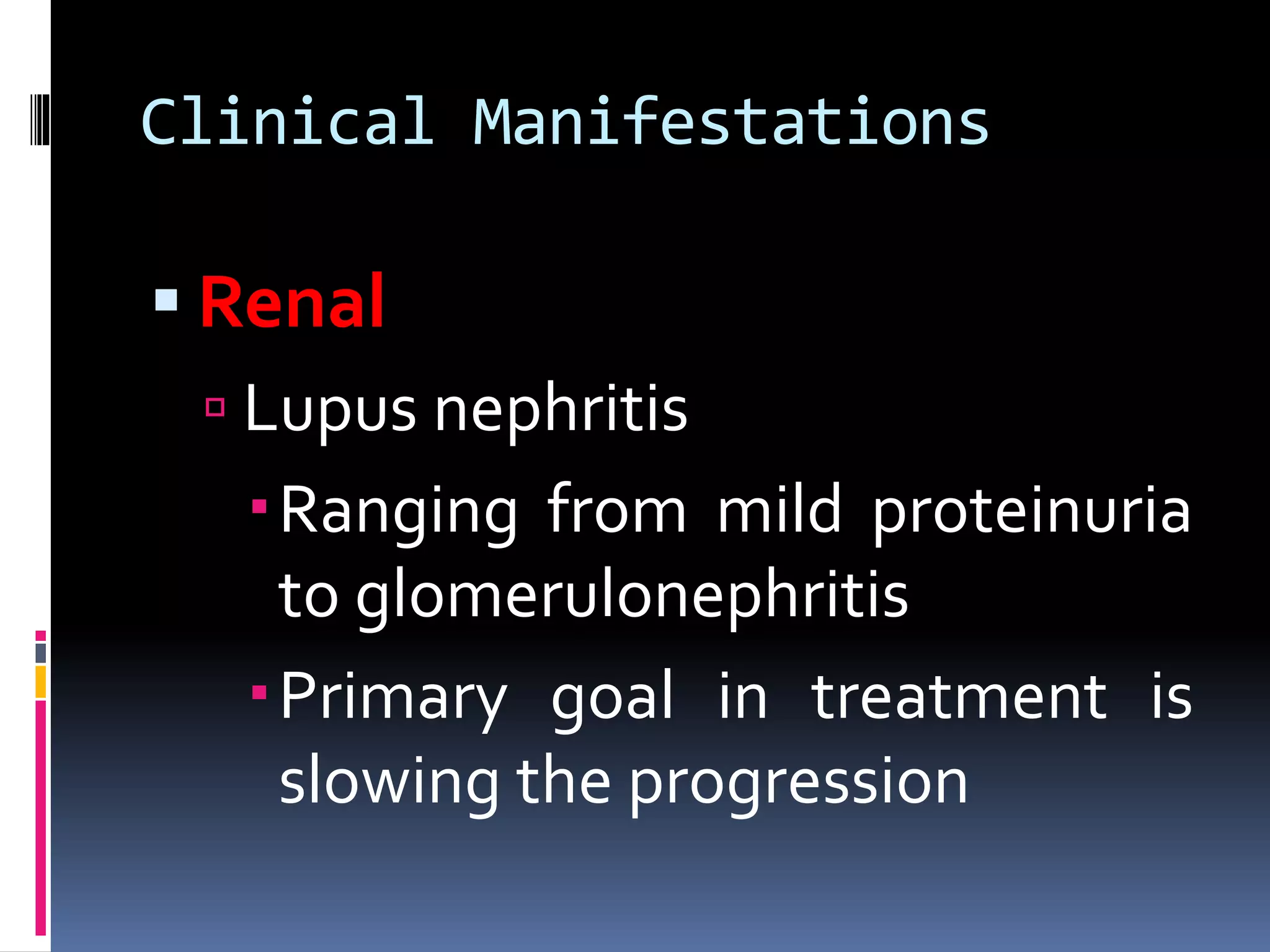
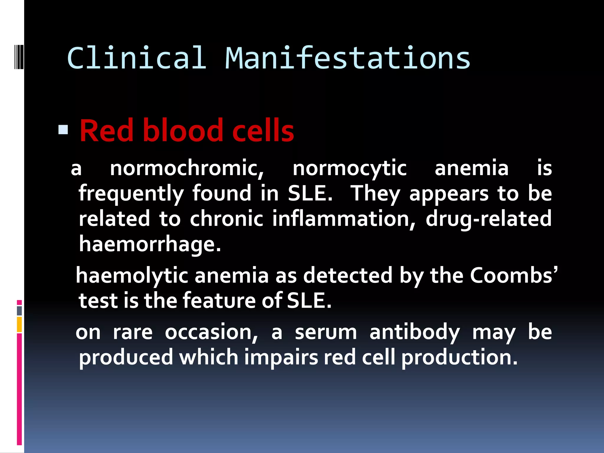
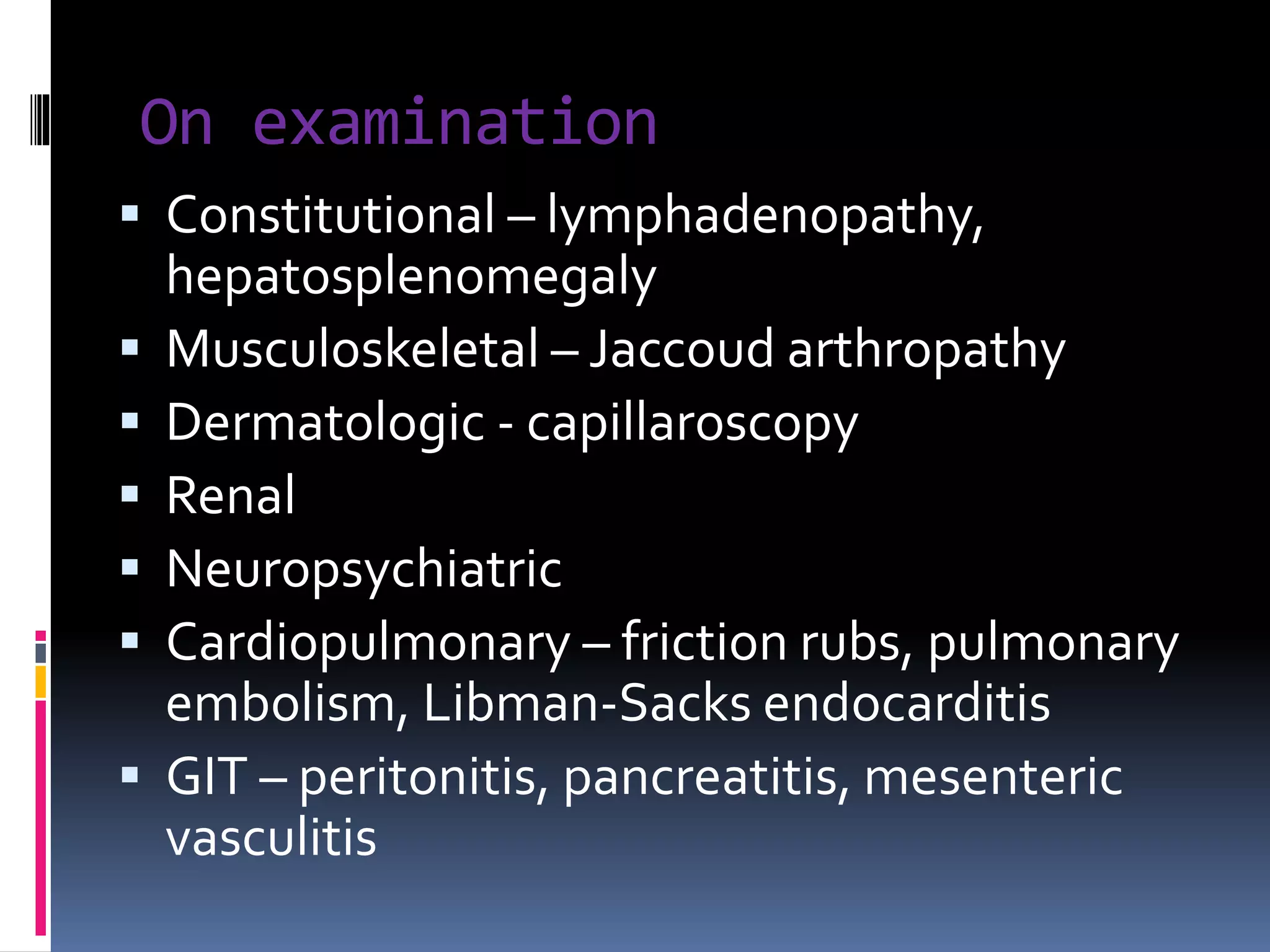
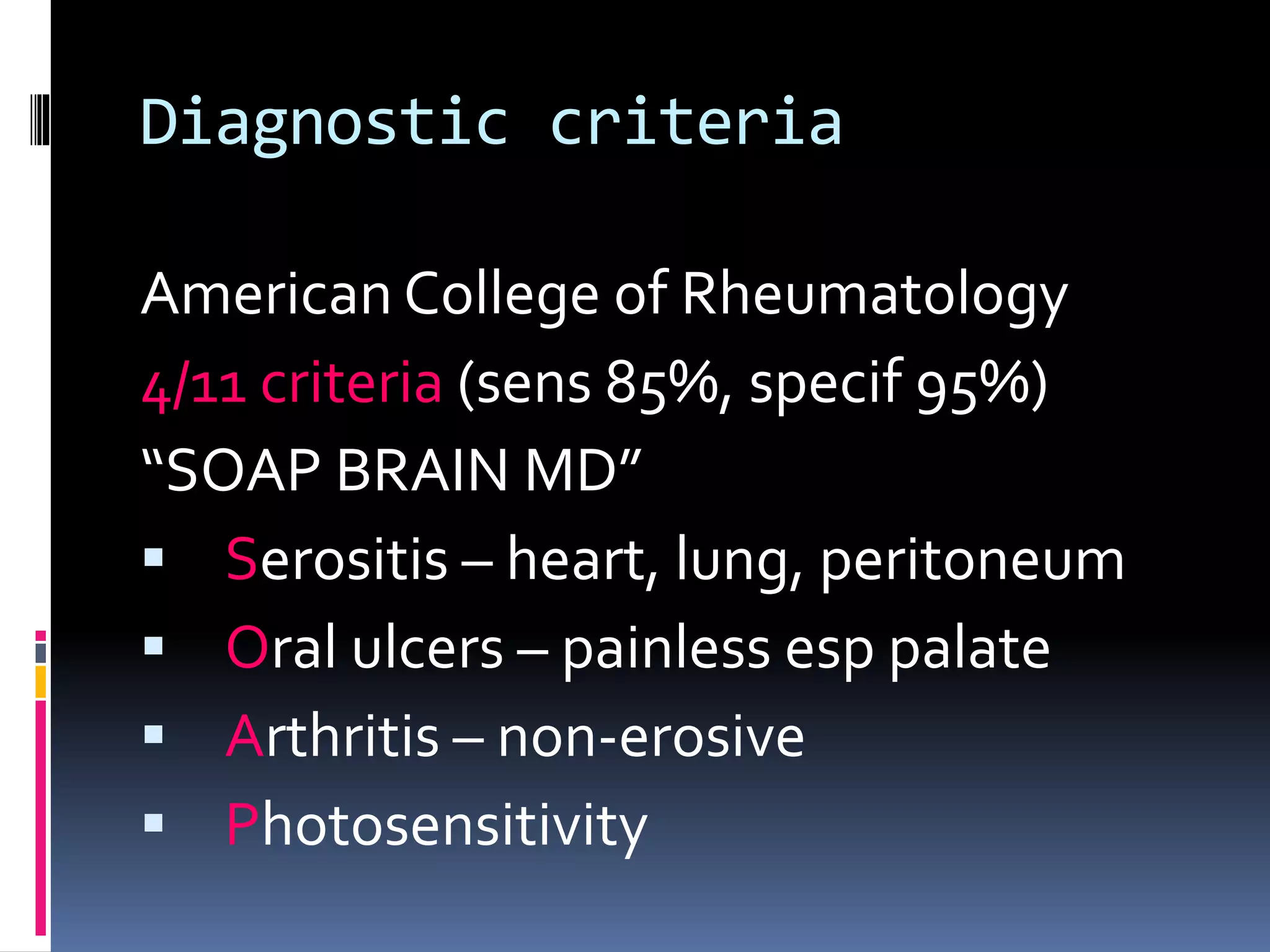
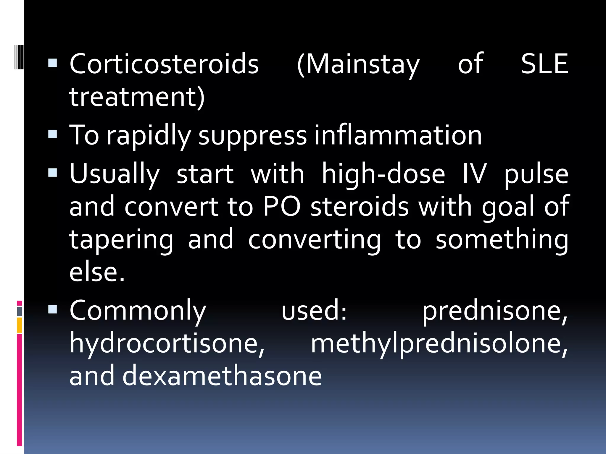
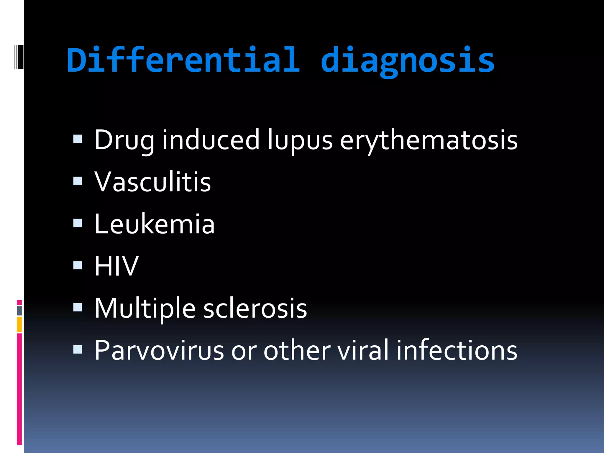
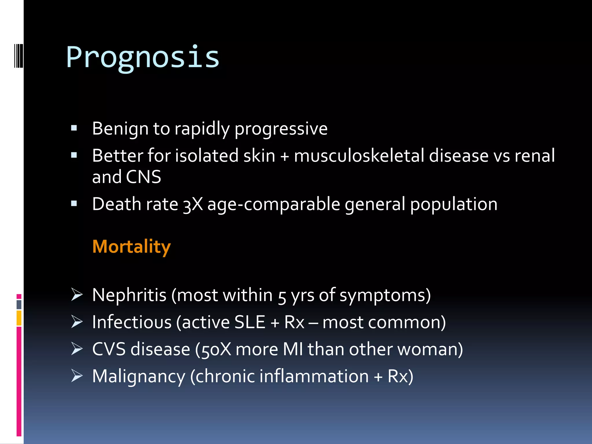
SYSTEMIC LUPUS
ERYTHEMATOSUS (SLE)
Dr. Angelo Smith M.D
WHPL
 Autoimmune disease that affects
multisystems
 1.5 million cases of lupus
 Prevalence of 17 to 48 per 100,000
population
 Women > Men - 9:1 ratio
 90% cases are women
 African Americans >Whites
 Onset usually between ages of 15 and
45 years, but
 Can occur in childhood or later in life
Clinical Manifestations
 Ranges from a relatively mild
disorder to rapidly progressing,
affecting many body systems.
 Chronic with relapsing and remitting
course.
 Most commonly affects the skin /
muscles, lining of lungs, heart,
nervous tissue, and kidneys
Etiology
 Etiology is unknown
 Most probable causes
 Genetic influence
 Hormones
 Environmental factors
 Certain medications
Pathogenesis of SLE
Pathophysiology
 Autoimmune reactions directed against
constituents of cell nucleus, DNA
 Antibody response related to B and
T cell hyperactivity
General symptoms
The most common symptoms listed as
initial complaints are fatigue, fever, and
weight loss.
 Fever: fever secondary to active disease
was recorded from 50% to 86%. No fever
curve or pattern is characteristic. It can be
difficult, but very important to distinguish
the fever of SLE from that caused by
complicating infections.
Clinical Manifestations
 Infection
 Increased susceptibility to
infections
 Fever should be considered
serious
 Infections such as pneumonia
are a common cause of death
 Fatigue is common in patients
with SLE, especially during
periods of disease activity. It is
also often the only symptom that
remains after treatment of acute
flares.
Low grade fever, anemia, or any
source of inflammation can result
in fatigue.
Clinical Manifestations
Dermatologic
 Cutaneous vascular lesions
 Photosensitivity
 Butterfly rash (Malar Rash)
 Oral/nasopharyngeal ulcers
 Alopecia
Malar Rash
Discoid Rash
 Maculopapular
eruption
Oral Ulcers
 Raynaud’s phenomenon is commonly found in
lupus. It lack specificity.
(a triphasic reaction of distal digits to cold or emotion, in
which the skin colour changes from white to blue to red)
 Vasculitic skin lesion
Clinical Manifestations
 Musculoskeletal (jaccoud
arthropathy)
 Polyarthralgia with morning stiffness
 A vascular necrosis
 Arthritis bilateral – hands / wrists /
knees
 Swan neck fingers
 Ulnar deviation
 Subluxation with hyperlaxity of joints
 Avacular necrosis of bone
Clinical Manifestations
 Cardiopulmonary
 Tachypnea
 Pleurisy
 Dysrhythmias
 Accelerated CAD
 Pericarditis
Pulmonary manifestations
 Pleurisy
it is the most common manifestation of
pulmonary involvement of SLE. The volume
of pleural effusions usually is small to
moderate and maybe unilateral or bilateral.
Large pleural effusion are uncommon. It
usually exudative in character.
Pleural effusions may also occur in SLE patients
with nephrotic syndrome, infection, cardiac failure.
 Lung
1) acute lupus pneumonitis: fever, dyspnea,
cough with scanty sputum, hemoptysis,
tachypnea and pleuritic chest pain.
2) pulmonary hemorrhage
3) chronic diffuse interstitial lung disease.
the diagnosis should not be made until
infectious processes such as viral pneumonia,
tuberculosis, and other bacterial, fungal and
pneumocystis carinii infection have been
completely excluded.
Cardiovascular manifestations
 Pericarditis is the most common cardiac
manifestation of SLE.
 Myocarditis (the clinical features of lupus
myocarditis resembles that of viral
myocarditis)
 Libman-Sacks endocarditis and valvular
disease
 Hypertension, cardiac failure
 SLE can be associated
with endocarditis.
 Shown here is
Libman-Sacks
endocarditis in which
there are many flat,
reddish-tan
vegetations spreading
over the mitral valve
and chordae.
Clinical Manifestations
 Renal
 Lupus nephritis
Ranging from mild proteinuria
to glomerulonephritis
Primary goal in treatment is
slowing the progression
 Haematuria
 Proteinure (>0.5g protein/d or 3+ )
 Cast
Lupus nephritis
Class I Minimal mesangial Normal light microscopy; abnormal
electron microscopy
Class II Mesangial
proliferative
Hypercellular on light microscopy
Class III Focal proliferative <50% glomeruli involved
Class IV Diffuse proliferative >50% glomeruli involved;
segmental/global
Class V Membranous Predominantly nephrotic disease
Class VI Advanced sclerosing Chronic lesions and sclerosis
Clinical Manifestations
 Nervous system
 Generalized/focal seizures
 Peripheral neuropathy
 Cognitive dysfunction
 Disorientation
 Memory and reasoning deficits
 Psychiatric symptoms – severe
depression / psychosis
Clinical Manifestations
 Red blood cells
a normochromic, normocytic anemia is
frequently found in SLE. They appears to be
related to chronic inflammation, drug-related
haemorrhage.
haemolytic anemia as detected by the Coombs’
test is the feature of SLE.
on rare occasion, a serum antibody may be
produced which impairs red cell production.
 Platelets
thrombocytopenia (<100*109/L) appears
to be mediated by anti-platelet
antibodies or/and anti-phospholipid
antibodies.
 White blood cell
leucopenia (<4.0*109/L), its cause is probably
a combination of destruction of white cells
by autoantibodies, decreased marrow
production, increased or marginal splenic
pooling, and complement activation.
it should also noted that the
immunosuppressive drugs used in the
treatment of SLE may cause a marked
leucopenia.
Gastrointestinal and hepatic
manifestation
 Esophagitis, dysphagia, nausea,
vomiting: (drug related in most cases)
 Chronic intestinal pseudo-obstruction,
mesenteric vasculitis, protein-losing
enteropathy
 Pancreatitis
 Lupus hepatitis
Diagnostic Studies
 No specific test
 SLE is diagnosed primarily on
criteria relating to
 patient history,
 physical examination, and
 laboratory findings
On examination
 Constitutional – lymphadenopathy,
hepatosplenomegaly
 Musculoskeletal – Jaccoud arthropathy
 Dermatologic - capillaroscopy
 Renal
 Neuropsychiatric
 Cardiopulmonary – friction rubs, pulmonary
embolism, Libman-Sacks endocarditis
 GIT – peritonitis, pancreatitis, mesenteric
vasculitis
Diagnostic Studies
 Antinuclear antibodies
 ANA and other antibodies indicate
autoimmune disease
 Anti-DNA and anti-Smith antibody tests most
specific for SLE
 LE prep can be positive with other rheumatoid
diseases
 ESR & CRP are indicative of inflammatory
activity
Radiological studies
 Joint x-rays: no erosions, periarticular osteopenia
+ soft tissue swelling
 CXR/CT chest: interstitial lung disease,
pneumonitis, pulmonary emboli, alveolar
hemorrhage
 CTBrain or Brain MRI ± angiography: lupus white
matter changes, vasculitis or stroke
 Echo: pericardial effusion, pulmonary
hypertension or Libman-Sacks endocarditis
Additional work-up
- Serum cr. and albumin
- CBC w/ diff
- U/A
- ESR
- Complement levels
- Renal profile if warranted
Invasive procedures
 LP – nonspecific ↑cells + protein, ↓
glucose
 Renal biopsy – prognosis and Rx
 Skin biopsy
Diagnostic criteria
American College of Rheumatology
4/11 criteria (sens 85%, specif 95%)
“SOAP BRAIN MD”
 Serositis – heart, lung, peritoneum
 Oral ulcers – painless esp palate
 Arthritis – non-erosive
 Photosensitivity
Diagnostic criteria
 Blood disorders - ↓RBC (Coombs +), PLT, WCC,
Lymphocytes
 Renal involvement – proteinuria /± casts
 ANA – titer > 1:160
 Immunologic phenomena – LE cells, anti-
dsDNA Ab, anti-Sm Ab, antiphospholipid Ab,
falseWR +
 Neurological disorders – seizures/ psychosis
 Malar rash – cheeks + nasal bridge
 Discoid rash – rimmed with scaling, follicular
plugging
Treatment
 Treatment plans are based on patient age, sex,
health, symptoms, and lifestyle and on disease
severity
 Fever, skin, musculoskeletal and serositis = milder
disease
 CNS and renal involvement – aggressive Rx
 Goals of treatment are to:
-prevent flares
-treat flares when they occur
-minimize organ damage and complications
Collaborative Care
 Drug therapy
 NSAIDs
 Antimalarial drugs
 Steroid-sparing drugs
 Corticosteroids
 Immunosuppressive drugs
Conservative management
 For those w/out major organ involvement.
 NSAIDs: to control pain, swelling, and fever
 Caution w/ NSAIDS though. SLE pts are at
increased risk for aseptic meningitis
 Antimalarials: Generally to treat fatigue joint
pain, skin rashes, and inflammation of the
lungs
 Commonly used: Hydroxycholorquine
 Used alone or in combination with other
drugs
 Corticosteroids (Mainstay of SLE
treatment)
 To rapidly suppress inflammation
 Usually start with high-dose IV pulse
and convert to PO steroids with goal of
tapering and converting to something
else.
 Commonly used: prednisone,
hydrocortisone, methylprednisolone,
and dexamethasone
Immunosuppressives
 Primarily for CNS/renal involvement
 Mycophenolate mofetil (cellcept)
 Azathioprine (imuran): requires several months
to be effective, effective in smaller percentage of
patients
 MTX: for treatment of dermatitis and arthritis,
not life-threatening disease
 Cyclosporine: used in steroid-resistant SLE, risk
of nephrotoxicity
 Cyclophosphamide (cytoxan) Almost all trials
performed on patients with nephritis
Differential diagnosis
 Drug induced lupus erythematosis
 Vasculitis
 Leukemia
 HIV
 Multiple sclerosis
 Parvovirus or other viral infections
Prognosis
 Benign to rapidly progressive
 Better for isolated skin + musculoskeletal disease vs renal
and CNS
 Death rate 3X age-comparable general population
Mortality
 Nephritis (most within 5 yrs of symptoms)
 Infectious (active SLE + Rx – most common)
 CVS disease (50X more MI than other woman)
 Malignancy (chronic inflammation + Rx)
SLE disease activity index (SLEDAI)
Clinical feature score
seizure , psychosis , organ brain syndrome 8
visual disturbance, cranial nerve disorder 8
lupus headache, cerebrovascular accidents, 8
vasculitis 8
arthritis 4
myositis 4
urinary casts, hematuria, proteinure, pyuria 4
rash, alopecia, mucosal ulcers, 2
pleurisy, pericarditis 2
low complement, increased DNA binding 2
fever 1
thrombocytopenia, leucopenia 1
Other therapy
 Plasma exchange
 Intravenous Immunoglobulin
 Stem cell transplantation
 Immune therapy ( anti-IL10, anti-CD20, and
immune tolerance therapy)

Systemic Lupus Erythematoses

  • 1.
  • 2.
     Autoimmune diseasethat affects multisystems  1.5 million cases of lupus  Prevalence of 17 to 48 per 100,000 population  Women > Men - 9:1 ratio  90% cases are women  African Americans >Whites  Onset usually between ages of 15 and 45 years, but  Can occur in childhood or later in life
  • 3.
    Clinical Manifestations  Rangesfrom a relatively mild disorder to rapidly progressing, affecting many body systems.  Chronic with relapsing and remitting course.  Most commonly affects the skin / muscles, lining of lungs, heart, nervous tissue, and kidneys
  • 5.
    Etiology  Etiology isunknown  Most probable causes  Genetic influence  Hormones  Environmental factors  Certain medications
  • 6.
  • 7.
    Pathophysiology  Autoimmune reactionsdirected against constituents of cell nucleus, DNA  Antibody response related to B and T cell hyperactivity
  • 15.
    General symptoms The mostcommon symptoms listed as initial complaints are fatigue, fever, and weight loss.  Fever: fever secondary to active disease was recorded from 50% to 86%. No fever curve or pattern is characteristic. It can be difficult, but very important to distinguish the fever of SLE from that caused by complicating infections.
  • 16.
    Clinical Manifestations  Infection Increased susceptibility to infections  Fever should be considered serious  Infections such as pneumonia are a common cause of death
  • 17.
     Fatigue iscommon in patients with SLE, especially during periods of disease activity. It is also often the only symptom that remains after treatment of acute flares. Low grade fever, anemia, or any source of inflammation can result in fatigue.
  • 20.
    Clinical Manifestations Dermatologic  Cutaneousvascular lesions  Photosensitivity  Butterfly rash (Malar Rash)  Oral/nasopharyngeal ulcers  Alopecia
  • 24.
  • 25.
  • 26.
  • 27.
  • 28.
     Raynaud’s phenomenonis commonly found in lupus. It lack specificity. (a triphasic reaction of distal digits to cold or emotion, in which the skin colour changes from white to blue to red)
  • 29.
  • 30.
    Clinical Manifestations  Musculoskeletal(jaccoud arthropathy)  Polyarthralgia with morning stiffness  A vascular necrosis  Arthritis bilateral – hands / wrists / knees  Swan neck fingers  Ulnar deviation  Subluxation with hyperlaxity of joints
  • 32.
  • 33.
    Clinical Manifestations  Cardiopulmonary Tachypnea  Pleurisy  Dysrhythmias  Accelerated CAD  Pericarditis
  • 34.
    Pulmonary manifestations  Pleurisy itis the most common manifestation of pulmonary involvement of SLE. The volume of pleural effusions usually is small to moderate and maybe unilateral or bilateral. Large pleural effusion are uncommon. It usually exudative in character. Pleural effusions may also occur in SLE patients with nephrotic syndrome, infection, cardiac failure.
  • 35.
     Lung 1) acutelupus pneumonitis: fever, dyspnea, cough with scanty sputum, hemoptysis, tachypnea and pleuritic chest pain. 2) pulmonary hemorrhage 3) chronic diffuse interstitial lung disease. the diagnosis should not be made until infectious processes such as viral pneumonia, tuberculosis, and other bacterial, fungal and pneumocystis carinii infection have been completely excluded.
  • 36.
    Cardiovascular manifestations  Pericarditisis the most common cardiac manifestation of SLE.  Myocarditis (the clinical features of lupus myocarditis resembles that of viral myocarditis)  Libman-Sacks endocarditis and valvular disease  Hypertension, cardiac failure
  • 37.
     SLE canbe associated with endocarditis.  Shown here is Libman-Sacks endocarditis in which there are many flat, reddish-tan vegetations spreading over the mitral valve and chordae.
  • 38.
    Clinical Manifestations  Renal Lupus nephritis Ranging from mild proteinuria to glomerulonephritis Primary goal in treatment is slowing the progression
  • 40.
     Haematuria  Proteinure(>0.5g protein/d or 3+ )  Cast
  • 41.
    Lupus nephritis Class IMinimal mesangial Normal light microscopy; abnormal electron microscopy Class II Mesangial proliferative Hypercellular on light microscopy Class III Focal proliferative <50% glomeruli involved Class IV Diffuse proliferative >50% glomeruli involved; segmental/global Class V Membranous Predominantly nephrotic disease Class VI Advanced sclerosing Chronic lesions and sclerosis
  • 43.
    Clinical Manifestations  Nervoussystem  Generalized/focal seizures  Peripheral neuropathy  Cognitive dysfunction  Disorientation  Memory and reasoning deficits  Psychiatric symptoms – severe depression / psychosis
  • 45.
    Clinical Manifestations  Redblood cells a normochromic, normocytic anemia is frequently found in SLE. They appears to be related to chronic inflammation, drug-related haemorrhage. haemolytic anemia as detected by the Coombs’ test is the feature of SLE. on rare occasion, a serum antibody may be produced which impairs red cell production.
  • 46.
     Platelets thrombocytopenia (<100*109/L)appears to be mediated by anti-platelet antibodies or/and anti-phospholipid antibodies.
  • 47.
     White bloodcell leucopenia (<4.0*109/L), its cause is probably a combination of destruction of white cells by autoantibodies, decreased marrow production, increased or marginal splenic pooling, and complement activation. it should also noted that the immunosuppressive drugs used in the treatment of SLE may cause a marked leucopenia.
  • 48.
    Gastrointestinal and hepatic manifestation Esophagitis, dysphagia, nausea, vomiting: (drug related in most cases)  Chronic intestinal pseudo-obstruction, mesenteric vasculitis, protein-losing enteropathy  Pancreatitis  Lupus hepatitis
  • 49.
    Diagnostic Studies  Nospecific test  SLE is diagnosed primarily on criteria relating to  patient history,  physical examination, and  laboratory findings
  • 50.
    On examination  Constitutional– lymphadenopathy, hepatosplenomegaly  Musculoskeletal – Jaccoud arthropathy  Dermatologic - capillaroscopy  Renal  Neuropsychiatric  Cardiopulmonary – friction rubs, pulmonary embolism, Libman-Sacks endocarditis  GIT – peritonitis, pancreatitis, mesenteric vasculitis
  • 51.
    Diagnostic Studies  Antinuclearantibodies  ANA and other antibodies indicate autoimmune disease  Anti-DNA and anti-Smith antibody tests most specific for SLE  LE prep can be positive with other rheumatoid diseases  ESR & CRP are indicative of inflammatory activity
  • 52.
    Radiological studies  Jointx-rays: no erosions, periarticular osteopenia + soft tissue swelling  CXR/CT chest: interstitial lung disease, pneumonitis, pulmonary emboli, alveolar hemorrhage  CTBrain or Brain MRI ± angiography: lupus white matter changes, vasculitis or stroke  Echo: pericardial effusion, pulmonary hypertension or Libman-Sacks endocarditis
  • 53.
    Additional work-up - Serumcr. and albumin - CBC w/ diff - U/A - ESR - Complement levels - Renal profile if warranted
  • 54.
    Invasive procedures  LP– nonspecific ↑cells + protein, ↓ glucose  Renal biopsy – prognosis and Rx  Skin biopsy
  • 56.
    Diagnostic criteria American Collegeof Rheumatology 4/11 criteria (sens 85%, specif 95%) “SOAP BRAIN MD”  Serositis – heart, lung, peritoneum  Oral ulcers – painless esp palate  Arthritis – non-erosive  Photosensitivity
  • 57.
    Diagnostic criteria  Blooddisorders - ↓RBC (Coombs +), PLT, WCC, Lymphocytes  Renal involvement – proteinuria /± casts  ANA – titer > 1:160  Immunologic phenomena – LE cells, anti- dsDNA Ab, anti-Sm Ab, antiphospholipid Ab, falseWR +  Neurological disorders – seizures/ psychosis  Malar rash – cheeks + nasal bridge  Discoid rash – rimmed with scaling, follicular plugging
  • 58.
    Treatment  Treatment plansare based on patient age, sex, health, symptoms, and lifestyle and on disease severity  Fever, skin, musculoskeletal and serositis = milder disease  CNS and renal involvement – aggressive Rx  Goals of treatment are to: -prevent flares -treat flares when they occur -minimize organ damage and complications
  • 59.
    Collaborative Care  Drugtherapy  NSAIDs  Antimalarial drugs  Steroid-sparing drugs  Corticosteroids  Immunosuppressive drugs
  • 60.
    Conservative management  Forthose w/out major organ involvement.  NSAIDs: to control pain, swelling, and fever  Caution w/ NSAIDS though. SLE pts are at increased risk for aseptic meningitis  Antimalarials: Generally to treat fatigue joint pain, skin rashes, and inflammation of the lungs  Commonly used: Hydroxycholorquine  Used alone or in combination with other drugs
  • 61.
     Corticosteroids (Mainstayof SLE treatment)  To rapidly suppress inflammation  Usually start with high-dose IV pulse and convert to PO steroids with goal of tapering and converting to something else.  Commonly used: prednisone, hydrocortisone, methylprednisolone, and dexamethasone
  • 62.
    Immunosuppressives  Primarily forCNS/renal involvement  Mycophenolate mofetil (cellcept)  Azathioprine (imuran): requires several months to be effective, effective in smaller percentage of patients  MTX: for treatment of dermatitis and arthritis, not life-threatening disease  Cyclosporine: used in steroid-resistant SLE, risk of nephrotoxicity  Cyclophosphamide (cytoxan) Almost all trials performed on patients with nephritis
  • 63.
    Differential diagnosis  Druginduced lupus erythematosis  Vasculitis  Leukemia  HIV  Multiple sclerosis  Parvovirus or other viral infections
  • 65.
    Prognosis  Benign torapidly progressive  Better for isolated skin + musculoskeletal disease vs renal and CNS  Death rate 3X age-comparable general population Mortality  Nephritis (most within 5 yrs of symptoms)  Infectious (active SLE + Rx – most common)  CVS disease (50X more MI than other woman)  Malignancy (chronic inflammation + Rx)
  • 66.
    SLE disease activityindex (SLEDAI) Clinical feature score seizure , psychosis , organ brain syndrome 8 visual disturbance, cranial nerve disorder 8 lupus headache, cerebrovascular accidents, 8 vasculitis 8 arthritis 4 myositis 4 urinary casts, hematuria, proteinure, pyuria 4 rash, alopecia, mucosal ulcers, 2 pleurisy, pericarditis 2 low complement, increased DNA binding 2 fever 1 thrombocytopenia, leucopenia 1
  • 67.
    Other therapy  Plasmaexchange  Intravenous Immunoglobulin  Stem cell transplantation  Immune therapy ( anti-IL10, anti-CD20, and immune tolerance therapy)