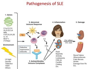
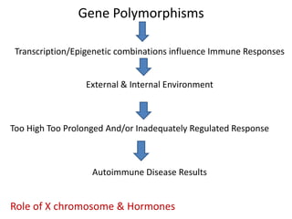
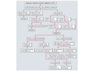
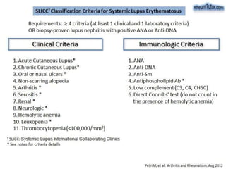
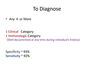
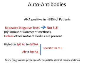
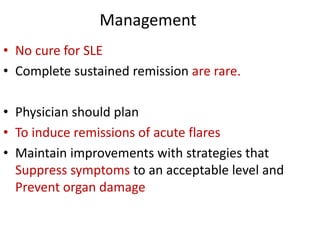
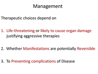
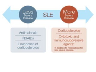
Systemic Lupus Erythematosis
Diagnosis & Treatment
Dr Suman Roy
PGT - Midnapore
Abnormal Immune Responses
Susceptibility Genes And Environmental Factors
• Activation of Innate Immunity
• Lowered Activation Thresholds Adaptive
• Abnormal Activation Pathways Immunity Cells
• Ineffective Regulatory CD4+ & CD8+ T Cells, B Cells
And Myeloid-derived Suppressor Cells
• Reduced Clearance Of Immune Complexes And
Apoptotic Cells
Self-antigens
Nucleosomal DNA/Protein; RNA/Protein in Sm, Ro La & Phospholipids
Recognized by Immune System in Surface Blebs of Apoptotic Cells
Autoantigens, Autoantibodies, Immune complexes
Inflammation & Ds
IFN1 & 2 TNF-α IL 17 & B Cell-maturation/Survival Cytokines
B Lymphocyte Stimulator (Blys/Baff) & IL-10.
Pathogenesis of SLE
SLE is a Multigene Disease
+ Environmental Factor
• Normal alleles of Multiple Gene
-- each contribute a small
-- if enough predisposing variations are present
Disease Results.
• Approx 45 predisposing genes have been
identified
Gene Polymorphisms
Transcription/Epigenetic combinations influence Immune Responses
External & Internal Environment
Too High Too Prolonged And/or Inadequately Regulated Response
Autoimmune Disease Results
Role of X chromosome & Hormones
Pathology
• Deposition of Ig at (DEJ)
• Injury to basal keratinocytes
• Inflammation dominated by T lymphocytes
-- DEJ
-- around blood vessels
-- dermal appendages.
• Clinically unaffected skin may also show Ig
deposition at the DEJ.
PATHOLOGY
• Histologic abnormalities in blood vessels
Shows
Leukocytoclastic Vasculitis
is most Common Patterns of vasculitis
BUT are not specific for SLE
• Lymph node biopsies are usually performed to rule out
Infection or Malignancies
• In SLE, they show nonspecific diffuse chronic
inflammation.
Diagnosis
Depends on
• C/F
• Auto-Antibody
To Diagnose
• Any 4 or More
1 Clinical Category
1 Immunologic Category
(Well documented at any time during individual’s history)
Specificity ~ 93%
Sensitivity ~ 92%
Auto-Antibodies
ANA positive in >98% of Patients
Repeated Negative Tests Not SLE
(By immunofluorescent method)
Unless other Autoantibodies are present
High-titer IgG Ab to dsDNA
specific for SLE
Ab to Sm-Ag
Favor diagnosis in presence of compatible clinical manifestations
If only antibodies +ve
Without Clinical symptoms
Should not be considered
Diagnostic for SLE
Although such persons are at increased risk.
Antiphospholipid antibodies
• Not specific for SLE
But their presence fulfills one classification criterion
• They identify patients at increased risk for
Venous or Arterial Clotting
Thrombocytopenia
Fetal Loss.
• There are three widely accepted tests that measure different
antibodies
 Anticardiolipin
 Anti-β2-glycoprotein
 Lupus anticoagulant
Antiphospholipid antibodies
• Women with child-bearing potential & SLE should
be screened for
• Antiphospholipid Antibodies
• Anti-Ro
Because
• Both antibodies have the potential to cause
Fetal harm
STANDARD TESTS FOR DIAGNOSIS
Screening tests for
• Complete Blood Count
• Platelet Count
• Urinalysis
• May Detect abnormalities that contribute to
DIAGNOSIS and influence Management Decisions
TESTS FOR FOLLOWING DISEASE COURSE
To See Any Organ Involvement
Urinalysis for
Hematuria
Proteinuria
Hemoglobin Levels
Platelet Counts
Serum Levels Of Creatinine or Albumin
Levels Of Anti-DNA & Anti-c1q Antibodies
Several Components of Complement
(C3 is most widely available) activated complement
products (including those that bind to the C4d receptor
on erythrocytes),
• IFN-inducible gene expression in peripheral blood cells,
serum levels of BLyS (B lymphocyte stimulator, also called
BAFF) and
• Urinary levels of TNF-like weak inducer of apoptosis
(TWEAK)
• Neutrophil gelatinase-associated lipocalin (NGAL)
• or monocyte chemotactic protein 1 (MCP-1)
None is uniformly agreed upon as a reliable indicator of flare
or of response to therapeutic interventions.
Interpretation of Clinical Manifestation
Fever Malaise Weight-Loss
Anorexia Fatigue
Jaccoud’s-like arthropathy.
These hand deformities resemble those that develop in patients with a history of rheumatic
fever and are caused by ligamentous and/or joint capsule laxity.
Deformities in the hands, such as ulnar drift at the metacarpophalangeal joints, swan neck
and boutonnière deformities, and hyperextension at the interphalangeal joint of the thumb,
closely resemble the deformities seen in rheumatoid arthritis.
Absence of erosions on radiographs and their reducibility distinguish this condition from the
deforming arthritis of rheumatoid arthritis.
Localized acute cutaneous lupus erythematosus
(malar rash, butterfly rash)
This lesion is characterized by macular or papular erythema in a malar
distribution, sparing the nasolabial folds.
Subacute Cutaneous manifestations
Chronic Cutaneous Manifestation
Discoid lupus erythematosus involving the dorsa of the hands. The
lesions spare the proximal interphalangeal joints, a characteristic feature
of lupus-specific rashes.
Bullous lupus erythematosus.
These lesions are a rare manifestation of lupus and are characterized by
nonscarring bullous lesions.
Lupus Panniculitis as an Initial Manifestation of SLE
HEMATOLOGIC MANIFESTATIONS
• Anemia -- normochromic normocytic reflecting
chronic illness.
• Hemolysis can be rapid in onset and severe,
requiring high-dose glucocorticoid therapy,
which is effective in most patients.
• Leukopenia is also common and almost always
consists of lymphopenia, not granulocytopenia
• Thrombocytopenia may be a recurring problem
• Lymphadenopathy
• Splenomegaly
• Hemolytic Anemia
VASCULAR OCCLUSIONS
• The Prevalence of TIA, strokes, and MI increased
• But not exclusive to SLE patients
• With Antibodies to Phospholipids (APLA)
which are associated with Hypercoagulability
and Acute Thrombotic Events
VASCULAR OCCLUSIONS
• Appropriate tests for antiphospholipid antibodies and
for sources of emboli should be ordered in such
patients
• To estimate the need for intensity of and duration of
Anti-inflammatory and/or Anticoagulant therapies.
• In SLE, Myocardial Infarctions are primarily
manifestations of accelerated atherosclerosis.
• The increased risk for vascular events is three- to
tenfold overall, and is highest in women <49 years old.
NEURO
Cognitive Dysfunction
including difficulties with Memory and Reasoning
HEADACHES are also common
When excruciating
• They often indicate SLE flare
• Seizures of any type often requires both
Antiseizure and Immunosuppressive therapies
• Psychosis can be the dominant manifestation
Renal involvement
• NEPHRITIS is usually the most serious manifestation of SLE
• Asymptomatic in most lupus patients
• URINALYSIS should be ordered in any person suspected of
having SLE.
• The classification of lupus nephritis is primarily histologic
• Renal biopsy is recommended for every SLE patient with
any clinical evidence of nephritis
• results are used to plan current and near-future therapies.
International Society of Nephrology(ISN)
and
The Renal Pathology Society (RPS)
classification
addition of “a” for active and “c” for chronic changes
gives information regarding the potential reversibility
The system focuses on glomerular disease
Although the presence of Tubular Interstitial and
Vascular Disease is important to clinical outcomes
Classification of Lupus Nephritis
(International Society of Nephrology and Renal Pathology Society)
Class I
Minimal Messangial
Normal Glomeruli
Messangial Immuno Deposie
Class II
Messangial Proliferative
Mesangial Hypercellularity
Matrix Expansion
Few Subepithelial & Subendothelial Deposits
Class III
Focal
Active Or Inactive
Focal , Segmental Or Global
Endo Or Extracapillary GN < 50% Of All Glomeruli
Class IV
Diffuse
Active Or Inactive
Focal , Segmental Or Global
Endo Or Extracapillary GN > 50% Of All Glomeruli
Class V
Membranous
Global Or Segmental Subepithelial Immune Deposits Or
Their Morphologic Sequelae
Class VI
Advance Sclerotic
>_ 90% Glomeruli Globally Sclerosed Without
Residual Activity
PULMONARY MANIFESTATION
• PLEURITIS with or without pleural effusion.
• When Mild, may respond to treatment NSAIDs
• When More Severe, patients require glucocorticoid
therapy.
• Pulmonary Infiltrates also occur as a manifestation of
active SLE
• Life-threatening pulmonary manifestations include
interstitial inflammation leading to fibrosis, shrinking
lung syndrome, and intraalveolar hemorrhage
CARDIAC MANIFESTATIONS
• PERICARDITIS
• responds to anti-inflammatory therapy and
infrequently leads to tamponade.
• More serious cardiac manifestations are
Myocarditis and Fibrinous Endocarditis of
Libman-Sacks.
• The Endocardial involvement can lead to valvular
insufficiencies, most commonly of the mitral or
aortic valves, or to embolic events.
OCULAR MANIFESTATIONS
• Sicca syndrome and nonspecific conjunctivitis
are common in SLE rarely threaten vision
Retinal Vasculitis and Optic Neuritis are serious
manifestations
Blindness can develop over days to weeks Aggressive
immunosuppression is recommended
Management
Management
• No cure for SLE
• Complete sustained remission are rare.
• Physician should plan
• To induce remissions of acute flares
• Maintain improvements with strategies that
Suppress symptoms to an acceptable level and
Prevent organ damage
Management
Therapeutic choices depend on
1. Life-threatening or likely to cause organ damage
justifying aggressive therapies
2. Whether Manifestations are potentially Reversible
3. To Preventing complications of Disease
• Thank You
Sle diagnosis &amp; treatment

Sle diagnosis &amp; treatment

  • 1.
    Systemic Lupus Erythematosis Diagnosis& Treatment Dr Suman Roy PGT - Midnapore
  • 2.
    Abnormal Immune Responses SusceptibilityGenes And Environmental Factors • Activation of Innate Immunity • Lowered Activation Thresholds Adaptive • Abnormal Activation Pathways Immunity Cells • Ineffective Regulatory CD4+ & CD8+ T Cells, B Cells And Myeloid-derived Suppressor Cells • Reduced Clearance Of Immune Complexes And Apoptotic Cells
  • 3.
    Self-antigens Nucleosomal DNA/Protein; RNA/Proteinin Sm, Ro La & Phospholipids Recognized by Immune System in Surface Blebs of Apoptotic Cells Autoantigens, Autoantibodies, Immune complexes Inflammation & Ds IFN1 & 2 TNF-α IL 17 & B Cell-maturation/Survival Cytokines B Lymphocyte Stimulator (Blys/Baff) & IL-10.
  • 4.
  • 5.
    SLE is aMultigene Disease + Environmental Factor • Normal alleles of Multiple Gene -- each contribute a small -- if enough predisposing variations are present Disease Results. • Approx 45 predisposing genes have been identified
  • 6.
    Gene Polymorphisms Transcription/Epigenetic combinationsinfluence Immune Responses External & Internal Environment Too High Too Prolonged And/or Inadequately Regulated Response Autoimmune Disease Results Role of X chromosome & Hormones
  • 7.
    Pathology • Deposition ofIg at (DEJ) • Injury to basal keratinocytes • Inflammation dominated by T lymphocytes -- DEJ -- around blood vessels -- dermal appendages. • Clinically unaffected skin may also show Ig deposition at the DEJ.
  • 8.
    PATHOLOGY • Histologic abnormalitiesin blood vessels Shows Leukocytoclastic Vasculitis is most Common Patterns of vasculitis BUT are not specific for SLE • Lymph node biopsies are usually performed to rule out Infection or Malignancies • In SLE, they show nonspecific diffuse chronic inflammation.
  • 9.
  • 12.
    To Diagnose • Any4 or More 1 Clinical Category 1 Immunologic Category (Well documented at any time during individual’s history) Specificity ~ 93% Sensitivity ~ 92%
  • 13.
    Auto-Antibodies ANA positive in>98% of Patients Repeated Negative Tests Not SLE (By immunofluorescent method) Unless other Autoantibodies are present High-titer IgG Ab to dsDNA specific for SLE Ab to Sm-Ag Favor diagnosis in presence of compatible clinical manifestations
  • 14.
    If only antibodies+ve Without Clinical symptoms Should not be considered Diagnostic for SLE Although such persons are at increased risk.
  • 15.
    Antiphospholipid antibodies • Notspecific for SLE But their presence fulfills one classification criterion • They identify patients at increased risk for Venous or Arterial Clotting Thrombocytopenia Fetal Loss. • There are three widely accepted tests that measure different antibodies  Anticardiolipin  Anti-β2-glycoprotein  Lupus anticoagulant
  • 16.
    Antiphospholipid antibodies • Womenwith child-bearing potential & SLE should be screened for • Antiphospholipid Antibodies • Anti-Ro Because • Both antibodies have the potential to cause Fetal harm
  • 17.
    STANDARD TESTS FORDIAGNOSIS Screening tests for • Complete Blood Count • Platelet Count • Urinalysis • May Detect abnormalities that contribute to DIAGNOSIS and influence Management Decisions
  • 18.
    TESTS FOR FOLLOWINGDISEASE COURSE To See Any Organ Involvement Urinalysis for Hematuria Proteinuria Hemoglobin Levels Platelet Counts Serum Levels Of Creatinine or Albumin Levels Of Anti-DNA & Anti-c1q Antibodies Several Components of Complement (C3 is most widely available) activated complement products (including those that bind to the C4d receptor on erythrocytes),
  • 19.
    • IFN-inducible geneexpression in peripheral blood cells, serum levels of BLyS (B lymphocyte stimulator, also called BAFF) and • Urinary levels of TNF-like weak inducer of apoptosis (TWEAK) • Neutrophil gelatinase-associated lipocalin (NGAL) • or monocyte chemotactic protein 1 (MCP-1) None is uniformly agreed upon as a reliable indicator of flare or of response to therapeutic interventions.
  • 20.
  • 21.
  • 22.
    Jaccoud’s-like arthropathy. These handdeformities resemble those that develop in patients with a history of rheumatic fever and are caused by ligamentous and/or joint capsule laxity. Deformities in the hands, such as ulnar drift at the metacarpophalangeal joints, swan neck and boutonnière deformities, and hyperextension at the interphalangeal joint of the thumb, closely resemble the deformities seen in rheumatoid arthritis. Absence of erosions on radiographs and their reducibility distinguish this condition from the deforming arthritis of rheumatoid arthritis.
  • 27.
    Localized acute cutaneouslupus erythematosus (malar rash, butterfly rash) This lesion is characterized by macular or papular erythema in a malar distribution, sparing the nasolabial folds.
  • 29.
  • 31.
  • 34.
    Discoid lupus erythematosusinvolving the dorsa of the hands. The lesions spare the proximal interphalangeal joints, a characteristic feature of lupus-specific rashes.
  • 37.
    Bullous lupus erythematosus. Theselesions are a rare manifestation of lupus and are characterized by nonscarring bullous lesions.
  • 38.
    Lupus Panniculitis asan Initial Manifestation of SLE
  • 41.
    HEMATOLOGIC MANIFESTATIONS • Anemia-- normochromic normocytic reflecting chronic illness. • Hemolysis can be rapid in onset and severe, requiring high-dose glucocorticoid therapy, which is effective in most patients. • Leukopenia is also common and almost always consists of lymphopenia, not granulocytopenia • Thrombocytopenia may be a recurring problem • Lymphadenopathy • Splenomegaly • Hemolytic Anemia
  • 42.
    VASCULAR OCCLUSIONS • ThePrevalence of TIA, strokes, and MI increased • But not exclusive to SLE patients • With Antibodies to Phospholipids (APLA) which are associated with Hypercoagulability and Acute Thrombotic Events
  • 43.
    VASCULAR OCCLUSIONS • Appropriatetests for antiphospholipid antibodies and for sources of emboli should be ordered in such patients • To estimate the need for intensity of and duration of Anti-inflammatory and/or Anticoagulant therapies. • In SLE, Myocardial Infarctions are primarily manifestations of accelerated atherosclerosis. • The increased risk for vascular events is three- to tenfold overall, and is highest in women <49 years old.
  • 44.
    NEURO Cognitive Dysfunction including difficultieswith Memory and Reasoning HEADACHES are also common When excruciating • They often indicate SLE flare • Seizures of any type often requires both Antiseizure and Immunosuppressive therapies • Psychosis can be the dominant manifestation
  • 45.
    Renal involvement • NEPHRITISis usually the most serious manifestation of SLE • Asymptomatic in most lupus patients • URINALYSIS should be ordered in any person suspected of having SLE. • The classification of lupus nephritis is primarily histologic • Renal biopsy is recommended for every SLE patient with any clinical evidence of nephritis • results are used to plan current and near-future therapies.
  • 46.
    International Society ofNephrology(ISN) and The Renal Pathology Society (RPS) classification addition of “a” for active and “c” for chronic changes gives information regarding the potential reversibility The system focuses on glomerular disease Although the presence of Tubular Interstitial and Vascular Disease is important to clinical outcomes
  • 47.
    Classification of LupusNephritis (International Society of Nephrology and Renal Pathology Society) Class I Minimal Messangial Normal Glomeruli Messangial Immuno Deposie Class II Messangial Proliferative Mesangial Hypercellularity Matrix Expansion Few Subepithelial & Subendothelial Deposits Class III Focal Active Or Inactive Focal , Segmental Or Global Endo Or Extracapillary GN < 50% Of All Glomeruli Class IV Diffuse Active Or Inactive Focal , Segmental Or Global Endo Or Extracapillary GN > 50% Of All Glomeruli Class V Membranous Global Or Segmental Subepithelial Immune Deposits Or Their Morphologic Sequelae Class VI Advance Sclerotic >_ 90% Glomeruli Globally Sclerosed Without Residual Activity
  • 50.
    PULMONARY MANIFESTATION • PLEURITISwith or without pleural effusion. • When Mild, may respond to treatment NSAIDs • When More Severe, patients require glucocorticoid therapy. • Pulmonary Infiltrates also occur as a manifestation of active SLE • Life-threatening pulmonary manifestations include interstitial inflammation leading to fibrosis, shrinking lung syndrome, and intraalveolar hemorrhage
  • 55.
    CARDIAC MANIFESTATIONS • PERICARDITIS •responds to anti-inflammatory therapy and infrequently leads to tamponade. • More serious cardiac manifestations are Myocarditis and Fibrinous Endocarditis of Libman-Sacks. • The Endocardial involvement can lead to valvular insufficiencies, most commonly of the mitral or aortic valves, or to embolic events.
  • 56.
    OCULAR MANIFESTATIONS • Siccasyndrome and nonspecific conjunctivitis are common in SLE rarely threaten vision Retinal Vasculitis and Optic Neuritis are serious manifestations Blindness can develop over days to weeks Aggressive immunosuppression is recommended
  • 57.
  • 58.
    Management • No curefor SLE • Complete sustained remission are rare. • Physician should plan • To induce remissions of acute flares • Maintain improvements with strategies that Suppress symptoms to an acceptable level and Prevent organ damage
  • 59.
    Management Therapeutic choices dependon 1. Life-threatening or likely to cause organ damage justifying aggressive therapies 2. Whether Manifestations are potentially Reversible 3. To Preventing complications of Disease
  • 113.