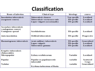
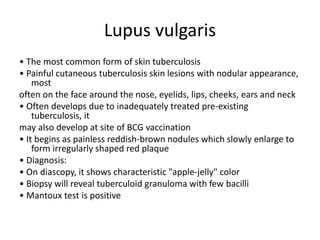
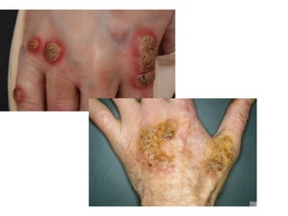
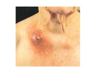
DEPARTMENT Of Dermatology, venereology
Name : Mohamed Mahmoud Abbass
Group : 7th
Course : 4th
Topic : CUTANEOUS TUBERCULOSIS
Introduction
• Cutaneous tuberculosis occurs rarely, despite a high and increasing
prevalence of tuberculosis worldwide. Mycobacterium tuberculosis,
Mycobacterrium bovis, and the Bacille Calmette-Guérin vaccine can cause
tuberculosis involving the skin
• Cutaneous tuberculosis can be acquired exogenously or endogenously and
present as a multitude of differing clinical morphologies
• Cutaneous TB- 1.5% of extrapulmonary TB
• Diagnosis of these lesions can be difficult, as they resemble many other
dermatological conditions that are often primarily considered.
• Commonest form :
in adults: Lupus Vulgaris
in childhood: Scrofuloderma and Lichen scrofulosorum
Immunopathology
• Skin biopsy specimens examined with monoclonal
antibodies
– intense expression of HLA-DR on keratinocytes
– moderate to high Langerhans cell hyperplasia
– infiltration by CD3+ pan-T cells as well as CD8+
and CD4+ T cells(CD4 > CD8)
• Predominant lymphocyte in dermal granulomas
– activated CD3+ T cell
– expressing MHC class II antigens & interleukin 2
receptor
Classification
Lupus vulgaris
• The most common form of skin tuberculosis
• Painful cutaneous tuberculosis skin lesions with nodular appearance,
most
often on the face around the nose, eyelids, lips, cheeks, ears and neck
• Often develops due to inadequately treated pre-existing
tuberculosis, it
may also develop at site of BCG vaccination
• It begins as painless reddish-brown nodules which slowly enlarge to
form irregularly shaped red plaque
• Diagnosis:
• On diascopy, it shows characteristic "apple-jelly" color
• Biopsy will reveal tuberculoid granuloma with few bacilli
• Mantoux test is positive
Verrucosa cutis (prosector’s wart)
• Rash of small, red papular nodules in the skin that may appear 2–4
weeks after inoculation by Mycobacterium tuberculosis in a previously
infected and immunocompetent individual.
• Entry point usually is the site of a trauma, wound or puncture in the
skin (eg- during an autopsy), the most frequent site for the wart are
the hands
• The diagnosis is confirmed by a skin biopsy and a positive culture
for acid-fast bacilli
• A Purified protein derivative (PPD) test may also result positive
Scrofuloderma
• Condition caused by tuberculous involvement
of the skin by direct extension, usually from
underlying tuberculous lymphadenitis.
• An asymptomatic reddish swelling which
breaks down to form sinuses, fistulate or
tuberculous ulcers
• Characteristic caseous material discharges
from lesions
• Most common sites – neck, chest
Miliary tuberculosis
• It is a rare haematogenous dissemination of tuberculosis
• Usually affects
– Young children
– Immunosuppressed patients
– Concurrent HIV infection
– Following viral infections
• Patient develops
– Crops of minute bluish papules, vesicles, pustules
– Erythematous nodules
– Haemorrhagic lesions
Tuberculid
• Hypersensitivity reaction to M. tuberculosis or its
products in patient with significant immunity
• Following criteria must be fulfilled to designate a
condition as tuberculid:
– Skin lesion must show tuberculoid histopathology
– Mycobacterium tuberculosis must not be
demonstrated in the lesion
– Tuberculin test must be strongly positive
– Treatment of underlying TB focus must lead to
resolution of skin lesion
Lichen scrofulosorum
• Second most common pattern of cutaneous TB in
children
• Rare tuberculid that presents as a lichenoid
eruption of
minute papules in children and adolescents with
tuberculosis
• The lesions are usually asymptomatic, closely
grouped, skin-colored to reddish-brown papules,
often perifollicular
• Mainly found on the abdomen, chest, back, and
proximal parts of the limbs
Diagnosis
• The diagnosis is usually on skin biopsy. Typical
tubercles are caseating epithelioid granulomas
that contain acid-fast bacilli. These are detected
by tissue staining, culture and polymerase chain
reaction
(PCR).
• Tuberculin skin test
• QFT-G
• X-ray
• Sputum culture
Treatment
• CTB treatment is the same as that for systemic TB
and consists of long, multidrug therapy
• The chemotherapeutic treatment of TB is divided
into two phases:
• an intensive or bactericidal phase, designed to
rapidly reduce the total body burden of
Mycobacterium tuberculosis
• a continuation or sterilizing phase
• The most commonly used drugs are isoniazid,
rifampin, pyrazinamide, and either ethambutol or
streptomycin
THANKS

Cutaneous tuberculosis ( SKIN TB )

  • 1.
    DEPARTMENT Of Dermatology,venereology Name : Mohamed Mahmoud Abbass Group : 7th Course : 4th Topic : CUTANEOUS TUBERCULOSIS
  • 2.
    Introduction • Cutaneous tuberculosisoccurs rarely, despite a high and increasing prevalence of tuberculosis worldwide. Mycobacterium tuberculosis, Mycobacterrium bovis, and the Bacille Calmette-Guérin vaccine can cause tuberculosis involving the skin • Cutaneous tuberculosis can be acquired exogenously or endogenously and present as a multitude of differing clinical morphologies • Cutaneous TB- 1.5% of extrapulmonary TB • Diagnosis of these lesions can be difficult, as they resemble many other dermatological conditions that are often primarily considered. • Commonest form : in adults: Lupus Vulgaris in childhood: Scrofuloderma and Lichen scrofulosorum
  • 3.
    Immunopathology • Skin biopsyspecimens examined with monoclonal antibodies – intense expression of HLA-DR on keratinocytes – moderate to high Langerhans cell hyperplasia – infiltration by CD3+ pan-T cells as well as CD8+ and CD4+ T cells(CD4 > CD8) • Predominant lymphocyte in dermal granulomas – activated CD3+ T cell – expressing MHC class II antigens & interleukin 2 receptor
  • 4.
  • 5.
    Lupus vulgaris • Themost common form of skin tuberculosis • Painful cutaneous tuberculosis skin lesions with nodular appearance, most often on the face around the nose, eyelids, lips, cheeks, ears and neck • Often develops due to inadequately treated pre-existing tuberculosis, it may also develop at site of BCG vaccination • It begins as painless reddish-brown nodules which slowly enlarge to form irregularly shaped red plaque • Diagnosis: • On diascopy, it shows characteristic "apple-jelly" color • Biopsy will reveal tuberculoid granuloma with few bacilli • Mantoux test is positive
  • 7.
    Verrucosa cutis (prosector’swart) • Rash of small, red papular nodules in the skin that may appear 2–4 weeks after inoculation by Mycobacterium tuberculosis in a previously infected and immunocompetent individual. • Entry point usually is the site of a trauma, wound or puncture in the skin (eg- during an autopsy), the most frequent site for the wart are the hands • The diagnosis is confirmed by a skin biopsy and a positive culture for acid-fast bacilli • A Purified protein derivative (PPD) test may also result positive
  • 9.
    Scrofuloderma • Condition causedby tuberculous involvement of the skin by direct extension, usually from underlying tuberculous lymphadenitis. • An asymptomatic reddish swelling which breaks down to form sinuses, fistulate or tuberculous ulcers • Characteristic caseous material discharges from lesions • Most common sites – neck, chest
  • 11.
    Miliary tuberculosis • Itis a rare haematogenous dissemination of tuberculosis • Usually affects – Young children – Immunosuppressed patients – Concurrent HIV infection – Following viral infections • Patient develops – Crops of minute bluish papules, vesicles, pustules – Erythematous nodules – Haemorrhagic lesions
  • 13.
    Tuberculid • Hypersensitivity reactionto M. tuberculosis or its products in patient with significant immunity • Following criteria must be fulfilled to designate a condition as tuberculid: – Skin lesion must show tuberculoid histopathology – Mycobacterium tuberculosis must not be demonstrated in the lesion – Tuberculin test must be strongly positive – Treatment of underlying TB focus must lead to resolution of skin lesion
  • 15.
    Lichen scrofulosorum • Secondmost common pattern of cutaneous TB in children • Rare tuberculid that presents as a lichenoid eruption of minute papules in children and adolescents with tuberculosis • The lesions are usually asymptomatic, closely grouped, skin-colored to reddish-brown papules, often perifollicular • Mainly found on the abdomen, chest, back, and proximal parts of the limbs
  • 17.
    Diagnosis • The diagnosisis usually on skin biopsy. Typical tubercles are caseating epithelioid granulomas that contain acid-fast bacilli. These are detected by tissue staining, culture and polymerase chain reaction (PCR). • Tuberculin skin test • QFT-G • X-ray • Sputum culture
  • 19.
    Treatment • CTB treatmentis the same as that for systemic TB and consists of long, multidrug therapy • The chemotherapeutic treatment of TB is divided into two phases: • an intensive or bactericidal phase, designed to rapidly reduce the total body burden of Mycobacterium tuberculosis • a continuation or sterilizing phase • The most commonly used drugs are isoniazid, rifampin, pyrazinamide, and either ethambutol or streptomycin
  • 22.