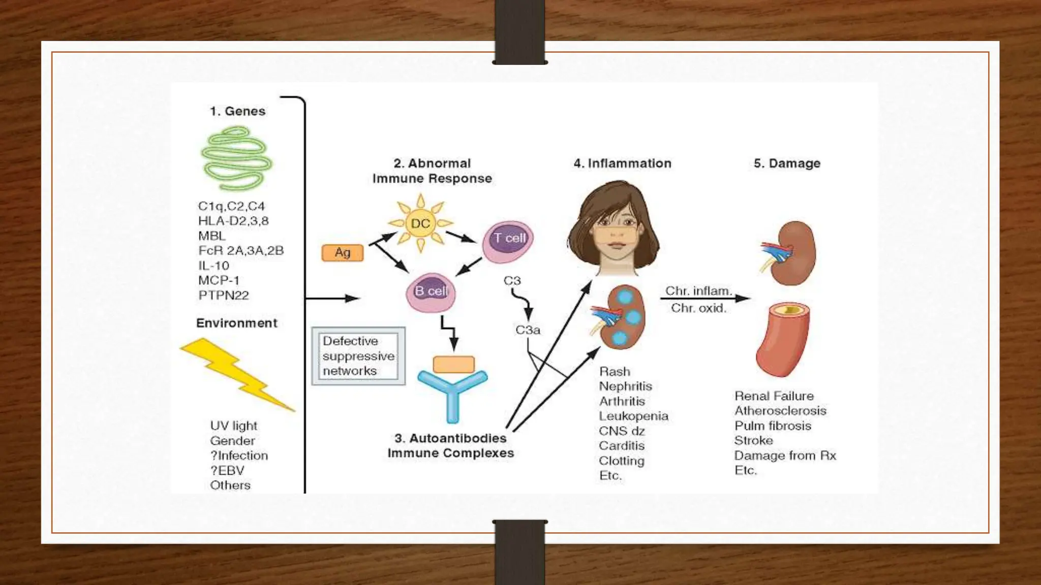
Systemic Lupus Erythematosus
Dr Ganesh G
DNB PG Resident
Department of General Medicine
District Hospital Chikkaballapur
Definition
• It is an autoimmune disease in
which organs & cells undergo
damage mediated by tissue
binding autoantibodies and
immune complexes
Epidemiology
• In the world, around 5 million people are affected with the SLE.
• Prevelance is higher in African-American , afro-carrebean women than
white men
• In India, 3 people per 1,00,000 population are affected with this
problem
• Usually occurs in young women b/w 20 to 30yrs
Risk factors
• Family history of SLE
• Smoking
• Infections like Epstein Barr Virus
• Persons who are using Immunosupressants and Biological drugs
• Stress
• Women
Etiology
• Epigenetic regulation of HLA-D2, D3, and D8 gene
• Environmental factors
• Hormones- Estrogen and Progesterone
• Abnormalities in immune cells and cytokines
Pathogenesis
Patients with risk factors
Exposed to triggering factors
Abnormal expression of genes responsible for immunity
• Activation of T cells and B cells
• Formation of inflammatory mediators like Interleukins, Cytokines,
Cytotoxins and TNF-α
Formation of auto antibodies
Antibody and antigen complex formation
Accumulation of above complex in multiple organs
Damage of multiple organs
• Autoantibody production – B cells of immune system produce antinuclear
antibodies that target the body’s own cells & tissues
• Immune complex formation – consists of autoantibodies bound to target
antigens , they circulate in blood and deposit in various tissues
• Complement avtivation – immune complexes formed in SLE activate
complement system with the release of C3a, C5a and formation of
Membrane attack complec which damage the cells directly
• Defective clearance of apoptotic cells –in SLE apoptotic cells may not be
cleared efficiently, the release of self antigens from dying cells can trigger
autoimmune response & contribute to production of autoantibodies
Pathophysiology
Pathophysiology continued…
• Inflammation and tissue damage – immune complexex, complement activation leads
to chronic inflammation with the release of mediators like cytokines, chemokines
contribute to tissue damage.
• Abnormal T-cell function – in SLE there is imbalance in T-cell subsets, increase in
autoreactive CD4+ T cells. It can recognize self antigens presented by antigen
presenting cells and provide help to B cells to produce autoantibodies. They also cause
tissue inflammation by release of IL-17
• Type 1 interferon (IFN) production – type 1 IFN-α play a crucial role in immune
response to viral infections. In SLE there is activation of plasmacytoid dendritic cells
that produce large amount of IFN-α in response to immune complex stimulation
which leads to further stimulation of immune system, enhance presentation of self
antigens.
Clinical Presentation
• Systemic – fatigue, malaise, fever,anorexia, nausea, weight loss
• Musculoskeletal- arthralgis, non-erosive polyarthritis, Jaccoud’s arthropathy
• Mucocutaneous – Malar rash, discoid rash, alopecia, Raynaud phenomenon, splinter hemorrhages
• Hematological – anemia, leukopenia, lymphopenia, thrombocytopenia,splenomegaly, hemolytic
anemia
• Neuropsychiatry – delirium, psychosis,seizures, polyneuropathy, mood disorders, stroke
(thromboembolic)
• Cardiopulmonary – pericarditis, myocarditis, Libman-Sacks endocarditis, congenital heart block
(neonatal lupus), shrinking lung syndrome
• Renal – proteinuria, nephrotic syndrome, renal failure
• Others- cutaneous vasculitis, antiphospholipid antibody syndrome, sicca syndrome
Renal manifestations in SLE
ISN/RPS Classification of LUPUS NEPHRITIS
Class 1 – minimal mesangial lupus nephritis
Class 2 – Mesangial proliferative LN
Class 3 – Focal Lupus nephritis
Class 4 – Diffuse lupus nephritis
Class 5 – Membranous lupus nephritis
Class 6 – Advanced sclerosing lupus nephritis
Complications
• Arthritis
• Pancytopenia
• Hypertension
• Stroke
• Pericarditis/ Myocarditis
• Lupus nephritis
• Anxiety
• Mood disorders
• Depression
• Neuropathy
• Pleuritis
• Vasculitis
Diagnosis
• Medical history
• Clinical presentation
• Complete blood count
• Lab tests based on complications
• Serology test for
• Auto antibodies (ANA)
• Antiphospholipid antibodies
• Complement proteins
Diagnosis Continued…..
• C Reactive protein
• Erythrocyte sedimentation rate
• Urine microscopy
• Chest x ray
• CT/MRI of brain/ lung/ chest- depending on effected organ
• ECG
• Renal biopsy
• Lumbar puncture
Diagnosis
SLE is characterized by production of autoantibodies , some are antinuclear, anticytoplasmic, some are specific
against surface antigens of of blood cells.
Autoantibodies in patients with SLE
• Antinuclear antibodies (ANA)
• Anti-double strand DNA
• Anti-Smith
• Anti Ribonuclear protein
• Anti-Ro
• Anti-La
• Anti Histone
• Antiphospholipid
• Antierythrocyte
• Antiplatelet
• Antineuronal
• Antiribosomal
• ANA is the best screening test, more than 90% patients show positive. However it is not specific
for SLE
• Anti – dsDNA and Anti Sm are relatively specific for SLE
• Rising levels of anti – ds DNA and low levels of complement C3 and C4 usually reflect disease
activity.
• ESR is raised in some patients
• CRP unlike ESR does not rise with disease activity unless there is arthritis or serositis.
• APTT is prolonged in patients with lupus anticoagulant (APLA)
• With active nephritis urine analysis show proteinuria, hematuria & cellular or granular casts.
Renal biopsy confirms renal involvement.
• Low levels of C3 & C4 indicate active disease especially nephritis.
• LE cell – phagocytic leukocyte (neutrophil or macrophage) that has engulfed the denatured
nucleus of an injured cell
American college of Rheumatology criteria for Systemic Lupus Erythematous
Entry criterion -
ANA at a titre of ≥ 80 on HEp – 2 lines or an equivalent positive test
Additive criteria
(A total score of 10 or more is considered to classify as SLE with entry criterion is a must)
Immunological domain & criteria weight
APLA - Anticardiolipin or
Anti β2 glycoprotein 1 or
Lupus anticoagulant
2
Complement proteins – Low C3 or C4 3
Low C3 & C4 4
SLE specific antibodies – Anti ds DNA antibody or 6
Anti Smith antibody
Clinical domain & criteria weight
Fever 2
Hematological - Leukopenia 3
Thrombocytopenia 4
Autoimmune hemolyses 4
Neuropsychiatry – delirium 2
Psychosis 3
Seizure 5
Mucucutaneous – non sparing alopacia 2
Oral ulcers 2
Subacute cutaneous or discoid lupus 4
Acute cutaneous lupus 6
Serosal – pleural or pericardial effusion 5
Acute pericarditis 6
Musculoskeletal – joint involvement 6
Renal – proteinuria > 0.5g/24h 4
Renal biopsy class II or V lupus nephritis 8
Renal biopsy class III or IV lupus nephritis 10
Note : additional criteria within
same domain will not be counted
Classify as SLE with a score of 10
or more (from both clinical &
immunological criteria)
along with Entry Criterion ANA
rising titre is a must
Non Pharmacological Treatment
• Protection from sun
• Exercise
• Smoking cessation
• Counselling
• Avoid exposure to live vaccines
Treatment of SLE
Initial management of SLE –
Sunscreen, SBE prophylaxis
Arthritis – NSAIDs, HCQ, if persistant add Methotrexate
Mucocutaneous symptoms , fatigue, alopecia – HCQ
Fever – indomethacin, if persistant ad Glucocorticoids
CNS symptoms – high dose steroids or IV cyclophosphamide
Renal involvement biopsy confirmed :
1. Class II – trial of moderate Glucocorticoids
2. Class III or IV – high dose Glucocorticoids , IV Cyclophosphamide, Mycophenolate mofetil,
azathioprine
3. Class V – trial of Glucocorticoids, MMF, cyclosporine
4. Class VI – close monitoring, dialysis, Renal transplant
Alveolitis – systemic GC + MMF or IV cyclophosphamide , then add rituximab or IV Ig, maintain with
azathioprine or mycophenolate
Antiphospholipid syndrome – anticoagulation, with or without HCQ, then add direct thrombin inhibitor.
Uncomplicated digital or cutaneous vasculitis – systemic GC, with or without HCQ, with or without
Methotrexate, then add Azathioprine or MMF then switch to IV cyclophosphamide.
 Immunosuppressive agents – azathioprine, methotrexate, cyclophosphamide,
mycophenolate mofetil are used in controlling severe disease. They are particularly useful
in patients with renal involvement..
 A combination of IV cyclophosphamide and steroids is most effective.
 Following IV cyclophosphamide, oral MMF is an alternative to maintain remission.
 Drugs targeting B-cell pathways such as Belimumab & rituximab are used in refractory
cases.
 New update : Antifrolumab, a human monoclonal antibody against IFN receptors
subunit1 was approved by US FDA for treatment of moderate to severe disease.
Drugs used in treatment of SLE
Drug Category Mode of action Dose Adverse effects
Prednisone Corticosteroids Control inflammation by
stabilizing lysosomes at
cellular level
0.1-1.5mg/kg/day-PO  Hypertension
 Diabetes
 Poor wound healing
 Osteoporosis
 Weight gain
Methyl Prednisolone Corticosteroids Control inflammation by
stabilizing lysosomes at
cellular level
100-1000mg-TID  Hypertension
 Diabetes
 Poor wound healing
 Osteoporosis
 Weight gain
Hydroxychloroquine Anti malarial drug Impairs complement
dependent antigen and
antibody reactions
200-400mg-PO-OD  Muscle weakness
 Leukopenia
 Thrombocytopenia
 Retinal damage
 Alopecia
Drugs used in treatment of SLE Continued..
Belimumab Biological agents Inhibit production of
antibodies from plasma cells
10mg/kg- IV-3 doses in 2 weeks
10mg/kg- IV-once in every 4 weeks
 Infusion reactions
 Diarrhea
 Pyrexia
 Nasopharyngitis
 Bronchitis
Cyclophosphomide Immunomodulator Produces irreversible T cell
apoptosis
500-1000mg-IV-once in every 6 months  Alopecia
 GI toxicity
 Leukopenia
 Amenorrhea
 Sterility
Mycophenolate
Mofetil
Immunosuppressant Inhibit T and B cell
production
0.5-3g/day-PO  Hyperglycaemia
 Hypercholesterolemia
 Hypomagnesaemia
 Dyspnoea
 Back pain
Azathioprine Immunomodulator Inhibit T and B cell
production
1.5-2mg/kg/day-PO  Leukopenia
 Infections
 Lymphoma
 Abdominal pain
 Alopecia
Drugs used in treatment of SLE Continued..
Methotrexate Immunomodulator Inhibit T cell replication 15-25mg-PO-Once
weekly
 Arachnoiditis
 Erythema
 Hyperurecemia
 Stomatitis
 Glossitis
Rituximab Monoclonal antibody Induces B cell lysis 500-1000mg- IV  Angioedema
 Pruritis
 Abdominal pain
 Leukopenia
 Infections
Aspirin NSAID Inhibit prostaglandin
synthesis
2.1-7.3g/day-OD-PO  Angioedema
 Bronchospasm
 CNS alteration
 Alopecia
 GI bleeding
Systemic lupus erythematosus, an autoimmune disorder

Systemic lupus erythematosus, an autoimmune disorder

  • 1.
    Systemic Lupus Erythematosus DrGanesh G DNB PG Resident Department of General Medicine District Hospital Chikkaballapur
  • 2.
    Definition • It isan autoimmune disease in which organs & cells undergo damage mediated by tissue binding autoantibodies and immune complexes
  • 3.
    Epidemiology • In theworld, around 5 million people are affected with the SLE. • Prevelance is higher in African-American , afro-carrebean women than white men • In India, 3 people per 1,00,000 population are affected with this problem • Usually occurs in young women b/w 20 to 30yrs
  • 4.
    Risk factors • Familyhistory of SLE • Smoking • Infections like Epstein Barr Virus • Persons who are using Immunosupressants and Biological drugs • Stress • Women
  • 5.
    Etiology • Epigenetic regulationof HLA-D2, D3, and D8 gene • Environmental factors • Hormones- Estrogen and Progesterone • Abnormalities in immune cells and cytokines
  • 6.
    Pathogenesis Patients with riskfactors Exposed to triggering factors Abnormal expression of genes responsible for immunity • Activation of T cells and B cells • Formation of inflammatory mediators like Interleukins, Cytokines, Cytotoxins and TNF-α Formation of auto antibodies Antibody and antigen complex formation Accumulation of above complex in multiple organs Damage of multiple organs
  • 7.
    • Autoantibody production– B cells of immune system produce antinuclear antibodies that target the body’s own cells & tissues • Immune complex formation – consists of autoantibodies bound to target antigens , they circulate in blood and deposit in various tissues • Complement avtivation – immune complexes formed in SLE activate complement system with the release of C3a, C5a and formation of Membrane attack complec which damage the cells directly • Defective clearance of apoptotic cells –in SLE apoptotic cells may not be cleared efficiently, the release of self antigens from dying cells can trigger autoimmune response & contribute to production of autoantibodies Pathophysiology
  • 8.
    Pathophysiology continued… • Inflammationand tissue damage – immune complexex, complement activation leads to chronic inflammation with the release of mediators like cytokines, chemokines contribute to tissue damage. • Abnormal T-cell function – in SLE there is imbalance in T-cell subsets, increase in autoreactive CD4+ T cells. It can recognize self antigens presented by antigen presenting cells and provide help to B cells to produce autoantibodies. They also cause tissue inflammation by release of IL-17 • Type 1 interferon (IFN) production – type 1 IFN-α play a crucial role in immune response to viral infections. In SLE there is activation of plasmacytoid dendritic cells that produce large amount of IFN-α in response to immune complex stimulation which leads to further stimulation of immune system, enhance presentation of self antigens.
  • 10.
    Clinical Presentation • Systemic– fatigue, malaise, fever,anorexia, nausea, weight loss • Musculoskeletal- arthralgis, non-erosive polyarthritis, Jaccoud’s arthropathy • Mucocutaneous – Malar rash, discoid rash, alopecia, Raynaud phenomenon, splinter hemorrhages • Hematological – anemia, leukopenia, lymphopenia, thrombocytopenia,splenomegaly, hemolytic anemia • Neuropsychiatry – delirium, psychosis,seizures, polyneuropathy, mood disorders, stroke (thromboembolic) • Cardiopulmonary – pericarditis, myocarditis, Libman-Sacks endocarditis, congenital heart block (neonatal lupus), shrinking lung syndrome • Renal – proteinuria, nephrotic syndrome, renal failure • Others- cutaneous vasculitis, antiphospholipid antibody syndrome, sicca syndrome
  • 12.
    Renal manifestations inSLE ISN/RPS Classification of LUPUS NEPHRITIS Class 1 – minimal mesangial lupus nephritis Class 2 – Mesangial proliferative LN Class 3 – Focal Lupus nephritis Class 4 – Diffuse lupus nephritis Class 5 – Membranous lupus nephritis Class 6 – Advanced sclerosing lupus nephritis
  • 13.
    Complications • Arthritis • Pancytopenia •Hypertension • Stroke • Pericarditis/ Myocarditis • Lupus nephritis • Anxiety • Mood disorders • Depression • Neuropathy • Pleuritis • Vasculitis
  • 15.
    Diagnosis • Medical history •Clinical presentation • Complete blood count • Lab tests based on complications • Serology test for • Auto antibodies (ANA) • Antiphospholipid antibodies • Complement proteins
  • 16.
    Diagnosis Continued….. • CReactive protein • Erythrocyte sedimentation rate • Urine microscopy • Chest x ray • CT/MRI of brain/ lung/ chest- depending on effected organ • ECG • Renal biopsy • Lumbar puncture
  • 17.
    Diagnosis SLE is characterizedby production of autoantibodies , some are antinuclear, anticytoplasmic, some are specific against surface antigens of of blood cells. Autoantibodies in patients with SLE • Antinuclear antibodies (ANA) • Anti-double strand DNA • Anti-Smith • Anti Ribonuclear protein • Anti-Ro • Anti-La • Anti Histone • Antiphospholipid • Antierythrocyte • Antiplatelet • Antineuronal • Antiribosomal
  • 18.
    • ANA isthe best screening test, more than 90% patients show positive. However it is not specific for SLE • Anti – dsDNA and Anti Sm are relatively specific for SLE • Rising levels of anti – ds DNA and low levels of complement C3 and C4 usually reflect disease activity. • ESR is raised in some patients • CRP unlike ESR does not rise with disease activity unless there is arthritis or serositis. • APTT is prolonged in patients with lupus anticoagulant (APLA) • With active nephritis urine analysis show proteinuria, hematuria & cellular or granular casts. Renal biopsy confirms renal involvement. • Low levels of C3 & C4 indicate active disease especially nephritis. • LE cell – phagocytic leukocyte (neutrophil or macrophage) that has engulfed the denatured nucleus of an injured cell
  • 19.
    American college ofRheumatology criteria for Systemic Lupus Erythematous Entry criterion - ANA at a titre of ≥ 80 on HEp – 2 lines or an equivalent positive test Additive criteria (A total score of 10 or more is considered to classify as SLE with entry criterion is a must) Immunological domain & criteria weight APLA - Anticardiolipin or Anti β2 glycoprotein 1 or Lupus anticoagulant 2 Complement proteins – Low C3 or C4 3 Low C3 & C4 4 SLE specific antibodies – Anti ds DNA antibody or 6 Anti Smith antibody
  • 20.
    Clinical domain &criteria weight Fever 2 Hematological - Leukopenia 3 Thrombocytopenia 4 Autoimmune hemolyses 4 Neuropsychiatry – delirium 2 Psychosis 3 Seizure 5 Mucucutaneous – non sparing alopacia 2 Oral ulcers 2 Subacute cutaneous or discoid lupus 4 Acute cutaneous lupus 6 Serosal – pleural or pericardial effusion 5 Acute pericarditis 6 Musculoskeletal – joint involvement 6 Renal – proteinuria > 0.5g/24h 4 Renal biopsy class II or V lupus nephritis 8 Renal biopsy class III or IV lupus nephritis 10 Note : additional criteria within same domain will not be counted Classify as SLE with a score of 10 or more (from both clinical & immunological criteria) along with Entry Criterion ANA rising titre is a must
  • 22.
    Non Pharmacological Treatment •Protection from sun • Exercise • Smoking cessation • Counselling • Avoid exposure to live vaccines
  • 23.
    Treatment of SLE Initialmanagement of SLE – Sunscreen, SBE prophylaxis Arthritis – NSAIDs, HCQ, if persistant add Methotrexate Mucocutaneous symptoms , fatigue, alopecia – HCQ Fever – indomethacin, if persistant ad Glucocorticoids CNS symptoms – high dose steroids or IV cyclophosphamide Renal involvement biopsy confirmed : 1. Class II – trial of moderate Glucocorticoids 2. Class III or IV – high dose Glucocorticoids , IV Cyclophosphamide, Mycophenolate mofetil, azathioprine 3. Class V – trial of Glucocorticoids, MMF, cyclosporine 4. Class VI – close monitoring, dialysis, Renal transplant Alveolitis – systemic GC + MMF or IV cyclophosphamide , then add rituximab or IV Ig, maintain with azathioprine or mycophenolate Antiphospholipid syndrome – anticoagulation, with or without HCQ, then add direct thrombin inhibitor. Uncomplicated digital or cutaneous vasculitis – systemic GC, with or without HCQ, with or without Methotrexate, then add Azathioprine or MMF then switch to IV cyclophosphamide.
  • 24.
     Immunosuppressive agents– azathioprine, methotrexate, cyclophosphamide, mycophenolate mofetil are used in controlling severe disease. They are particularly useful in patients with renal involvement..  A combination of IV cyclophosphamide and steroids is most effective.  Following IV cyclophosphamide, oral MMF is an alternative to maintain remission.  Drugs targeting B-cell pathways such as Belimumab & rituximab are used in refractory cases.  New update : Antifrolumab, a human monoclonal antibody against IFN receptors subunit1 was approved by US FDA for treatment of moderate to severe disease.
  • 25.
    Drugs used intreatment of SLE Drug Category Mode of action Dose Adverse effects Prednisone Corticosteroids Control inflammation by stabilizing lysosomes at cellular level 0.1-1.5mg/kg/day-PO  Hypertension  Diabetes  Poor wound healing  Osteoporosis  Weight gain Methyl Prednisolone Corticosteroids Control inflammation by stabilizing lysosomes at cellular level 100-1000mg-TID  Hypertension  Diabetes  Poor wound healing  Osteoporosis  Weight gain Hydroxychloroquine Anti malarial drug Impairs complement dependent antigen and antibody reactions 200-400mg-PO-OD  Muscle weakness  Leukopenia  Thrombocytopenia  Retinal damage  Alopecia
  • 26.
    Drugs used intreatment of SLE Continued.. Belimumab Biological agents Inhibit production of antibodies from plasma cells 10mg/kg- IV-3 doses in 2 weeks 10mg/kg- IV-once in every 4 weeks  Infusion reactions  Diarrhea  Pyrexia  Nasopharyngitis  Bronchitis Cyclophosphomide Immunomodulator Produces irreversible T cell apoptosis 500-1000mg-IV-once in every 6 months  Alopecia  GI toxicity  Leukopenia  Amenorrhea  Sterility Mycophenolate Mofetil Immunosuppressant Inhibit T and B cell production 0.5-3g/day-PO  Hyperglycaemia  Hypercholesterolemia  Hypomagnesaemia  Dyspnoea  Back pain Azathioprine Immunomodulator Inhibit T and B cell production 1.5-2mg/kg/day-PO  Leukopenia  Infections  Lymphoma  Abdominal pain  Alopecia
  • 27.
    Drugs used intreatment of SLE Continued.. Methotrexate Immunomodulator Inhibit T cell replication 15-25mg-PO-Once weekly  Arachnoiditis  Erythema  Hyperurecemia  Stomatitis  Glossitis Rituximab Monoclonal antibody Induces B cell lysis 500-1000mg- IV  Angioedema  Pruritis  Abdominal pain  Leukopenia  Infections Aspirin NSAID Inhibit prostaglandin synthesis 2.1-7.3g/day-OD-PO  Angioedema  Bronchospasm  CNS alteration  Alopecia  GI bleeding