Secondary Structure Prediction of proteins

Secondary structure prediction has been around for almost a quarter of a century. The early methods suffered from a lack of data. Predictions were performed on single sequences rather than families of homologous sequences, and there were relatively few known 3D structures from which to derive parameters. Probably the most famous early methods are those of Chou & Fasman, Garnier, Osguthorbe & Robson (GOR) and Lim. Although the authors originally claimed quite high accuracies (70-80 %), under careful examination, the methods were shown to be only between 56 and 60% accurate (see Kabsch & Sander, 1984 given below). An early problem in secondary structure prediction had been the inclusion of structures used to derive parameters in the set of structures used to assess the accuracy of the method. Some good references on the subject:

Recommended
More Related Content
What's hot
What's hot (20)
Viewers also liked
Viewers also liked (15)
Similar to Secondary Structure Prediction of proteins
Similar to Secondary Structure Prediction of proteins (20)
More from Vijay Hemmadi
More from Vijay Hemmadi (20)
Recently uploaded
Recently uploaded (20)
Secondary Structure Prediction of proteins
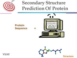
- 1. Secondary Structure Prediction Of Protein Protein Sequence + Structure VIJAY
- 2. INRODUCTION Primary structure (Amino acid sequence) ↓ Secondary structure (α-helix, β-sheet) ↓ Tertiary structure (Three-dimensional structure formed by assembly of secondary structures) ↓ Quaternary structure (Structure formed by more than one polypeptide chains)
- 3. Secondary Structure Defined as the local conformation of protein backbone Primary Structure —folding— Secondary Structure a helix and b sheet Secondary Structure Regular Secondary Structure (a-helices, b-sheets) Irregular Secondary Structure (Tight turns, Random coils, bulges)
- 5. a helix •common confirmation. •spiral structure •Tightly packed coiled polypeptide backbone, with extending side chains •Spontaneous •stabilized by H-bonding between amide hydrogens and carbonyl oxygens of peptide bonds. •R-groups lie on the exterior of the helix and perpendicular to its axis. •complete turn of helix —3.6 aminoacyl residues with distance 0.54 nm e.g. the keratins- entirely α-helical Myoglobin- 80% helical
- 6. •Glycine and Proline , bulky amino acids, charged amino acids favor disruption of the helix.
- 7. b sheet •β-sheets are composed of 2 or more different regions of stretches of at least 5-10 amino acids. •The folding and alignment of stretches of the polypeptide backbone aside one another to form β-sheets is stabilized by H-bonding between amide hydrogens and carbonyl oxygens •the peptide backbone of the β sheet is highly extended. •R groups of adjacent residues point in opposite directions. • β-sheets are either parallel or antiparallel
- 10. What is secondary structure prediction? Given a protein sequence (primary structure) 1st step in prediction of protein structure. Technique concerned with determination of secondary structure of given polypeptide by locating the Coils Alpha Helix Beta Strands in plypeptide GHWIATRGQLIREAYEDYRHFSSECPFIP Predict its secondary structure content (C=Coils H=Alpha Helix E=Beta Strands) CEEEEECHHHHHHHHHHHCCCHHCCCCCC
- 11. Why secondary structure prediction? o secondary structure —tertiary structure prediction o Protein function prediction o Protein classification o Predicting structural change o detection and alignment of remote homology between proteins o on detecting transmembrane regions, solvent-accessibleresidues, and other important features of molecules o Detection of hydrophobic region and hydrophilic region
- 12. Prediction methods o Statistical method o Chou-Fasman method, GOR I-IV o Nearest neighbors o NNSSP, SSPAL o Neural network o PHD, Psi-Pred, J-Pred o Support vector machine (SVM) o HMM
- 13. Chou-Fasman algorithm Chou and fasman in 1978 It is based on assigning a set of prediction value to amino acid residue in polypeptide and applying an algorithm to the conformational parameter and positional frequency. conformational parameter for each amino acid is calculated by considering the relative frequency of each 20 amino acid in proteins By this C=Coils H=Alpha Helix E=Beta Strands are determined Also called preference parameter
- 14. • A table of prediction value or preference parameter for each of 20 amino acid in alpha helix ,beta plate and turn already calculated and standardised. • To obtain the prediction value the frequency of amino acids( i) in structure is divided by of all residences in protein (s) • i/s • The resulting structural parameter of p(alpha),p(beta),p(turn)vary —0.5 to 1.5 for 20 amino acid
- 16. Window is scanned to find a short sequence of amino acid that has high probability to form one type of structure When 4 out of 6 amino acid have high probability >1.03 the – alpha helix 3 out of 5 amino acid with probability >1.03-beta RULES
- 17. ALGORITHM o Note preference parameter for 20 aa in peptide o Scan the window and identify the region where 4 out of 6 contiguous residue have p(alpha helix) >1.00 o Continue scanning in both the direction until the 4 contiguous residue that have an average p(alpha helix)<1.00,end of helix o If segment is longer than 5aa and p(alpha helix)>p(beta sheet )-segment –completely alpha helix o scan different segment and identify - alpha helix
- 18. Identify the region where 3 out of 5 aa have the value of p( beta sheet) >1.00 ,region is predicted as beta sheet Continue scanning both the direction until 4 residue that have p( beta sheet) <1.00 End of beta sheet average p( beta sheet) >105 and p( beta sheet) >p(alpha helix) than consider complete segment as b pleated sheet
- 19. If any region is over lapping than consider it as alpha helix if average p(alpha helix)>p(beta sheet ) Or beta sheet if p(alpha helix)<p(beta sheet ) To identify turn P(t)=f(j)f(j+1)f(j+2)f(j+3) J=residual number
- 20. result Accuracy: ~50% ~60% helix alanine,glutamine,leucine,methionine Helix breaking proline and glycine Beta sheet isoleucine,valine,tyrosine Beta breaking proline,aspargine,glutamine Turn contains proline(30%),serine(14%),lysine, aspargine(10%) Glycine(19%),aspartic acid (`18%),serine(13%),tyrosine(11%) http://www.accelrys.com/product/gcg-wisconsin- package/program-list.html
- 21. Out put of Chou-Fasman
- 23. GOR METHOD • GOR(Garnier,Osguthorpe,Robson)1978 • Chou fasman method is based on assumption that each amino acid individually influence the 2ry structure of sequence • GOR is based on, amino acid flanking the central amino acid will influence the 2ry structure • Consider a peptide central amino acid side amino acid • It assume that amino acid up to 8 residue on sides will influence the 2ry structure of central residue • 4th version • 64% accurate
- 24. ALGORITHUM •It uses the sliding window of 17 amino acid •The side amino acid sequence and alignment is determined to predict secondary structure of central sequence •Good for helix than sheet because beta sheet has more inter sequence hydrogen bonding •36.5% accurate for beta sheet •input any amino acid sequence •Output tells about secondary structure
- 26. NEAREST NEIGHBOUR METHOD o Based on ,short homologues sequences of amino acids have the same secondary structure o It predicts secondary structure of central homologues segment by neighbour homologues sequences o By using structural database find some secondary structure of sequence which may be homologues to our target sequence o Naturally evolved proteins with 35% identical amino acid sequence will have same secondary structure o Find some sequence which may match with target sequence o Scoring matrix,MSA
- 27. “Singleton” score matrix Helix Sheet Loop Buried Inter Exposed Buried Inter Exposed Buried Inter Exposed ALA -0.578 -0.119 -0.160 0.010 0.583 0.921 0.023 0.218 0.368 ARG 0.997 -0.507 -0.488 1.267 -0.345 -0.580 0.930 -0.005 -0.032 ASN 0.819 0.090 -0.007 0.844 0.221 0.046 0.030 -0.322 -0.487 ASP 1.050 0.172 -0.426 1.145 0.322 0.061 0.308 -0.224 -0.541 CYS -0.360 0.333 1.831 -0.671 0.003 1.216 -0.690 -0.225 1.216 GLN 1.047 -0.294 -0.939 1.452 0.139 -0.555 1.326 0.486 -0.244 GLU 0.670 -0.313 -0.721 0.999 0.031 -0.494 0.845 0.248 -0.144 GLY 0.414 0.932 0.969 0.177 0.565 0.989 -0.562 -0.299 -0.601 HIS 0.479 -0.223 0.136 0.306 -0.343 -0.014 0.019 -0.285 0.051 ILE -0.551 0.087 1.248 -0.875 -0.182 0.500 -0.166 0.384 1.336 LEU -0.744 -0.218 0.940 -0.411 0.179 0.900 -0.205 0.169 1.217 LYS 1.863 -0.045 -0.865 2.109 -0.017 -0.901 1.925 0.474 -0.498 MET -0.641 -0.183 0.779 -0.269 0.197 0.658 -0.228 0.113 0.714 PHE -0.491 0.057 1.364 -0.649 -0.200 0.776 -0.375 -0.001 1.251 PRO 1.090 0.705 0.236 1.249 0.695 0.145 -0.412 -0.491 -0.641 SER 0.350 0.260 -0.020 0.303 0.058 -0.075 -0.173 -0.210 -0.228 THR 0.291 0.215 0.304 0.156 -0.382 -0.584 -0.012 -0.103 -0.125 TRP -0.379 -0.363 1.178 -0.270 -0.477 0.682 -0.220 -0.099 1.267 TYR -0.111 -0.292 0.942 -0.267 -0.691 0.292 -0.015 -0.176 0.946 VAL -0.374 0.236 1.144 -0.912 -0.334 0.089 -0.030 0.309 0.998
- 29. Neural Network Method •Prediction is done by utilizing the information of different DATABASE •Linear sequence 3D structure of Polypeptide
- 30. Neural network Input signals are summed and turned into zero or one 3. J1 J2 J3 J4 Feed-forward multilayer network Input layer Hidden layer Output layer neurons
- 31. Enter sequences Compare Prediction to Reality AdjustWeights Neural network training
- 32. Simple Neural Network With Hidden Layer outi f ij 2 J f jk 1 Jk kin j Simple neural network with hidden layer
- 33. A C D E F G H I K L M N P Q R S T V W Y . H E L D (L) R (E) Q (E) G (E) F (E) V (E) P (E) A (H) A (H) Y (H) V (E) K (E) K (E) Neural network for secondary structure
- 34. Summary Introduction What is secondary structure prediction Why Chou-Fasman method GOR I-IV Nearest neighbors Neural network
- 37. Suggested reading: Chapter 15 in “Current Topics in Computational Molecular Biology, edited by Tao Jiang, Ying Xu, and Michael Zhang. MIT Press. 2002.” Bioinformatics by Cynthia and per jambeck Bioinformatics by S.C.RASTOGI Bioinformatics By Andreas Optional reading: Review by Burkhard Rost: http://cubic.bioc.columbia.edu/papers/2003_r ev_dekker/paper.html Reference