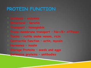
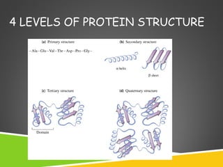
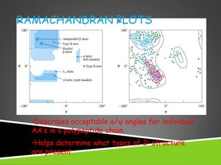
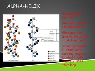
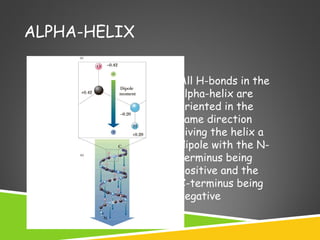
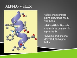
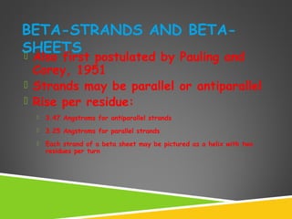
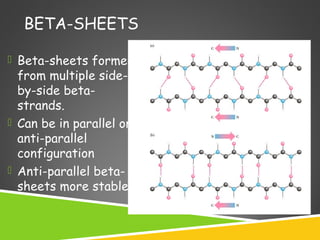
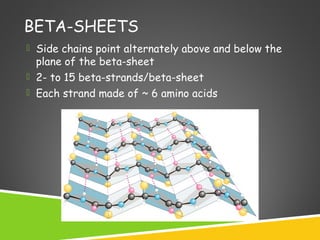
2. TERMINOLOGY
Conformation – spatial arrangement of atoms in a protein
Native conformation – conformation of functional protein
3.
4.
5.
6. PROTEIN CLASSIFICATION
Simple – composed only of amino acid residues
Conjugated – contain prosthetic groups
(metal ions, co-factors, lipids, carbohydrates)
Example: Hemoglobin – Heme
7. PROTEIN CLASSIFICATION
• One polypeptide chain - monomeric protein
• More than one - multimeric protein
• Homomultimer - one kind of chain
• Heteromultimer - two or more different
chains
(e.g. Hemoglobin is a heterotetramer. It has
two alpha chains and two beta chains.)
8. PROTEIN CLASSIFICATION
Fibrous –
1) polypeptides arranged in long strands or sheets
2) water insoluble (lots of hydrophobic AA’s)
3) strong but flexible
4) Structural (keratin, collagen)
Globular –
1) polypeptide chains folded into spherical or globular
form
2) water soluble
3) contain several types of secondary structure
4) diverse functions (enzymes, regulatory proteins)
15. 2O
STRUCTURE RELATED TO PEPTIDE
BACKBONE
•Double bond nature of peptide
bond cause planar geometry
•Free rotation at N - αC and αC-
carbonyl C bonds
•Angle about the C(alpha)-N bond
is denoted phi (φ)
•Angle about the C(alpha)-C bond is
denoted psi (ψ)
•The entire path of the peptide
backbone is known if all phi and psi
angles are specified
18. ALPHA-HELIX
• First proposed by Linus Pauling and
Robert Corey in 1951
• Identified in keratin by Max Perutz
• A ubiquitous component of proteins
• Stabilized by H-bonds
19. ALPHA-HELIX
•Residues per
turn: 3.6
•Rise per residue:
1.5 Angstroms
•Rise per turn
(pitch): 3.6 x 1.5A
= 5.4 Angstroms
•amino hydrogen
H-bonds with
carbonyl oxygen
located 4 AA’s
away forms 13
atom loop
Right handed
helix
20. ALPHA-HELIX
All H-bonds in the
alpha-helix are
oriented in the
same direction
giving the helix a
dipole with the N-
terminus being
positive and the
C-terminus being
negative
21. ALPHA-HELIX
•Side chain groups
point outwards from
the helix
•AA’s with bulky side
chains less common in
alpha-helix
•Glycine and proline
destabilizes alpha-
helix
22. AMPHIPATHIC ALPHA-HELICES
+
One side of the helix (dark) has mostly hydrophobic
AA’s
Two amphipathic helices can associate through
hydrophobic interactions
23. BETA-STRANDS AND BETA-
SHEETS Also first postulated by Pauling and
Corey, 1951
Strands may be parallel or antiparallel
Rise per residue:
3.47 Angstroms for antiparallel strands
3.25 Angstroms for parallel strands
Each strand of a beta sheet may be pictured as a helix with two
residues per turn
24. BETA-SHEETS
Beta-sheets formed
from multiple side-
by-side beta-
strands.
Can be in parallel or
anti-parallel
configuration
Anti-parallel beta-
sheets more stable
25. BETA-SHEETS
Side chains point alternately above and below the
plane of the beta-sheet
2- to 15 beta-strands/beta-sheet
Each strand made of ~ 6 amino acids
26. LOOPS AND TURNS
Loops
Loops usually contain hydrophillic residues.
Found on surfaces of proteins
Connect alpha-helices and beta-sheets
Turns
Loops with < 5 AA’s are called turns
Beta-turns are common
27. BETA-TURNS
allows the peptide chain to reverse direction
carbonyl C of one residue is H-bonded to the amide
proton of a residue three residues away
proline and glycine are prevalent in beta turns