Comet assay
•Download as PPTX, PDF•
26 likes•16,669 views

complete description about comet assay

Recommended
More Related Content
What's hot
What's hot (20)
Viewers also liked
Viewers also liked (20)
Similar to Comet assay
Similar to Comet assay (20)
More from Vijay Hemmadi
More from Vijay Hemmadi (20)
Recently uploaded
Recently uploaded (20)
Comet assay
- 2. Introduction • Single cell gel electrophoresis (SCGE) or the Comet assay is a versatile, sensitive yet simple and economical technique used to measure DNA damage and repair in individual cells. The comet assay helps to measure the single/ double strand DNA breaks, alkali labile sites (apurinic / apyrimidinic sites), DNA cross-links, base / base-pair damages and apoptotic nuclei in the cell • In 1984, Ostling and Johnson demonstrated migration of DNA strands from nuclei which were exposed to an electric field under neutral conditions. Later, in 1988, Singh and his co-workers modified and optimized this process using alkaline conditions which substantially increased is specificity and reproducibility. This method was developed to measure low levels of strand breaks with high sensitivity. • The most widely used method for assessment of DNA damage is the alkaline comet assay. Detection of DNA damage at the level of an individual eukaryotic cell warrants high significance in the fields of toxicology, pharmaceuticals, genotoxicity testing, environmental/ human bio-monitoring, diagnosis of genetic disorders.
- 3. Principle • The Comet Assay is based on the ability of negatively charged loops/fragments of DNA to be drawn through an agarose gel in response to an electric field • The extent of DNA migration depends directly on the DNA damage present in the cells. It should be noted that DNA lesions consisting of strand breaks after treatment with alkali either alone or in combination with certain enzymes (e.g.endonucleases) increases DNA migration, whereas DNA-DNA and DNA-protein cross-links result in retarded DNA migration compared to those in concurrent controls • Treatment of agarose-embedded cells (lymphocytes) with hypertonic lysis solution and non- ionic detergent removes their cell membranes, cytoplasm, nucleoplasm and dissolves nucleosomes. Subsequently, when the leftover nucleoid is treated with high alkaline solution, DNA supercoils unwind/ relax thereby exposing the alkali labile sites (apurinic / apyrimidinic sites) which appears as breaks. Such breaks migrate towards the anode when exposed to current during electrophoresis thereby producing a ‘comet’-like appearance. Use of high concentration of alkali substantially improves the resolving power of the assay without affecting its sensitivity.
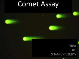
- 4. Specific genotoxic chemicals cause single strand breaks in DNA molecules The Single Cell Gel Electrophoresis (Comet Assay) can illuminate the extent of the damage Pictures: Edler, Kim, Park, Thielmann, 2002.
- 5. Materials required • Dimethyl sulfoxide (DMSO) - HiMedia (MB058) • Disodium EDTA HiMedia ( GM1370) • Ethidium Bromide HiMedia (MB071) • Lymphocyte Separation medium HiMedia (LSM 1077) • Phosphate buffered saline ( PBS) HiMedia ( TS1006) • Sodium Chloride (NaCl) HiMedia (MB023) • Sodium hydroxide (NaOH) HiMedia (MB095) • Triton X-100 HiMedia (RM845) • Tris HCL HiMedia (MB030) • Normal Melting Point Agarose (NMA) HiMedia (MB0175) • Low Melting Point Agarose ( LMPA) HiMedia (MB080) • Methanol Merck • Coverslips (no.1, 24x60 mm) HiMedia • Microcentrifuge tubes Tarsons ( 500010) • Micropipettor and tips Thermo & Tarsons • Microscope slides with label HiMedia
- 6. • Consumables: Glass sides (75x25mm)- plain/ frosted, glass cover slips ( 24x60mm), glass beakers, glass conical flask, glass measuring cylinder, staining trough, staining amber, squeeze bottles , blunt foreceps, slide tray, micropipette, micropipette tips, micro centrifuge tubes, pasture pipette, slide storage box, aluminum foil. • Instruments : Electronic weighing balance ( minimum 0.001 g sensitivity) , centrifuge, microwave oven, magnetic stirrer, pH meter, vortex mixer, refrigerator, water distillation unit, horizontal submarine gel electrophoresis system with power pack, platform rocker, bright- field light microscope / epifluorescence microscope, charge-coupled device camera, computer with image analysis software , autoclave • Chemicals and Solutions: • Lysis solution • Electrophoresis Buffer • PBS Buffer • Ethidium Bromide • Tris Buffer Gel electrophoresis Epifluorescence microscope
- 7. Chemical Preparation Lysis Buffer: (Stock) pH- 10 • 2.5 M – NaCl 100 mM- EDTA • 10 mM – Tris HCl • In 1 Liter distilled water Working solution – Stock buffer 445 ml + DMSO 10%- 50ml + Titron X (1%) – 5ml PBS Solution: pH- 7.4 • 140 mM – NaCl • 10 mM – NaH2 PO4 -850 ml DH2O • 10 mM – Na2HPO4 -150 ml DH2O Electrophoresis Buffer: pH -13 • 300 mM - NaOH • 1mM - EDTA • In 1 liter distilled water
- 8. Neutralization Buffer: pH -7.4 • 400 mM in distilled water make up to 1 L • EtBr:10 mg in 50 ml distilled water • Normal saline 0.89 % Preparation Of Normal Melting Agarose: NMA • 1% NMA dissolved in 100 ml of 0.89% Normal saline Preparation of Low Melting Agarose: • 1% LMA dissolved in 100 ml PBS solution Preparation of Precoated Slide: In grease glass slide was coated with NMA by dipping the slide into melted NMA and back side of the slide should be wipe clearly. Allow the slide to dry over night
- 9. Staining solution EtBr- Ethidium bromide (EtBr, 10 X stock -20 µg / mL): add 10 mg in 50mL distilled water, store at room temperature. For 1X Stock – mix 1 mL with 9 ml distilled water Lymphocytes separation- • Anticoagulant treated blood 3 ml mix with 3 ml RPMI medium or with PBS solution (1:1 ratio) • Add LSM ( 3 ml) to the new centrifuge tube ( 15 ml capacity). • Carefully layer the diluted blood sample ( 4 ml) on LSM ( Lymphocyte separation medium) • When layering the sample do not mix with LSM. • Centrifuge at 400 x g for 30-40 minutes at 18-20 C. • Draw off the upper layer using a clean Pasteur pipette, leaving the lymphocyte layer undisturbed at the interface. Care should be taken not to disturb the lymphocyte layer
- 10. Procedure Comet assay- Major Steps • Agarose gel and sample preparation • Alkaline lysis • Electrophoresis • Computer image analysis • The cell suspension ( 100 µL ) should mixed with 200 µL. of 1 % LMA ( 37 C) prepared in PBS, pH -7.4 and immediately pipetted onto a 1 % NMA (prepared in distilled water ) precoated and overnight dried slides. • The slides should placed on a chilled plate for 10 min to allow complete polymerization of agarose and then immersed in freshly made lysis solution ( 2.5 NaCl, 100m M Tris, NaOH to , pH 10.0, 10 % DMSO and 1% Triton X-100) at room temperature for 2 h to remove cellular proteins • The slides should rinsed with distilled water for 15 min. After repeating the distilled water wash 4 times, the slides should placed in a horizontal electrophoresis tank containing electrophoresis buffer ( 300 mM NaOH and 1 mM EDTA , pH - 13) for 45min , allowing salty equilibration and further DNA unwinding before electrophoresis and run the electrophoresis at 20V, 300 mA for 25 min
- 11. Contd. • Drain the electrophoresis buffer from the tank slowly for 15 min • The slides should wash 2 times (5 min with 0.4 M Tris- HCl (pH - 7.5) at 4 C. After the second neutralization wash, the slides should dried at room temperature and kept in refrigerator in a sealed container until analysis. • The dried slide should stained with EtBr (10 µg/ ml) and examine under 250x using a fluorescent microscope. • All slides should code and examine blindly. A total of 50 cells from each of the 2 replicate slides should examined per sample. The length and width of DNA mass should measure using an ocular micrometer disk and the DNA length/ width ratios should calculated Visualization: All slides should be scored by one person to avoid inters corer variability. Undamaged cells had the appearance of the comet • Tail moment (TM) analyzer as comet assay parameter and image should analyze using comet image analyzer and data capture software. • % DNA in tail is linearly related to DNA break frequency up to about 80 % in tail, and this defines the useful range of the assay.
- 12. How does the comet assay work? Using any nucleated cell
- 14. Quality control and Quality assurance • The use of positive and negative (untreated) controls is recommended. Chemical inducers of genetic damage are commonly utilized, and these can be classed as oxidizers such as hydrogen peroxide and free –radical initiator. Hydrogen peroxide – The molarity of the hydrogen peroxide stock was checked regularly for accuracy as the molarity changes with storage. Due to the variability of hydrogen peroxide, a dose (recommended use at anywhere between 1 mM and 1 µM for 30 min prior o harvesting the cells) was established based on the current stock. Precautions / Health and safety warning • Handle EtBr with adequate precaution as it is known carcinogen. Perform the procedure step1-3 under yellow/ dimmed light .This is to prevent any DNA damage. The longer exposure to alkali will greater the expression of alkali-liable damage.
- 15. Calculation: • The control gels (no enzyme treatment) provide an estimate of the background of DNA strand breaks (SB). The enzyme-treated gels reveal strand breaks and oxidised bases (SB + OX). Assuming a linear dose response, whether working in % DNA in tail or in arbitrary units, subtraction of (SB) from (SB + OX) gives a measure of oxidised pyrimidines/altered purines. Calibration : • Ionising radiation produces strand breaks in DNA with known efficiency. If the breaks introduced in cells by different doses of X-rays are detected with the comet assay, a standard curve can be drawn, with break frequency expressed as Gray-equivalents, or as breaks per unit length of DNA
- 16. References • Alok Dhawan,PhD.,MNASc, Mahima Bajpayee, MSc. , Alok Kumar Pandey, MSc, Devender Parmar, PhD. Development Toxicology Division , Industrail Toxicology Research Centre. ITRC-THE SCGE/ COMET ASSAY PROTOCOL • N.P.Singh , M.T.McCoy, R.R.Tice , E.L.Schneider : A simple technique for quantitation of low levels of DNA damage in individual cells. Exp. Cell Res. 175( (1988) 184-191. • S. Nandhakumar, S.Parasuraman, M.M.Shanmugam, K.Ramachandra Rao, Prakash Chand and B.Vishnu Bhat. Journal of Pharmacother, 2011 Apr-Jun; 2 (2): 107-111.