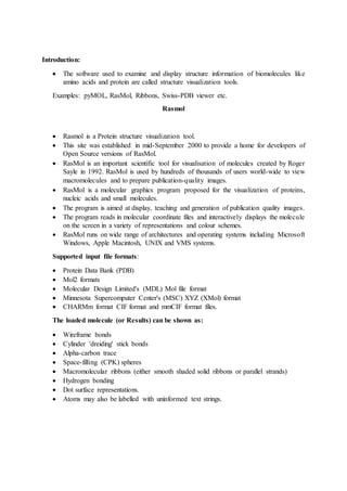
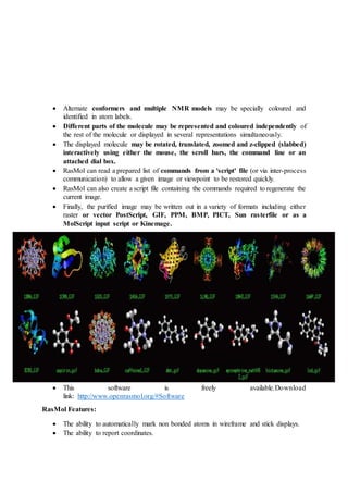
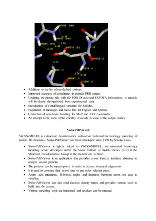
Introduction:
 The software used to examine and display structure information of biomolecules like
amino acids and protein are called structure visualization tools.
Examples: pyMOL, RasMol, Ribbons, Swiss-PDB viewer etc.
Rasmol
 Rasmol is a Protein structure visualization tool.
 This site was established in mid-September 2000 to provide a home for developers of
Open Source versions of RasMol.
 RasMol is an important scientific tool for visualisation of molecules created by Roger
Sayle in 1992. RasMol is used by hundreds of thousands of users world-wide to view
macromolecules and to prepare publication-quality images.
 RasMol is a molecular graphics program proposed for the visualization of proteins,
nucleic acids and small molecules.
 The program is aimed at display, teaching and generation of publication quality images.
 The program reads in molecular coordinate files and interactively displays the molecule
on the screen in a variety of representations and colour schemes.
 RasMol runs on wide range of architectures and operating systems including Microsoft
Windows, Apple Macintosh, UNIX and VMS systems.
Supported input file formats:
 Protein Data Bank (PDB)
 Mol2 formats
 Molecular Design Limited's (MDL) Mol file format
 Minnesota Supercomputer Center's (MSC) XYZ (XMol) format
 CHARMm format CIF format and mmCIF format files.
The loaded molecule (or Results) can be shown as:
 Wireframe bonds
 Cylinder 'dreiding' stick bonds
 Alpha-carbon trace
 Space-filling (CPK) spheres
 Macromolecular ribbons (either smooth shaded solid ribbons or parallel strands)
 Hydrogen bonding
 Dot surface representations.
 Atoms may also be labelled with uninformed text strings.
 Alternate conformers and multiple NMR models may be specially coloured and
identified in atom labels.
 Different parts of the molecule may be represented and coloured independently of
the rest of the molecule or displayed in several representations simultaneously.
 The displayed molecule may be rotated, translated, zoomed and z-clipped (slabbed)
interactively using either the mouse, the scroll bars, the command line or an
attached dial box.
 RasMol can read a prepared list of commands from a 'script' file (or via inter-process
communication) to allow a given image or viewpoint to be restored quickly.
 RasMol can also create a script file containing the commands required to regenerate the
current image.
 Finally, the purified image may be written out in a variety of formats including either
raster or vector PostScript, GIF, PPM, BMP, PICT, Sun rasterfile or as a
MolScript input script or Kinemage.
 This software is freely available.Download
link: http://www.openrasmol.org/#Software
RasMol Features:
 The ability to automatically mark non bonded atoms in wireframe and stick displays.
 The ability to report coordinates.
 Additions to the list of pre-defined colours.
 Improved accuracy of coordinates in pseudo-PDB output.
 Updating the picture title with the PDB ID code and EXPDTA information, so models
will be clearly distinguished from experimental data.
 Introduction of a multilingual structure for RasMol.
 Population of messages and menu lists for English and Spanish.
 Correction of coordinate handling for Mol2 and XYZ coordinates
 An attempt to fix some of the chirality reversals in some of the output modes.
Swiss-PdbViewer
SWISS-MODEL is a structural bioinformatics web-server dedicated to homology modeling of
protein 3D structures. Swiss-PdbViewer has been developed since 1994 by Nicolas Guex.
 Swiss-PdbViewer is tightly linked to SWISS-MODEL, an automated homology
modeling server developed within the Swiss Institute of Bioinformatics (SIB) at the
Structural Bioinformatics Group at the Biozentrum in Basel.
 Swiss-PdbViewer is an application that provides a user friendly interface allowing to
analyze several proteins.
 The proteins can be superimposed in order to deduce structural alignments
 It is used to compare their active sites or any other relevant parts.
 Amino acid mutations, H-bonds, angles and distances between atoms are easy to
visualize.
 Swiss-PdbViewer can also read electron density maps, and provides various tools to
build into the density.
 Various modeling tools are integrated and residues can be mutated.
Homology modeling is currently the most accurate method to generate reliable three-
dimensional protein structure models.
Homology (or comparative) modelling methods make use of experimental protein structures
("templates") to build models for evolutionary related proteins ("targets").
Today, SWISS-MODEL consists of three tightly integrated components:
(1) The SWISS-MODEL pipeline - a suite of software tools and databases for automated
protein structure modelling
(2) The SWISS-MODEL Workspace - a web-based graphical user workbench,
(3) The SWISS-MODEL Repository - a continuously updated database of homology models
for a set of model organism proteomes of high biomedical interest.
The Pipeline
SWISS-MODEL pipeline comprises the four main steps that are involved in building a
homology model of a given protein structure:
 Identification of structural template(s). BLAST and HHblits are used to identify
templates.
 The templates are stored in the SWISS-MODEL Template Library (SMTL), which is
derived from PDB.
 Alignment of target sequence and template structure(s).
 Model building and energy minimization.
 Assessment of the model's quality using QMEAN, a statistical potential of mean force.
The Workspace
.In this mode the input is a project file that can be generated by the DeepView (Swiss Pdb
Viewer) visualization and structural analysis tool, to allow the user to examine and manipulate
the target-template alignment in its structural context.
In all three cases the output is a PDB file with atom coordinates of the model or a DeepView
project file.
The four main steps of homology modelling may be repeated iteratively until a satisfactory
model is achieved.
The SWISS-MODEL Workspace is accessible via the ExPASy web server, or it can be used as
part of the program DeepView (Swiss Pdb-Viewer).
The Repository:
The SWISS-MODEL Repository provides access to an up-to-date collection of annotated
three-dimensional protein models for a set of model organisms of high general interest. SWISS-
MODEL Repository is integrated with several external resources, such as UniProt, InterPro,
STRING, and Nature PSI SBKB.
New developments of the SWISS-MODEL expert system feature
(1) automated modelling of homo-oligomeric assemblies
(2) modelling of essential metal ions and biologically relevant ligands in protein structures
(3) local (per-residue) model reliability estimates based on the QMEAN local score function
(4) mapping of UniProt features to models.

Rasmol

  • 1.
    Introduction:  The softwareused to examine and display structure information of biomolecules like amino acids and protein are called structure visualization tools. Examples: pyMOL, RasMol, Ribbons, Swiss-PDB viewer etc. Rasmol  Rasmol is a Protein structure visualization tool.  This site was established in mid-September 2000 to provide a home for developers of Open Source versions of RasMol.  RasMol is an important scientific tool for visualisation of molecules created by Roger Sayle in 1992. RasMol is used by hundreds of thousands of users world-wide to view macromolecules and to prepare publication-quality images.  RasMol is a molecular graphics program proposed for the visualization of proteins, nucleic acids and small molecules.  The program is aimed at display, teaching and generation of publication quality images.  The program reads in molecular coordinate files and interactively displays the molecule on the screen in a variety of representations and colour schemes.  RasMol runs on wide range of architectures and operating systems including Microsoft Windows, Apple Macintosh, UNIX and VMS systems. Supported input file formats:  Protein Data Bank (PDB)  Mol2 formats  Molecular Design Limited's (MDL) Mol file format  Minnesota Supercomputer Center's (MSC) XYZ (XMol) format  CHARMm format CIF format and mmCIF format files. The loaded molecule (or Results) can be shown as:  Wireframe bonds  Cylinder 'dreiding' stick bonds  Alpha-carbon trace  Space-filling (CPK) spheres  Macromolecular ribbons (either smooth shaded solid ribbons or parallel strands)  Hydrogen bonding  Dot surface representations.  Atoms may also be labelled with uninformed text strings.
  • 2.
     Alternate conformersand multiple NMR models may be specially coloured and identified in atom labels.  Different parts of the molecule may be represented and coloured independently of the rest of the molecule or displayed in several representations simultaneously.  The displayed molecule may be rotated, translated, zoomed and z-clipped (slabbed) interactively using either the mouse, the scroll bars, the command line or an attached dial box.  RasMol can read a prepared list of commands from a 'script' file (or via inter-process communication) to allow a given image or viewpoint to be restored quickly.  RasMol can also create a script file containing the commands required to regenerate the current image.  Finally, the purified image may be written out in a variety of formats including either raster or vector PostScript, GIF, PPM, BMP, PICT, Sun rasterfile or as a MolScript input script or Kinemage.  This software is freely available.Download link: http://www.openrasmol.org/#Software RasMol Features:  The ability to automatically mark non bonded atoms in wireframe and stick displays.  The ability to report coordinates.
  • 3.
     Additions tothe list of pre-defined colours.  Improved accuracy of coordinates in pseudo-PDB output.  Updating the picture title with the PDB ID code and EXPDTA information, so models will be clearly distinguished from experimental data.  Introduction of a multilingual structure for RasMol.  Population of messages and menu lists for English and Spanish.  Correction of coordinate handling for Mol2 and XYZ coordinates  An attempt to fix some of the chirality reversals in some of the output modes. Swiss-PdbViewer SWISS-MODEL is a structural bioinformatics web-server dedicated to homology modeling of protein 3D structures. Swiss-PdbViewer has been developed since 1994 by Nicolas Guex.  Swiss-PdbViewer is tightly linked to SWISS-MODEL, an automated homology modeling server developed within the Swiss Institute of Bioinformatics (SIB) at the Structural Bioinformatics Group at the Biozentrum in Basel.  Swiss-PdbViewer is an application that provides a user friendly interface allowing to analyze several proteins.  The proteins can be superimposed in order to deduce structural alignments  It is used to compare their active sites or any other relevant parts.  Amino acid mutations, H-bonds, angles and distances between atoms are easy to visualize.  Swiss-PdbViewer can also read electron density maps, and provides various tools to build into the density.  Various modeling tools are integrated and residues can be mutated.
  • 4.
    Homology modeling iscurrently the most accurate method to generate reliable three- dimensional protein structure models. Homology (or comparative) modelling methods make use of experimental protein structures ("templates") to build models for evolutionary related proteins ("targets"). Today, SWISS-MODEL consists of three tightly integrated components: (1) The SWISS-MODEL pipeline - a suite of software tools and databases for automated protein structure modelling (2) The SWISS-MODEL Workspace - a web-based graphical user workbench, (3) The SWISS-MODEL Repository - a continuously updated database of homology models for a set of model organism proteomes of high biomedical interest. The Pipeline SWISS-MODEL pipeline comprises the four main steps that are involved in building a homology model of a given protein structure:  Identification of structural template(s). BLAST and HHblits are used to identify templates.  The templates are stored in the SWISS-MODEL Template Library (SMTL), which is derived from PDB.  Alignment of target sequence and template structure(s).  Model building and energy minimization.  Assessment of the model's quality using QMEAN, a statistical potential of mean force. The Workspace .In this mode the input is a project file that can be generated by the DeepView (Swiss Pdb Viewer) visualization and structural analysis tool, to allow the user to examine and manipulate the target-template alignment in its structural context. In all three cases the output is a PDB file with atom coordinates of the model or a DeepView project file. The four main steps of homology modelling may be repeated iteratively until a satisfactory model is achieved. The SWISS-MODEL Workspace is accessible via the ExPASy web server, or it can be used as part of the program DeepView (Swiss Pdb-Viewer). The Repository: The SWISS-MODEL Repository provides access to an up-to-date collection of annotated three-dimensional protein models for a set of model organisms of high general interest. SWISS- MODEL Repository is integrated with several external resources, such as UniProt, InterPro, STRING, and Nature PSI SBKB. New developments of the SWISS-MODEL expert system feature (1) automated modelling of homo-oligomeric assemblies
  • 5.
    (2) modelling ofessential metal ions and biologically relevant ligands in protein structures (3) local (per-residue) model reliability estimates based on the QMEAN local score function (4) mapping of UniProt features to models.