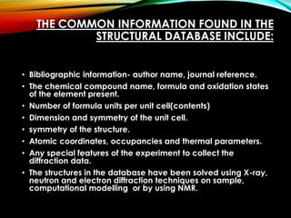
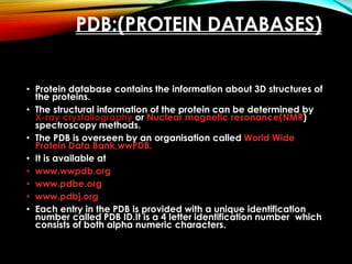
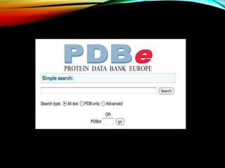
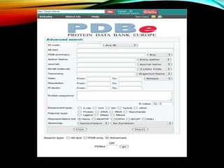
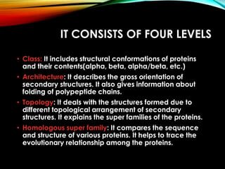
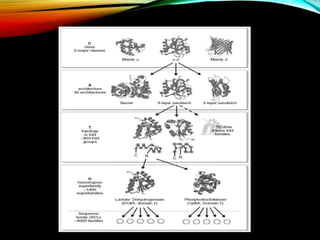
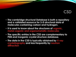
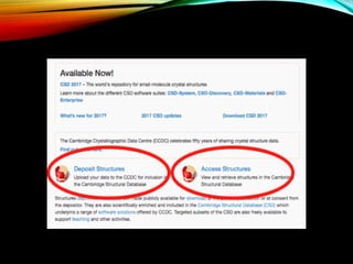
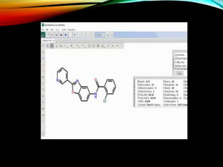
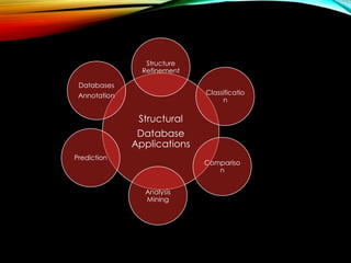
Structural databases like PDB, CSD, and CATH contain 3D structural information of proteins, small molecules, and macromolecules determined through techniques like X-ray crystallography and NMR spectroscopy. These databases provide bibliographic data, atomic coordinates, and other details for each entry. PDB contains protein structures, CSD contains organic and metal-organic structures, and CATH classifies protein domains hierarchically. Structural databases have wide applications in structure prediction, analysis, mining, comparison, classification, structure refinement, and database annotation.
Sequence alig Sequence Alignment Pairwise alignment:-naveed ul mushtaq
Sequence Alignment Pairwise alignment:- Global Alignment and Local AlignmentTwo types of alignment Progressive Programs for multiple sequence alignment BLOSUM Point accepted mutation (PAM)PAM VS BLOSUM
Sequence alig Sequence Alignment Pairwise alignment:-naveed ul mushtaq
Sequence Alignment Pairwise alignment:- Global Alignment and Local AlignmentTwo types of alignment Progressive Programs for multiple sequence alignment BLOSUM Point accepted mutation (PAM)PAM VS BLOSUM
INTRODUCTION.
NCBI.
EMBL.
DDBJ.
CONCLUSION.
REFERENSE.
The National Center for Biotechnology Information (NCBI) is part of the United States National Library of Medicine (NLM), a branch of the National Institutes of Health.
The NCBI is located in Bethesda, Maryland and was founded in 1988 through legislation sponsored by Senator Claude Pepper.
The NCBI houses a series of databases relevant to biotechnology and biomedicine. Major databases include GenBank for DNA sequences and PubMed, a bibliographic database for the biomedical literature.
All these databases are available online through the Entrez search engine.
Scoring system is a set of values for qualifying the set of one residue being substituted by another in an alignment.
It is also known as substitution matrix.
Scoring matrix of nucleotide is relatively simple.
A positive value or a high score is given for a match & negative value or a low score is given for a mismatch.
Scoring matrices for amino acids are more complicated because scoring has to reflect the physicochemical properties of amino acid residues.
The Protein Data Bank (PDB) is a database for the three-dimensional structural data of large biological molecules, such as proteins and nucleic acids. This presentation deals with what, why, how, where and who of PDB. In this presentation we have also included briefing about various file formats available in PDB with emphasis on PDB file format
An integrated publicly accessible bioinformatics resource to support genomic/proteomic research and scientific discovery.
Established in 1984, by the National Biomedical Research Foundation (NBRF) Georgetown University Medial Center, Washington D.C., USA.
It is the source of annotated protein databases and analysis tools for the researchers.
Serve as primary resource for the exploration of protein information.
Accessible by text search for entry and list retrieval, and also BLAST search and peptide match.
This presentation gives you a detailed information about the swiss prot database that comes under UniProtKB. It also covers TrEMBL: a computer annotated supplement to Swiss-Prot.
It includes the information related to a bioinformatics tool BLAST (Basic Local Alignment Search Tool), BLAST is in-silico hybridisation to find regions of similarity between biological sequences. The program compares nucleotide or protein sequences to sequence databases and calculates the statistical significance. This presentation too contains the input - output format, Blast process and its types .
Biological databases are libraries of biological sciences, collected from scientific experiments, published literature, high-throughput experiment technology, and computational analysis. They contain information from research areas including genomics, proteomics, metabolomics, microarray gene expression, and phylogenetics
The iCSS CompTox Dashboard is a publicly accessible dashboard provided by the National Center for Computation Toxicology at the US-EPA. It serves a number of purposes, including providing a chemistry database underpinning many of our public-facing projects (e.g. ToxCast and ExpoCast). The available data and searches provide a valuable path to structure identification using mass spectrometry as the source data. With an underlying database of over 720,000 chemicals, the dashboard has already been used to assist in identifying chemicals present in house dust. However, it can also be applied to many other purposes, e.g., the identification of agrochemicals in waste streams. This presentation will provide a review of the EPA’s platform and underlying algorithms used for the purpose of compound identification using high-resolution mass spectrometry data. In order to examine its performance for structure identification, especially in terms of rank-ordering database hits, we have compared it with the ChemSpider database, a well-regarded public database that has become one of the community standards for structure identification. The study has shown that the CompTox Dashboard outperforms ChemSpider in terms of structure identification and ranking providing improved outcomes for mass spectrometry analysis of “known unknowns”.
Earliest Galaxies in the JADES Origins Field: Luminosity Function and Cosmic ...Sérgio Sacani
We characterize the earliest galaxy population in the JADES Origins Field (JOF), the deepest
imaging field observed with JWST. We make use of the ancillary Hubble optical images (5 filters
spanning 0.4−0.9µm) and novel JWST images with 14 filters spanning 0.8−5µm, including 7 mediumband filters, and reaching total exposure times of up to 46 hours per filter. We combine all our data
at > 2.3µm to construct an ultradeep image, reaching as deep as ≈ 31.4 AB mag in the stack and
30.3-31.0 AB mag (5σ, r = 0.1” circular aperture) in individual filters. We measure photometric
redshifts and use robust selection criteria to identify a sample of eight galaxy candidates at redshifts
z = 11.5 − 15. These objects show compact half-light radii of R1/2 ∼ 50 − 200pc, stellar masses of
M⋆ ∼ 107−108M⊙, and star-formation rates of SFR ∼ 0.1−1 M⊙ yr−1
. Our search finds no candidates
at 15 < z < 20, placing upper limits at these redshifts. We develop a forward modeling approach to
infer the properties of the evolving luminosity function without binning in redshift or luminosity that
marginalizes over the photometric redshift uncertainty of our candidate galaxies and incorporates the
impact of non-detections. We find a z = 12 luminosity function in good agreement with prior results,
and that the luminosity function normalization and UV luminosity density decline by a factor of ∼ 2.5
from z = 12 to z = 14. We discuss the possible implications of our results in the context of theoretical
models for evolution of the dark matter halo mass function.
This pdf is about the Schizophrenia.
For more details visit on YouTube; @SELF-EXPLANATORY;
https://www.youtube.com/channel/UCAiarMZDNhe1A3Rnpr_WkzA/videos
Thanks...!
Observation of Io’s Resurfacing via Plume Deposition Using Ground-based Adapt...Sérgio Sacani
Since volcanic activity was first discovered on Io from Voyager images in 1979, changes
on Io’s surface have been monitored from both spacecraft and ground-based telescopes.
Here, we present the highest spatial resolution images of Io ever obtained from a groundbased telescope. These images, acquired by the SHARK-VIS instrument on the Large
Binocular Telescope, show evidence of a major resurfacing event on Io’s trailing hemisphere. When compared to the most recent spacecraft images, the SHARK-VIS images
show that a plume deposit from a powerful eruption at Pillan Patera has covered part
of the long-lived Pele plume deposit. Although this type of resurfacing event may be common on Io, few have been detected due to the rarity of spacecraft visits and the previously low spatial resolution available from Earth-based telescopes. The SHARK-VIS instrument ushers in a new era of high resolution imaging of Io’s surface using adaptive
optics at visible wavelengths.
Slide 1: Title Slide
Extrachromosomal Inheritance
Slide 2: Introduction to Extrachromosomal Inheritance
Definition: Extrachromosomal inheritance refers to the transmission of genetic material that is not found within the nucleus.
Key Components: Involves genes located in mitochondria, chloroplasts, and plasmids.
Slide 3: Mitochondrial Inheritance
Mitochondria: Organelles responsible for energy production.
Mitochondrial DNA (mtDNA): Circular DNA molecule found in mitochondria.
Inheritance Pattern: Maternally inherited, meaning it is passed from mothers to all their offspring.
Diseases: Examples include Leber’s hereditary optic neuropathy (LHON) and mitochondrial myopathy.
Slide 4: Chloroplast Inheritance
Chloroplasts: Organelles responsible for photosynthesis in plants.
Chloroplast DNA (cpDNA): Circular DNA molecule found in chloroplasts.
Inheritance Pattern: Often maternally inherited in most plants, but can vary in some species.
Examples: Variegation in plants, where leaf color patterns are determined by chloroplast DNA.
Slide 5: Plasmid Inheritance
Plasmids: Small, circular DNA molecules found in bacteria and some eukaryotes.
Features: Can carry antibiotic resistance genes and can be transferred between cells through processes like conjugation.
Significance: Important in biotechnology for gene cloning and genetic engineering.
Slide 6: Mechanisms of Extrachromosomal Inheritance
Non-Mendelian Patterns: Do not follow Mendel’s laws of inheritance.
Cytoplasmic Segregation: During cell division, organelles like mitochondria and chloroplasts are randomly distributed to daughter cells.
Heteroplasmy: Presence of more than one type of organellar genome within a cell, leading to variation in expression.
Slide 7: Examples of Extrachromosomal Inheritance
Four O’clock Plant (Mirabilis jalapa): Shows variegated leaves due to different cpDNA in leaf cells.
Petite Mutants in Yeast: Result from mutations in mitochondrial DNA affecting respiration.
Slide 8: Importance of Extrachromosomal Inheritance
Evolution: Provides insight into the evolution of eukaryotic cells.
Medicine: Understanding mitochondrial inheritance helps in diagnosing and treating mitochondrial diseases.
Agriculture: Chloroplast inheritance can be used in plant breeding and genetic modification.
Slide 9: Recent Research and Advances
Gene Editing: Techniques like CRISPR-Cas9 are being used to edit mitochondrial and chloroplast DNA.
Therapies: Development of mitochondrial replacement therapy (MRT) for preventing mitochondrial diseases.
Slide 10: Conclusion
Summary: Extrachromosomal inheritance involves the transmission of genetic material outside the nucleus and plays a crucial role in genetics, medicine, and biotechnology.
Future Directions: Continued research and technological advancements hold promise for new treatments and applications.
Slide 11: Questions and Discussion
Invite Audience: Open the floor for any questions or further discussion on the topic.
(May 29th, 2024) Advancements in Intravital Microscopy- Insights for Preclini...Scintica Instrumentation
Intravital microscopy (IVM) is a powerful tool utilized to study cellular behavior over time and space in vivo. Much of our understanding of cell biology has been accomplished using various in vitro and ex vivo methods; however, these studies do not necessarily reflect the natural dynamics of biological processes. Unlike traditional cell culture or fixed tissue imaging, IVM allows for the ultra-fast high-resolution imaging of cellular processes over time and space and were studied in its natural environment. Real-time visualization of biological processes in the context of an intact organism helps maintain physiological relevance and provide insights into the progression of disease, response to treatments or developmental processes.
In this webinar we give an overview of advanced applications of the IVM system in preclinical research. IVIM technology is a provider of all-in-one intravital microscopy systems and solutions optimized for in vivo imaging of live animal models at sub-micron resolution. The system’s unique features and user-friendly software enables researchers to probe fast dynamic biological processes such as immune cell tracking, cell-cell interaction as well as vascularization and tumor metastasis with exceptional detail. This webinar will also give an overview of IVM being utilized in drug development, offering a view into the intricate interaction between drugs/nanoparticles and tissues in vivo and allows for the evaluation of therapeutic intervention in a variety of tissues and organs. This interdisciplinary collaboration continues to drive the advancements of novel therapeutic strategies.
Introduction:
RNA interference (RNAi) or Post-Transcriptional Gene Silencing (PTGS) is an important biological process for modulating eukaryotic gene expression.
It is highly conserved process of posttranscriptional gene silencing by which double stranded RNA (dsRNA) causes sequence-specific degradation of mRNA sequences.
dsRNA-induced gene silencing (RNAi) is reported in a wide range of eukaryotes ranging from worms, insects, mammals and plants.
This process mediates resistance to both endogenous parasitic and exogenous pathogenic nucleic acids, and regulates the expression of protein-coding genes.
What are small ncRNAs?
micro RNA (miRNA)
short interfering RNA (siRNA)
Properties of small non-coding RNA:
Involved in silencing mRNA transcripts.
Called “small” because they are usually only about 21-24 nucleotides long.
Synthesized by first cutting up longer precursor sequences (like the 61nt one that Lee discovered).
Silence an mRNA by base pairing with some sequence on the mRNA.
Discovery of siRNA?
The first small RNA:
In 1993 Rosalind Lee (Victor Ambros lab) was studying a non- coding gene in C. elegans, lin-4, that was involved in silencing of another gene, lin-14, at the appropriate time in the
development of the worm C. elegans.
Two small transcripts of lin-4 (22nt and 61nt) were found to be complementary to a sequence in the 3' UTR of lin-14.
Because lin-4 encoded no protein, she deduced that it must be these transcripts that are causing the silencing by RNA-RNA interactions.
Types of RNAi ( non coding RNA)
MiRNA
Length (23-25 nt)
Trans acting
Binds with target MRNA in mismatch
Translation inhibition
Si RNA
Length 21 nt.
Cis acting
Bind with target Mrna in perfect complementary sequence
Piwi-RNA
Length ; 25 to 36 nt.
Expressed in Germ Cells
Regulates trnasposomes activity
MECHANISM OF RNAI:
First the double-stranded RNA teams up with a protein complex named Dicer, which cuts the long RNA into short pieces.
Then another protein complex called RISC (RNA-induced silencing complex) discards one of the two RNA strands.
The RISC-docked, single-stranded RNA then pairs with the homologous mRNA and destroys it.
THE RISC COMPLEX:
RISC is large(>500kD) RNA multi- protein Binding complex which triggers MRNA degradation in response to MRNA
Unwinding of double stranded Si RNA by ATP independent Helicase
Active component of RISC is Ago proteins( ENDONUCLEASE) which cleave target MRNA.
DICER: endonuclease (RNase Family III)
Argonaute: Central Component of the RNA-Induced Silencing Complex (RISC)
One strand of the dsRNA produced by Dicer is retained in the RISC complex in association with Argonaute
ARGONAUTE PROTEIN :
1.PAZ(PIWI/Argonaute/ Zwille)- Recognition of target MRNA
2.PIWI (p-element induced wimpy Testis)- breaks Phosphodiester bond of mRNA.)RNAse H activity.
MiRNA:
The Double-stranded RNAs are naturally produced in eukaryotic cells during development, and they have a key role in regulating gene expression .
2. INTRODUCTION:
• Structural databases are the essential tools for all
crystallographic works.
• They are used in the process of producing, solving
,refining and publishing the structure of a new material.
3. THE COMMON INFORMATION FOUND IN THE
STRUCTURAL DATABASE INCLUDE:
• Bibliographic information- author name, journal reference.
• The chemical compound name, formula and oxidation states
of the element present.
• Number of formula units per unit cell(contents)
• Dimension and symmetry of the unit cell.
• symmetry of the structure.
• Atomic coordinates, occupancies and thermal parameters.
• Any special features of the experiment to collect the
diffraction data.
• The structures in the database have been solved using X-ray,
neutron and electron diffraction techniques on sample,
computational modelling or by using NMR.
4. PDB:(PROTEIN DATABASES)
• Protein database contains the information about 3D structures of
the proteins.
• The structural information of the protein can be determined by
X-ray crystallography or Nuclear magnetic resonance(NMR)
spectroscopy methods.
• The PDB is overseen by an organisation called World Wide
Protein Data Bank,wwPDB.
• It is available at
• www.wwpdb.org
• www.pdbe.org
• www.pdbj.org
• Each entry in the PDB is provided with a unique identification
number called PDB ID.It is a 4 letter identification number which
consists of both alpha numeric characters.
5.
6.
7. PDB FILE FORMAT:
The PDB file format is the standard file format for protein
structure file. It describes how molecules are held together in
3-D Structure of a protein.
• The file contain hundreds or thousands of lines called
records. Each record provides a different set of information
like
• HEADER: This reocord contains file name, date of submission
and the PDB ID of the molecule.
• TITLE: This record contains the title of the PDB entry.
• COMPND: This record includes the protein name.
• SOURCE: This record contains the name of the organism in
which the particular protein is obtained.
• KEYWDS: This record contains the keywords that describes
about the protein.
8. PDB FILE FORMAT:
• EXPDTA: This record contains the method used for the
protein structure experiment.
• AUTHOR: This record contains the name of the
contributors who put the data into the database.
• REVDATA: This record contains the revision date of the
data related to protein.(Date of modification)
• JRNL: This record contains the journal details of the
literature about the protein
• REMARK: This record contains the remarks about the
protein structure.
• DBREF: This record contains the reference to the protein
in the sequence databases.
9. PDB FILE FORMAT:
• SEQRES: This record contains information about the
amino acid sequence of protein.
• HET: This record contains details about the non protein
substances in the protein.
• HETNAM: This record contain the compound name of
the non protein substances.
• HETSYN: This record contains the identical compound
name for the non protein substances.
• FORMUL: This record contain the chemical formula of
the non protein substances.
• HELIX: This record holds the recognition of helical
substructures.
10. PDB FILE FORMAT:
• LINK: This record holds the recognition of inter-residue bonds.
• ATOM: This record contains the atomic coordinates for the
structure.
• HEATM: This record contains the atomic coordinate record for
non protein substances.
• CONECT: This record contains the details about the bonds
involved in non protein atoms.
• MASTER: This record contains the details about the number of
REMARK records, HET records, HELIX records, CONECT records
and SEQRES records, etc.
• END: This record represent the end of the file.
•
11.
12. THE PDB FORMAT
• 123456789+123456789+123456789+123456789+123456789+123456789+123456789+123456789+
• HEADER RETINOIC-ACID TRANSPORT 28-SEP-94 1CBS 1CBS 2
• COMPND CELLULAR RETINOIC-ACID-BINDING PROTEIN TYPE II COMPLEXED 1CBS 3
• COMPND 2 WITH ALL-TRANS-RETINOIC ACID (THE PRESUMED PHYSIOLOGICAL 1CBS 4
• COMPND 3 LIGAND) 1CBS 5
• SOURCE HUMAN (HOMO SAPIENS) 1CBS 6
• SOURCE 2 EXPRESSION SYSTEM: (ESCHERICHIA COLI) BL21 (DE3) 1CBS 7
• SOURCE 3 PLASMID: PET-3A 1CBS 8
• SOURCE 4 GENE: HUMAN CRABP-II 1CBS 9
• AUTHOR G.J.KLEYWEGT,T.BERGFORS,T.A.JONES 1CBS 10
• REVDAT 1 26-JAN-95 1CBS 0 1CBS 11
• -------------------------------------------------------------------------------------------------------------------------------------------
13. CATH:
• The CATH means Class, Architecture,Topology and
homologouus super family database for proteins
• It was created by Janet Thornton and colleagues at the
university college London.
• It is available at
http://www.biochem.ucl.ac.uk/bsm/cath
• http://www.cathdb.info
• It is a protein classification tool
14. IT CONSISTS OF FOUR LEVELS
• Class: It includes structural conformations of proteins
and their contents(alpha, beta, alpha/beta, etc.)
• Architecture: It describes the gross orientation of
secondary structures. It also gives information about
folding of polypeptide chains.
• Topology: It deals with the structures formed due to
different topological arrangement of secondary
structures. It explains the super families of the proteins.
• Homologous super family: It compares the sequence
and structure of various proteins. It helps to trace the
evolutionary relationship among the proteins.
15.
16. CATH
• The CATH aims to provide official releases of protein
structures every 12 months
• It is a free publicly available online resource.
• The latest version of CATH contains 1,14,215
domains,2178 homologous superfamilies,1110 fold
groups.
17.
18. THE CATH SERVER
• The CATH have recently set up a server which allows
the user to submit the co-ordinates of the newly
determined structure for automatic classification in
CATH.
• DOMAIN BOUNDARIES AND SEQUENCE COMPARISON
• CATH contains a detective program which is good for
identifying multidomain proteins.
• The results from the detective are returned to the user in
less than a minutes.
• Identified domains are scanned against non identical
representatives from CATH using a global sequence
alignment method
19. CATH SERVER
• If a sequence match 95% then the domain is identical
to one in CATH.
• If a sequence match less than 30% then the structures
are compared with all the sequence families (s-level).
• ASSESING STRUCTURAL SIMILARITY:
• TOPSCAN compares the secondary strucutres in each
fold family to identify the possible fold families to which
the new structures belong.
• Subsequently the fast version of structure comparison
SSAP scans represetatives from all the families
• Structural pairs having a ssap score more than 80 are
possible homologues while the score with 70-80 don’t
have no sequence or functional similiarity.
• Finally the SSAP structural alignment is displayed using a
graphical display package.
20.
21.
22. CSD
• The cambridge structural Database is both a repository
and a validated resource for 3-D structural data of
molecules containing carbon and hydrogen.
• It is used to know about the structures of organic,
metal-organic and organometallic molecules
• The specific entries in the CSD are complementary to
PDB and Inorganic crystal structure database.
• The data in the CSD is typically obtained by X-ray
crystallography and less frequently by neutron
diffraction
23. CSD
• The data in the CSD is submitted by crystallographers and
chemists from all over the world.
• The CSD is maintained by an incorporated company called
Cambridge Crystallographic Data centre, CCDC
• The CCDC are publicly available for download at the point of
publication.
• The CSD is updated with about 50,000 new structures each
year and are freely available to support teaching and other
activities
• The CSD is available at
• www.ccdc.cam.ac.uk
• webcsd.ccdc.cam.ac.uk