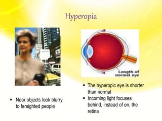
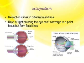
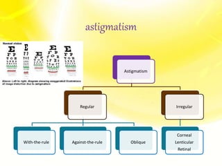
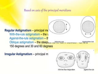
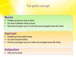
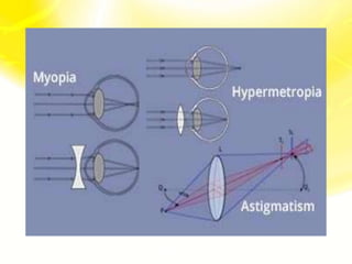
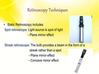
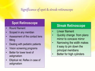
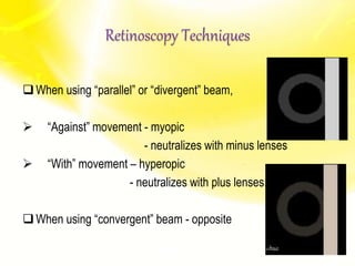
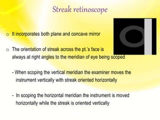
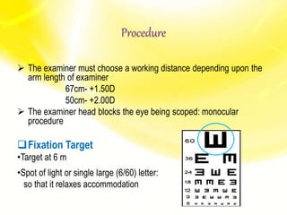
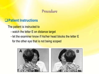
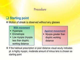
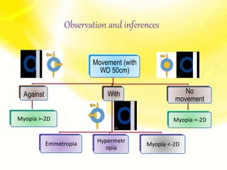
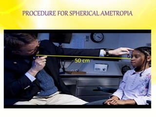
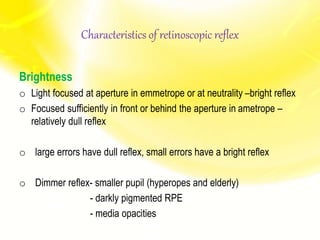
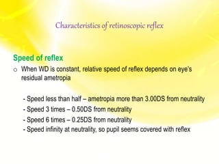
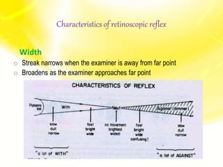
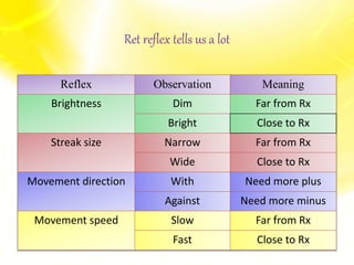
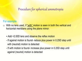
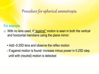
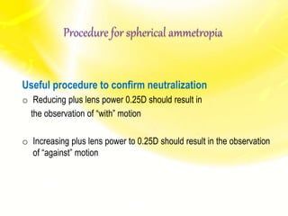
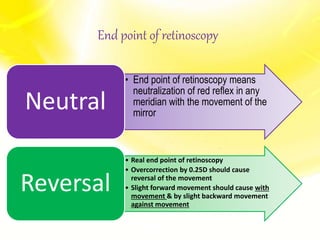
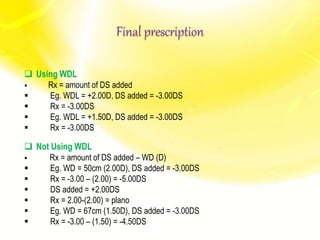
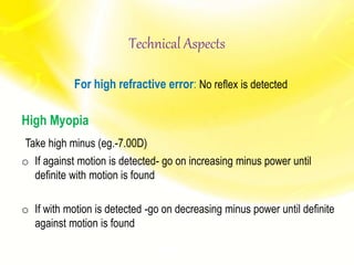
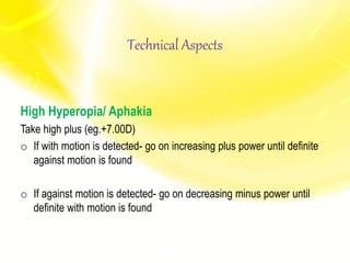
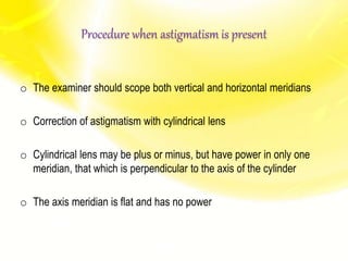
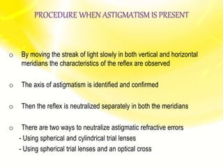
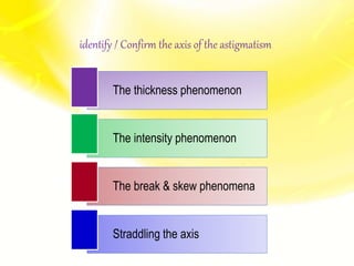
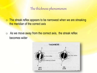
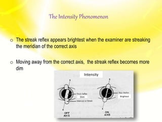
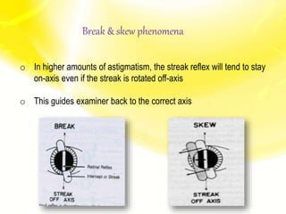
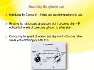
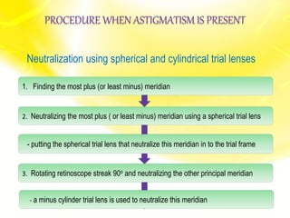
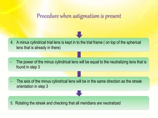
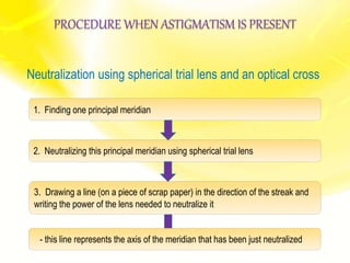
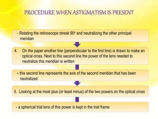
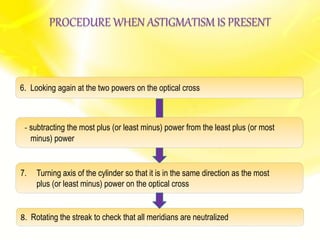
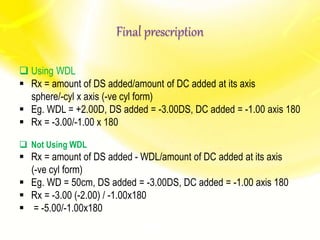
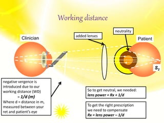
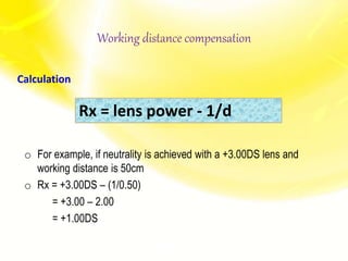
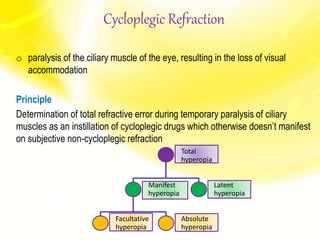
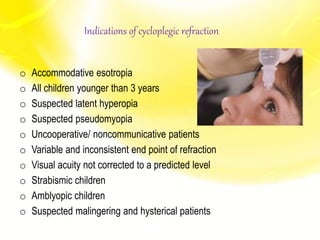
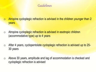
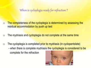
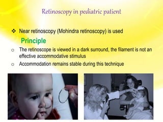
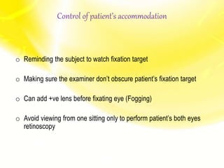
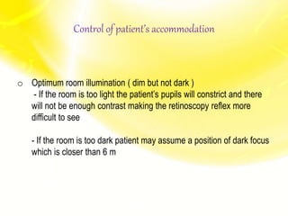
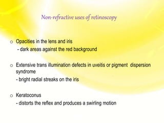
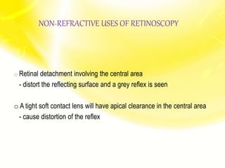
DIRECT DOWNLOAD LINK ❤❤https://healthkura.com/retinoscopy/❤❤
Dear viewers Check Out my other piece of works at ❤❤❤ https://healthkura.com ❤❤❤
Retinoscopy and Objective Refraction and Subjective Refraction in spherical ametropia and astigmatism
Retinoscopy (Principle & Techniques of Retinoscopy) and objective refraction, Subjective Refracition
Best presentation about retinoscopy and objective refraction techniques, and basis of subjective refraction. If you want to master the technique of retinoscopy, this presentation can be your guidance and partner in your journey to retinoscopy, objective refraction and subjective refraction.
Presentation Layout:
Retinoscope, types of retinoscope and uses of retinoscope
-Introduction to retinoscopy and objective refraction
-Retinoscopy
- In spherical ametropia
- In astigmatism
- Others: strabismus, amblyopia, pediatric pt.,
cycloplegic refraction
-Static and Dynamic Retinoscopy
-Problems seeing reflex during retinoscopy
-Errors in retinoscopy
Objective of retinoscopy and objective refraction
-To locate the far point of the eye conjugate to the retina
- Myopia or hyperopia
-Bring far point to the infinity by using appropriate lenses
- Determines amount of ametropia by retinoscopy and objective refraction
References:
-Clinical Procedures in Optometry by Eskridge, Amos and Bartlett ,
-Primary Care Optometry by Grosvenor T.,
-Borish’s Clinical Refraction by Benjamin W. J.,
-Theory And Practice Of Optics And Refraction by AK Khurana
-Retinoscopy-Student Manual by ICEE Refractive Error Training Package (2009)
-Clinical Optics and Refraction By Andrew Keirl, Caroline Christie
-Clinical Refraction Guide - A Kumar Bhootra
-Clinical Procedures in Primary Eye Care by David B. Elliott
-Internet
Follow me to get in touch with optometric and ophthalmic updates.
Detailed instumentaion and use of manual Lensometer and just a outline of automated lensometer.
I have used the picture of manual lensometer with out the parts describtion because i have explained orally by showing the picture..
Hope u all like it and may help you in learning better. :)
Detailed instumentaion and use of manual Lensometer and just a outline of automated lensometer.
I have used the picture of manual lensometer with out the parts describtion because i have explained orally by showing the picture..
Hope u all like it and may help you in learning better. :)
what is Duochrome Test, Why do we take Red and Green color only,
What is the Principal of Duochrome Test, Why Hyperopic Pt sees green better than red and vice versa
It describes about the procedure of Hess charting. it serves as a great tool to understand the concepts involved. Suitable for optometry course. This is not a routine procedure but an important procedure which is used in diagnosis.
these slides explain the objective refraction in optometry , and describes its types and its measurement , and it gives you in details the types of Retinoscopy.
Techniques of refraction is the process of calculation of glass power.drbrijeshbhu
Refractive errors are most common cause of ocular morbidity. It affects all age groups, and ethnic profiles. There is no g nder discrimination. Most common symptoms are blur vission along with pain in eye ,headache and tiredness. Refraction is process of determination of eye and currect it with power glass power or contact lens power. It can subjective or objective.
This presentation explain about retinoscope, the instrument, its history, its types, the procedure and different cases also the advantages and disadvantages of the instrument and the working lens
Dark Room Procedures for undergraduates(MB,BS) in the field of Ophthalmology are explained in simple terms in this presentation. Series of lectures taken at Central Park Medical College Lahore Pakistan.
Retinoscope is an objective refraction instrument used to
determine the spherocylindrical refractive error, as well as
observe optical aberrations, irregularities, and opacities.
The technique is called Retinoscopy/Skiascopy/Shadow Test
Passive Therapy in Management of Amblyopia (healthkura.com)Bikash Sapkota
DIRECT DOWNLOAD LINK ❤❤https://healthkura.com/lazy-eye-amblyopia/❤❤
Dear viewers Check Out my other piece of works at ❤❤❤ https://healthkura.com ❤❤
Passive Therapy in Management of Amblyopia
. Passive Therapy
The patient experiences a change in visual stimulation without any conscious effort
- Proper refractive correction
- Occlusion
- Penalization
Active Vision Therapy in Management of Amblyopia (healthkura.com)Bikash Sapkota
DIRECT DOWNLOAD LINK ❤❤https://healthkura.com/lazy-eye-amblyopia/❤❤
In the request of my viewers, I have compiled my works here in a website. Visit this website (healthkura.com) to freely download this presentation along with other tons of presentations. Some useful links are given here.____Remember___healthkura.com
Active Vision Therapy in Management of Amblyopia
- Pleoptics
- Near activities
- Active stimulation therapy using CAM vision stimulator
- Syntonic phototherapy
- Role of perceptual learning
- Binocular stimulation
- Software-based active treatments
- Exposure to dark
- Pharmacological Therapy
Interventions to Reduce Myopia Progression in Children (Journal Club) (health...Bikash Sapkota
DIRECT DOWNLOAD LINK ❤❤https://healthkura.com/reduce-myopia/❤❤
Dear viewers Check Out my other piece of works at___ https://healthkura.com
Interventions to Reduce Myopia Progression in Children (Journal Club)
Objectives:
- To discuss about the different interventions to reduce myopia progression in children
- To determine the effectiveness of different interventions to slow down the progression of myopia in children
Interventions to Reduce Myopia Progression:
Environmental Considerations
- Time Spent Outdoors
- Near-Vision Activities
Spectacles & Contact Lenses
- Gas-Permeable Contact Lens Wear
- Bifocal & Multifocal Spectacles
- Soft Bifocal Contact Lenses
- Orthokeratology
Pharmacological Therapies
- Antimuscarinic Agents: Atropine & Pirenzepine
Under Correction of Myopia
Dear viewers Check Out my other piece of works at___ https://healthkura.com
Data Collection (Methods/ Tools/ Techniques), Primary & Secondary Data, Assessment of Qualitative Data, Qualitative & Quantitative Data, Data Processing
Presentation Contents:
- Introduction to data
- Classification of data
- Collection of data
- Methods of data collection
- Assessment of qualitative data
- Processing of data
- Editing
- Coding
- Tabulation
- Graphical representation
If anyone is really interested about research related topics particularly on data collection, this presentation will be the best reference.
For Further Reading
- Biostatistics by Prem P. Panta
- Fundamentals of Research Methodology and Statistics by Yogesh k. Singh
- Research Design by J. W. Creswell
- Internet
DIRECT DOWNLOAD LINK ❤❤https://healthkura.com/vision-therapy/❤❤
Dear viewers Check Out my other piece of works at___ https://healthkura.com
Vision Training/ Vision Therapy (Active/ Passive Vision Therapy)/ Sports Vision/ Computer Vision Syndrome
Contents:
-Vision Training
Overview
Misconception
Tips for success
Office Vision Training
Home Vision Training
Conditions treated by vision training
Sports Vision Training
Computer Vision Syndrome
Controversy
Summary
Summary
• Vision training is active therapy as it requires conscious participation by the pt.
• The achievement of the final goal occurs slowly and progressively
• VT is not a substitute to lenses or surgical therapy, it is an additional treatment
• Variety of BSV related conditions can be treated with VT
• The underlying neuropsychophysiological mechanisms affected by VT are still
under intense investigation
• VT is the most controversial subject in eyecare profession
Current Trend in Management of Amblyopia (Amblyopia Therapy)/ Amblyopia Treat...Bikash Sapkota
DIRECT DOWNLOAD LINK ❤❤https://healthkura.com/lazy-eye-amblyopia/❤❤
Dear viewers, to download this presentation visit___ https://healthkura.com/lazy-eye-amblyopia/
Current Trend in Management of Amblyopia. Latest as well as old methods of amblyopia management which include active and passive therapies. Amblyopia Therapy/ Amblyopia Treatment
What would be the perfect amblyopia therapy?
Effective
Good compliance
Acceptable to pts. and parent
Quick
Safe
Easy to administer
Cost effective
Well maintained
..............
Summary
Amblyopia occurs due to abnormal visual experience early in life
Proper optical correction alone is necessary for short period of time (6-8 weeks)
before initiation of other therapy
Part time occlusion of better eye is mainstay of treatment since 18th century to till
now
For severe and moderate amblyopia, 6 hrs and 2 hrs of patching is advised
respectively
Atropine is also used in children with poor compliance
Trial of patching can be given in patients as old as 17 yrs
Perceptual learning and pharmacological manipulation have shown areas of
amblyopia treatment beyond the critical period of visual development
Binocular stimulation, software based treatments and other methods do not have
promising result to replace the patching therapy till date
Most of the active therapy methods have good results when used together with
patching therapy
DIRECT DOWNLOAD LINK ❤❤https://healthkura.com/ocular-ultrasound/❤❤
Dear viewers Check Out my other piece of works at___ https://healthkura.com
Ocular Ultrasonography (Ocular USG/ Ophthalmic USG), ophthalmic ultrasound/ ophthalmic ultrasonography/ ocular ultrasound/ Ultrasound of eye and orbit
PRESENTATION LAYOUT
Introduction
History
Physics
Principles & instrumentation
Terminologies
Indications & contraindications
Methods - A-Scan - B-Scan
Interpretation
Definition
Ultrasound Waves are acoustic waves that have frequencies greater than 20 KHz
The human ear can respond to an audible frequency range, roughly 20 Hz - 20 kHz
......................
For Further Reading
Clinical Procedures in Optometry by J. D. Barlett, J. B. Eskridge & J. F. Amos
Ophthalmic Ultrasound: A Diagnostic Atlas by C. W. DiBernardo & E. F. Greenberg Internet
Accommodation/ Accommodation of Eye, Measurement of Accommodation of Eye (hea...Bikash Sapkota
CLICK HERE TO DOWNLOAD FULL PPT ❤❤ https://healthkura.com/measurement-of-accommodation-of-eye/ ❤❤
Dear viewers Check Out my other piece of works at ❤❤❤ https://healthkura.com ❤❤❤
Measurement of Accommodation of eye:
Amplitude, Facility,
Relative Accommodation, Fatigue, Lag,
Dynamic Retinoscopy
Presentation Layout:
-Introduction to accommodation of eye
-Mechanism
-Components
-Measurement of accommodation of eye
- Amplitude
- Facility
- Relative accommodation
- Lag
-Dynamic Retinoscopy
Accommodation
-dioptric adjustment of the crystalline lens of the eye
- to obtain clear vision for a given target of regard
-process by which the refractive power of eye is altered
- to ensure a clear retinal image
For further reading
-Clinical Procedures in Optometry by J.D. Bartlett, J.B. Eskridge, J.F. Amos
-Primary Care Optometry by Theodere Grosvenor
-Borish’s Clinical Refraction by W.J. Benjamin
-Clinical Procedures for Ocular examination by Carlson et al
-American Academy of Ophthalmology
-Optometric Clinical Practice Guideline by American Optometric Association
-Internet
Follow me to get in touch with optometric and ophthalmic updates
Presbyopia/ Methods of Presbyopic Addition Determination (healthkura.com)Bikash Sapkota
DIRECT DOWNLOAD LINK ❤❤https://healthkura.com/presbyopia-near-addition/❤❤
Dear viewers Check Out my other piece of works at ❤❤❤ https://healthkura.com ❤❤❤
Presbyopia and techniques of measurement
A fantastic presentation in the topic "Presbyopia and techniques of measurement"
A detailed information about presbyopia, techniques of presbyopic add determination and different correction methods.
Informative slide presentation on presbyopia for ophthalmology residents, ophthalmologists, optometrists, ophthalmic assistants, ophthalmic technicians, ophthalmic nurses, medical students, medical professors, teaching guides.
Presentation Contents:
--Introduction to presbyopia
-Types of presbyopia
-Risk factors
-Symptoms and signs
-Refractive error and presbyopia
-Methods of determining near add.
-Management of presbyopia
In a nutshell..
- The evaluation and management of presbyopia are important because significant functional deficits can occur when the condition is left untreated
- Undercorrected or uncorrected presbyopia can cause significant visual disability and have a negative impact on the pt.'s quality of life
- Finally, every tentative addition should be adjusted according to the particular needs of the patient
For Further Reading:
-Clinical Procedures in Optometry by J.D. Bartlett, J.B. Eskridge, J.F. Amos
-Primary Care Optometry by Theodere Grosvenor
-Borish’s Clinical Refraction by W.J. Benjamin
-Clinical Procedures for Ocular examination by Carlson et al
-American Academy of Ophthalmology
-Optometric Clinical Practice Guideline by American Optometric Association
-Internet
Follow me to get in touch with optometric and ophthalmic updates.
Direct Download Link ❤❤https://healthkura.com/antibacterial-agents/❤❤
Dear viewers Check Out my other piece of works at ❤❤❤ https://healthkura.com ❤❤❤
Antibacterial Agents/ antibiotics (Ocular Pharmacology)
PRESENTATION LAYOUT
Introduction to antimicrobial drugs
Classification of antimicrobial drugs
Antibacterial drugs:
- Classification
- Indications
- Side effects
Antibacterial Resistance
Antimicrobial drugs are chemotherapeutic drugs
Two categories: – Antibiotics : Antimicrobial drugs produced by microorganisms
– Synthetic drugs : Antimicrobial drugs synthesized in the lab
..............................................
For Further Reading
oTextbook of microbiology by Ananthanarayan & Paniker
o Essentials of Medical Pharmacology KD Tripathi
o Basic & Clinical Pharmacology by Bertram G. Katzung
o Ophthalmic Drugs by Graham Hopkins and Richard Pearson
o Internet
Direct Download Link ❤❤https://healthkura.com/eye-ppt/28/❤❤
Dear viewers Check Out my other piece of works at ❤❤❤ https://healthkura.com/eye-ppt/❤❤❤
anatomy of optic nerve and its blood supply and clinical corelation
Presentation Layout: optic nerve anatomy
Embryology of optic nerve
Introduction
Parts of optic nerve
Blood supply
Clinical significance
For Further Reading
Wolff’s Anatomy of the eye and orbit by Bron, Tripathi and Tripathi
Anatomy and Physiology of eye by A.K. Khurana 2nd edition
Comprehensive Ophthalmology by A.K. Khurana 5th edition
AAO- Fundamentals & Principles of Ophthalmology : sec 2
Walsh and Hoyt’s Clinical Ophthalmology
Internet
Polarization of Light and its Application (healthkura.com)Bikash Sapkota
Download link ❤❤https://healthkura.com/eye-ppt/29/❤❤
Dear viewers Check Out my other piece of works at ❤❤❤ https://healthkura.com/eye-ppt/ ❤❤❤
polarization of light & its application.
PRESENTATION LAYOUT
Concept of Polarization
Types of Polarization
Methods of achieving Polarization
Applications of Polarization
POLARIZATION
Transforming unpolarized light into polarized light
Restriction of electric field vector E in a particular plane so that vibration occurs in a single plane
Characteristic of transverse wave
Longitudinal waves can’t be polarized; direction of their oscillation is along the direction of propagation.............
For Further Reading
•Optics by Tunnacliffe
•Optics and Refraction by A.K. Khurana
•Principle of Physics, Ayam Publication
•Internet
TEST BANK for Operations Management, 14th Edition by William J. Stevenson, Ve...kevinkariuki227
TEST BANK for Operations Management, 14th Edition by William J. Stevenson, Verified Chapters 1 - 19, Complete Newest Version.pdf
Couples presenting to the infertility clinic- Do they really have infertility...Sujoy Dasgupta
Dr Sujoy Dasgupta presented the study on "Couples presenting to the infertility clinic- Do they really have infertility? – The unexplored stories of non-consummation" in the 13th Congress of the Asia Pacific Initiative on Reproduction (ASPIRE 2024) at Manila on 24 May, 2024.
The prostate is an exocrine gland of the male mammalian reproductive system
It is a walnut-sized gland that forms part of the male reproductive system and is located in front of the rectum and just below the urinary bladder
Function is to store and secrete a clear, slightly alkaline fluid that constitutes 10-30% of the volume of the seminal fluid that along with the spermatozoa, constitutes semen
A healthy human prostate measures (4cm-vertical, by 3cm-horizontal, 2cm ant-post ).
It surrounds the urethra just below the urinary bladder. It has anterior, median, posterior and two lateral lobes
It’s work is regulated by androgens which are responsible for male sex characteristics
Generalised disease of the prostate due to hormonal derangement which leads to non malignant enlargement of the gland (increase in the number of epithelial cells and stromal tissue)to cause compression of the urethra leading to symptoms (LUTS
These simplified slides by Dr. Sidra Arshad present an overview of the non-respiratory functions of the respiratory tract.
Learning objectives:
1. Enlist the non-respiratory functions of the respiratory tract
2. Briefly explain how these functions are carried out
3. Discuss the significance of dead space
4. Differentiate between minute ventilation and alveolar ventilation
5. Describe the cough and sneeze reflexes
Study Resources:
1. Chapter 39, Guyton and Hall Textbook of Medical Physiology, 14th edition
2. Chapter 34, Ganong’s Review of Medical Physiology, 26th edition
3. Chapter 17, Human Physiology by Lauralee Sherwood, 9th edition
4. Non-respiratory functions of the lungs https://academic.oup.com/bjaed/article/13/3/98/278874
Report Back from SGO 2024: What’s the Latest in Cervical Cancer?bkling
Are you curious about what’s new in cervical cancer research or unsure what the findings mean? Join Dr. Emily Ko, a gynecologic oncologist at Penn Medicine, to learn about the latest updates from the Society of Gynecologic Oncology (SGO) 2024 Annual Meeting on Women’s Cancer. Dr. Ko will discuss what the research presented at the conference means for you and answer your questions about the new developments.
Lung Cancer: Artificial Intelligence, Synergetics, Complex System Analysis, S...Oleg Kshivets
RESULTS: Overall life span (LS) was 2252.1±1742.5 days and cumulative 5-year survival (5YS) reached 73.2%, 10 years – 64.8%, 20 years – 42.5%. 513 LCP lived more than 5 years (LS=3124.6±1525.6 days), 148 LCP – more than 10 years (LS=5054.4±1504.1 days).199 LCP died because of LC (LS=562.7±374.5 days). 5YS of LCP after bi/lobectomies was significantly superior in comparison with LCP after pneumonectomies (78.1% vs.63.7%, P=0.00001 by log-rank test). AT significantly improved 5YS (66.3% vs. 34.8%) (P=0.00000 by log-rank test) only for LCP with N1-2. Cox modeling displayed that 5YS of LCP significantly depended on: phase transition (PT) early-invasive LC in terms of synergetics, PT N0—N12, cell ratio factors (ratio between cancer cells- CC and blood cells subpopulations), G1-3, histology, glucose, AT, blood cell circuit, prothrombin index, heparin tolerance, recalcification time (P=0.000-0.038). Neural networks, genetic algorithm selection and bootstrap simulation revealed relationships between 5YS and PT early-invasive LC (rank=1), PT N0—N12 (rank=2), thrombocytes/CC (3), erythrocytes/CC (4), eosinophils/CC (5), healthy cells/CC (6), lymphocytes/CC (7), segmented neutrophils/CC (8), stick neutrophils/CC (9), monocytes/CC (10); leucocytes/CC (11). Correct prediction of 5YS was 100% by neural networks computing (area under ROC curve=1.0; error=0.0).
CONCLUSIONS: 5YS of LCP after radical procedures significantly depended on: 1) PT early-invasive cancer; 2) PT N0--N12; 3) cell ratio factors; 4) blood cell circuit; 5) biochemical factors; 6) hemostasis system; 7) AT; 8) LC characteristics; 9) LC cell dynamics; 10) surgery type: lobectomy/pneumonectomy; 11) anthropometric data. Optimal diagnosis and treatment strategies for LC are: 1) screening and early detection of LC; 2) availability of experienced thoracic surgeons because of complexity of radical procedures; 3) aggressive en block surgery and adequate lymph node dissection for completeness; 4) precise prediction; 5) adjuvant chemoimmunoradiotherapy for LCP with unfavorable prognosis.
Ethanol (CH3CH2OH), or beverage alcohol, is a two-carbon alcohol
that is rapidly distributed in the body and brain. Ethanol alters many
neurochemical systems and has rewarding and addictive properties. It
is the oldest recreational drug and likely contributes to more morbidity,
mortality, and public health costs than all illicit drugs combined. The
5th edition of the Diagnostic and Statistical Manual of Mental Disorders
(DSM-5) integrates alcohol abuse and alcohol dependence into a single
disorder called alcohol use disorder (AUD), with mild, moderate,
and severe subclassifications (American Psychiatric Association, 2013).
In the DSM-5, all types of substance abuse and dependence have been
combined into a single substance use disorder (SUD) on a continuum
from mild to severe. A diagnosis of AUD requires that at least two of
the 11 DSM-5 behaviors be present within a 12-month period (mild
AUD: 2–3 criteria; moderate AUD: 4–5 criteria; severe AUD: 6–11 criteria).
The four main behavioral effects of AUD are impaired control over
drinking, negative social consequences, risky use, and altered physiological
effects (tolerance, withdrawal). This chapter presents an overview
of the prevalence and harmful consequences of AUD in the U.S.,
the systemic nature of the disease, neurocircuitry and stages of AUD,
comorbidities, fetal alcohol spectrum disorders, genetic risk factors, and
pharmacotherapies for AUD.
New Directions in Targeted Therapeutic Approaches for Older Adults With Mantl...i3 Health
i3 Health is pleased to make the speaker slides from this activity available for use as a non-accredited self-study or teaching resource.
This slide deck presented by Dr. Kami Maddocks, Professor-Clinical in the Division of Hematology and
Associate Division Director for Ambulatory Operations
The Ohio State University Comprehensive Cancer Center, will provide insight into new directions in targeted therapeutic approaches for older adults with mantle cell lymphoma.
STATEMENT OF NEED
Mantle cell lymphoma (MCL) is a rare, aggressive B-cell non-Hodgkin lymphoma (NHL) accounting for 5% to 7% of all lymphomas. Its prognosis ranges from indolent disease that does not require treatment for years to very aggressive disease, which is associated with poor survival (Silkenstedt et al, 2021). Typically, MCL is diagnosed at advanced stage and in older patients who cannot tolerate intensive therapy (NCCN, 2022). Although recent advances have slightly increased remission rates, recurrence and relapse remain very common, leading to a median overall survival between 3 and 6 years (LLS, 2021). Though there are several effective options, progress is still needed towards establishing an accepted frontline approach for MCL (Castellino et al, 2022). Treatment selection and management of MCL are complicated by the heterogeneity of prognosis, advanced age and comorbidities of patients, and lack of an established standard approach for treatment, making it vital that clinicians be familiar with the latest research and advances in this area. In this activity chaired by Michael Wang, MD, Professor in the Department of Lymphoma & Myeloma at MD Anderson Cancer Center, expert faculty will discuss prognostic factors informing treatment, the promising results of recent trials in new therapeutic approaches, and the implications of treatment resistance in therapeutic selection for MCL.
Target Audience
Hematology/oncology fellows, attending faculty, and other health care professionals involved in the treatment of patients with mantle cell lymphoma (MCL).
Learning Objectives
1.) Identify clinical and biological prognostic factors that can guide treatment decision making for older adults with MCL
2.) Evaluate emerging data on targeted therapeutic approaches for treatment-naive and relapsed/refractory MCL and their applicability to older adults
3.) Assess mechanisms of resistance to targeted therapies for MCL and their implications for treatment selection
2. Presentation Layout
Introduction
Retinoscopy
- In spherical ametropia
- In astigmatism
- Others: strabismus, amblyopia, pediatric pt.,
cycloplegic refraction
Problems seeing reflex during retinoscopy
Errors in retinoscopy
3. Emmetropia: An unaccommodated eye brings parallel rays from a distant
object to a sharp focus on the retina
Ametropia: Not emmetropic due to refractive error
introduction
4. Myopia
Far objects are blurry
for nearsighted people
The myopic eye is longer
than normal
Incoming light focuses in
front of, instead of directly
on, the retina
5. Hyperopia
Near objects look blurry
to farsighted people
The hyperopic eye is shorter
than normal
Incoming light focuses
behind, instead of on, the
retina
6. astigmatism
• Refraction varies in different meridians
• Rays of light entering the eye can’t converge to a point
focus but form focal lines
8. Based on axis of the principal meridians
Regular Astigmatism – principal meridians are perpendicular
With-the-rule astigmatism – the vertical meridian is steepest
Against-the-rule astigmatism – the horizontal meridian is steepest
Oblique astigmatism – the steepest curve lies in between 120 and
150 degrees and 30 and 60 degrees
Irregular Astigmatism – principal meridians are not perpendicular
9. With accommodation relaxed:
Simple Astigmatism
Simple hyperopic astigmatism – first focal line is on retina, while the
second is located behind the retina
Simple myopic astigmatism – first focal line is in front of the retina,
while the second is on the retina
Compound Astigmatism
Compound hyperopic astigmatism – both focal lines are located
behind the retina
Compound myopic astigmatism – both focal lines are located in
front of the retina
Mixed Astigmatism – focal lines are on both sides of the retina
Based on focus of the principal meridians
10.
11. Etiology
Regular Astigmatism
Corneal: abnormalities of curvature (common)
Lenticular (rare)
Curvatural- abnormalities of curvature of lens as seen in lenticonus
Positional- tilting or oblique placement of lens, subluxation
Retinal- oblique placement of macula (rare)
Irregular astigmatism
• Corneal: scars, keratoconus, flap complications, marginal degeneration
• Lenticular: cataract maturation
12. To locate the far point of the eye conjugate to the retina
- Myopia or hyperopia
Bring far point to the infinity by using appropriate lenses
- Determines amount of ametropia
Objective of retinoscopy
13. Far point concept
Myopia
Parallel rays focus in front of retina
Far point is between infinity and eye
Minus lens diverges rays on to the retina and conjugate fovea with infinity
Hyperopia
Parallel rays focus behind retina
Far point is beyond infinity
Plus lens converges rays on to retina and conjugate fovea with infinity
Astigmatism
Have two far points
16. Prerequisites for retinoscopy
Cylindrical lenses( Plus & Minus)
0.25-2.00D in increments of 0.25D
2.50-6.00D in increments of 0.50D
Prisms up to 10 D
Additional two of 15 & 20
Accessories
Plano lens, Opaque disc
Pinhole, Stenopaeic disc
Maddox rod
Red & green glasses
Spherical lenses( Plus & Minus)
0.12D
0.25-4.00D in increments of 0.25D
4.50-6.00D in increments of 0.50D
7.00-14.00D in increments of 1.00D
16.00 to 20.00D in increments of 2D
2. A trial set
1. A dark room: 6m long or 3m long with plane mirror
3. Phoropter
4. Distance vision chart
5. Near vision chart
6. Retinoscope
17. Retinoscopy Techniques
• Static Retinoscopy includes
Spot retinoscope: Light source is spot of light
- Plane mirror effect
Streak retinoscope: The bulb provides a beam in the form of a
streak rather than a spot
- Plane mirror effect
- Concave mirror effect
18. Significance of spot & streak retinoscope
• Round filament
• Scoped in any meridian
• Assessment of the contact lens
fitting
• Dealing with pediatric patients
• Vision screening programs
• Better for lower level of
astigmatism
• Elliptical ret. Reflex in case of
astigmatism
• Linear filament
• Quickly change from plano
mirror to concave mirror
• Narrowing the width makes
it easy to pin down the
principal meridians
• Better for high cylinders
Spot Retinoscope
Streak Retinoscope
19. When using “parallel” or “divergent” beam,
“Against” movement - myopic
- neutralizes with minus lenses
“With” movement – hyperopic
- neutralizes with plus lenses.
When using “convergent” beam - opposite
Retinoscopy Techniques
20. Streak retinoscope
o It incorporates both plane and concave mirror
o The orientation of streak across the pt.’s face is
always at right angles to the meridian of eye being scoped
- When scoping the vertical meridian the examiner moves the
instrument vertically with streak oriented horizontally
- In scoping the horizontal meridian the instrument is moved
horizontally while the streak is oriented vertically
21. Procedure
The examiner must choose a working distance depending upon the
arm length of examiner
67cm- +1.50D
50cm- +2.00D
The examiner head blocks the eye being scoped: monocular
procedure
Fixation Target
•Target at 6 m
•Spot of light or single large (6/60) letter:
so that it relaxes accommodation
22. working lens to compensate for the working distance
Advantages
– Instant identification of myope or hyperope
– Working lens might help relax accommodation
– No need for mental arithmetic to allow for working distance
Disadvantages
– Too much blur does not necessarily relax accommodation
– Working lens adds extra reflections to the view
23. Patient Instructions
The patient is instructed to
- watch the letter E on distance target
- let the examiner know if his/her head blocks the letter E
for the other eye that is not being scoped
Procedure
24. o “Keep looking at the target”
o “Please tell me if my head gets in the way and you cannot see
the target anymore”
o “The target might be blurry- don’t worry about that, but just
relax and keep looking in that direction”
o “Please keep both of your eyes open”
Patient Instructions
25. Starting point
Motion of streak is observed without any glasses
With movement
Hyperopia
Emmetropia
Low myopia (myopia
less than dioptric
working distance
Against movement
Myopia greater than
dioptric working
distance
If the habitual prescription or poor distance visual acuity indicates
pt. is highly myopic, moderate amount of minus lens is chosen as
starting point
Procedure
27. o Patient sits at a distance of 50cm from the examiner
o Patient is asked to fix at a distance target to relax accommodation
o Divergent beam is used
o Light is thrown on the patient’s eye from retinoscope
o By moving the streak of light slowly the characteristics of the reflex are
observed
o Then the reflex is neutralized
o Examiner must examine the patient’s right eye by his/her
right eye using retinoscope in right hand & vice versa
PROCEDURE FOR SPHERICAL AMETROPIA
50 cm
28. Characteristics of retinoscopic reflex
Brightness
o Light focused at aperture in emmetrope or at neutrality –bright reflex
o Focused sufficiently in front or behind the aperture in ametrope –
relatively dull reflex
o large errors have dull reflex, small errors have a bright reflex
o Dimmer reflex- smaller pupil (hyperopes and elderly)
- darkly pigmented RPE
- media opacities
29. Speed of reflex
o When WD is constant, relative speed of reflex depends on eye’s
residual ametropia
- Speed less than half – ametropia more than 3.00DS from neutrality
- Speed 3 times – 0.50DS from neutrality
- Speed 6 times – 0.25DS from neutrality
- Speed infinity at neutrality, so pupil seems covered with reflex
Characteristics of retinoscopic reflex
30. Width
o Streak narrows when the examiner is away from far point
o Broadens as the examiner approaches far point
Characteristics of retinoscopic reflex
31. Ret reflex tells us a lot
Reflex Observation Meaning
Brightness Dim Far from Rx
Bright Close to Rx
Streak size Narrow Far from Rx
Wide Close to Rx
Movement direction With Need more plus
Against Need more minus
Movement speed Slow Far from Rx
Fast Close to Rx
32. For example
o With no lens used, if “with” motion is seen in both the vertical and
horizontal meridians using the plane mirror:
▪ Add +2.00D lens and observe the reflex motion
- If against motion is found- reduce plus power in 0.25D step until
with (neutral) motion is detected
- If with motion is found- increase plus power in 0.25D step until
against (neutral) motion is detected
Procedure for spherical ammetropia
33. For example
o With no lens used, if “against” motion is seen in both the vertical
and horizontal meridians using the plane mirror:
▪ Add -0.25D lens and observe the reflex motion
- If against motion is found- increase minus power in 0.25D step
until with (neutral) motion is detected
Procedure for spherical ammetropia
34. Useful procedure to confirm neutralization
o Reducing plus lens power 0.25D should result in
the observation of “with” motion
o Increasing plus lens power to 0.25D should result in the observation
of “against” motion
Procedure for spherical ammetropia
35. End point of retinoscopy
• End point of retinoscopy means
neutralization of red reflex in any
meridian with the movement of the
mirror
Neutral
• Real end point of retinoscopy
• Overcorrection by 0.25D should cause
reversal of the movement
• Slight forward movement should cause with
movement & by slight backward movement
against movement
Reversal
37. Technical Aspects
For high refractive error: No reflex is detected
High Myopia
Take high minus (eg.-7.00D)
o If against motion is detected- go on increasing minus power until
definite with motion is found
o If with motion is detected -go on decreasing minus power until definite
against motion is found
38. High Hyperopia/ Aphakia
Take high plus (eg.+7.00D)
o If with motion is detected- go on increasing plus power until definite
against motion is found
o If against motion is detected- go on decreasing minus power until
definite with motion is found
Technical Aspects
39. Procedure when astigmatism is present
o The examiner should scope both vertical and horizontal meridians
o Correction of astigmatism with cylindrical lens
o Cylindrical lens may be plus or minus, but have power in only one
meridian, that which is perpendicular to the axis of the cylinder
o The axis meridian is flat and has no power
40. o By moving the streak of light slowly in both vertical and horizontal
meridians the characteristics of the reflex are observed
o The axis of astigmatism is identified and confirmed
o Then the reflex is neutralized separately in both the meridians
o There are two ways to neutralize astigmatic refractive errors
- Using spherical and cylindrical trial lenses
- Using spherical trial lenses and an optical cross
PROCEDURE WHEN ASTIGMATISM IS PRESENT
41. identify / Confirm the axis of the astigmatism
The thickness phenomenon
The intensity phenomenon
The break & skew phenomena
Straddling the axis
42. The thickness phenomenon
o The streak reflex appears to be narrowest when we are streaking
the meridian of the correct axis
o As we move away from the correct axis, the streak reflex
becomes wider
43. The Intensity Phenomenon
o The streak reflex appears brightest when the examiner are streaking
the meridian of the correct axis
o Moving away from the correct axis, the streak reflex becomes more
dim
Intensity
Dim Brightest
44. o In higher amounts of astigmatism, the streak reflex will tend to stay
on-axis even if the streak is rotated off-axis
o This guides examiner back to the correct axis
Break & skew phenomena
45. Straddling the cylinder axis
o Introduced by Copeland – finding and bracketing astigmatic axis
o Rotating the retinoscopy streak such that it becomes align 450
oblique to the axis of correcting cylinder, to either side
o Comparing the speed of rotation and alignment of fundus reflex
streak with correcting cylinder axis
46. Neutralization using spherical and cylindrical trial lenses
PROCEDURE WHEN ASTIGMATISM IS PRESENT
1. Finding the most plus (or least minus) meridian
- putting the spherical trial lens that neutralize this meridian in to the trial frame
2. Neutralizing the most plus ( or least minus) meridian using a spherical trial lens
3. Rotating retinoscope streak 90o and neutralizing the other principal meridian
- a minus cylinder trial lens is used to neutralize this meridian
47. 5. Rotating the streak and checking that all meridians are neutralized
- The axis of the minus cylindrical lens will be in the same direction as the streak
orientation in step 3
- The power of the minus cylindrical lens will be equal to the neutralizing lens that is
found in step 3
4. A minus cylindrical trial lens is kept in to the trial frame ( on top of the spherical
lens that is already in there)
Procedure when astigmatism is present
48. Neutralization using spherical trial lens and an optical cross
2. Neutralizing this principal meridian using spherical trial lens
1. Finding one principal meridian
3. Drawing a line (on a piece of scrap paper) in the direction of the streak and
writing the power of the lens needed to neutralize it
- this line represents the axis of the meridian that has been just neutralized
PROCEDURE WHEN ASTIGMATISM IS PRESENT
49. 4. On the paper another line (perpendicular to the first line) is drawn to make an
optical cross. Next to this second line the power of the lens needed to
neutralize this meridian is written
- this second line represents the axis of the second meridian that has been
neutralized
5. Looking at the most plus (or least minus) of the two powers on the optical cross
- a spherical trial lens of this power is kept in the trial frame
- Rotating the retinoscope streak 90o and neutralizing the other principal
meridian
PROCEDURE WHEN ASTIGMATISM IS PRESENT
50. 7. Turning axis of the cylinder so that it is in the same direction as the most
plus (or least minus) power on the optical cross
8. Rotating the streak to check that all meridians are neutralized
- subtracting the most plus (or least minus) power from the least plus (or most
minus) power
6. Looking again at the two powers on the optical cross
PROCEDURE WHEN ASTIGMATISM IS PRESENT
51. Final prescription
Using WDL
Rx = amount of DS added/amount of DC added at its axis
sphere/-cyl x axis (-ve cyl form)
Eg. WDL = +2.00D, DS added = -3.00DS, DC added = -1.00 axis 180
Rx = -3.00/-1.00 x 180
Not Using WDL
Rx = amount of DS added - WDL/amount of DC added at its axis
(-ve cyl form)
Eg. WD = 50cm, DS added = -3.00DS, DC added = -1.00 axis 180
Rx = -3.00 (-2.00) / -1.00x180
= -5.00/-1.00x180
52. Clinician
S2
Patient
Working distance
neutrality
negative vergence is
introduced due to our
working distance (WD)
= 1/d (m)
Where d = distance in m,
measured between your
ret and patient’s eye
added lenses
To get the right prescription
we need to compensate
Rx = lens power – 1/d
So to get neutral, we needed:
lens power = Rx + 1/d
53. Working distance compensation
Calculation
o For example, if neutrality is achieved with a +3.00DS lens and
working distance is 50cm
o Rx = +3.00DS – (1/0.50)
= +3.00 – 2.00
= +1.00DS
Rx = lens power - 1/d
54. RADICAL RETINOSCOPY
o Due to small pupils/cataract/other media opacities: faint retinoscopic
reflex
o The practitioner finds easy as moving closer to the patient
o Involves a WD as close as 20 cm/or even 10cm
Eg: if possible at 20 cm WD then +5.00D is subtracted from lens
power
55. Retinoscopy in amblyopia
o If, during retinoscopy, the fixating eye is the amblyopic eye, it may not
see the fixation target (if best corrected VA <6/60)
o The examiner may have to move further to the temporal side of the
tested eye
so that it can see the fixation target
(although this increases the angle of obliquity)
56. o The pt. is asked to alter gaze to another fixation target (or close that
eye) so that the tested eye is better positioned
o Where eccentric fixation is present with strabismus, the examiner must
decide whether to refract the fovea or the eccentric fixating point on the
fundus
Retinoscopy in strabismus
57. Cycloplegic Refraction
o paralysis of the ciliary muscle of the eye, resulting in the loss of visual
accommodation
Principle
Determination of total refractive error during temporary paralysis of ciliary
muscles as an instillation of cycloplegic drugs which otherwise doesn’t manifest
on subjective non-cycloplegic refraction
Total
hyperopia
Manifest
hyperopia
Facultative
hyperopia
Absolute
hyperopia
Latent
hyperopia
58. Indications of cycloplegic refraction
o Accommodative esotropia
o All children younger than 3 years
o Suspected latent hyperopia
o Suspected pseudomyopia
o Uncooperative/ noncommunicative patients
o Variable and inconsistent end point of refraction
o Visual acuity not corrected to a predicted level
o Strabismic children
o Amblyopic children
o Suspected malingering and hysterical patients
59. o Atropine cycloplegic refraction is advised in the children younger than 2
years
o Atropine cycloplegic refraction is advised in esotropic children
(accommodative type) up to 4 years
o After 4 years, cyclopentolate cycloplegic refraction is advised up to 25-
30 years
o Above 30 years, amplitude and lag of accommodation is checked and
cycloplegic refraction is advised
Guidelines
60. When is cycloplegia ready for refraction ?
o The completeness of the cycloplegia is determined by assessing the
residual accommodation by push up test
o The mydriasis and cycloplegia do not complete at the same time
o The cycloplegia is completed prior to mydriasis (in cyclopentolate)
- when there is complete mydriasis the cycloplegia is considered to be
complete for the refraction
61. Post mydriatic treatment (PMT)
o Assessment of the finding of cyclorefraction by subjective means
after the effect of cycloplegia is eliminated
o Ciliary tonus should be subtracted
(Ciliary tonus being +0.50 to +0.75D in case of cyclopentolate)
62. Retinoscopy in pediatric patient
Near retinoscopy (Mohindra retinoscopy) is used
Principle
o The retinoscope is viewed in a dark surround, the filament is not an
effective accommodative stimulus
o Accommodation remains stable during this technique
63. Indications for near retinoscopy
o A child is anxious about the instillation of the drops
o A child is at risk for an adverse effect to cycloplegic drops (low weight,
neurologically impaired)
o Previous adverse effect to cycloplegic drugs
64. Procedure
o All the room lights are extinguished
o The child is encouraged to fixate the retinoscope light by calling their
name and talking reassuringly
o Retinoscopy is performed monocularly at the working distance of 50 cm
65. compensation
o Most patients exhibits anomalous myopia during near retinoscopy
o To compensate for this effect, tonus factor of + 0.75D is applied
o The total adjustment factor used is a combination of the working
distance allowance and the tonus factor
i.e. -2.00D + 0.75D= -1.25D
66. Scissors (fish mouth) reflex
Due to
• large pupil diameter (aberrations)
• Irregular astigmatism
• Irregular retina
• Tilted lens
• Corneal scar
Neutralized by lens that provides more or less equal thickness
and brightness to the opposing reflex
Problems seeing the retinoscopic reflex
67. PROBLEMS SEEING THE RETINOSCOPIC REFLEX
o High refractive error
o Large pupils (or dilated pupils)
Observation
- “With” movement in the central part of the ret. reflex
- “Against” movement in the peripheral part of the ret. reflex
Retinoscopy Technique
- Central part of the ret. reflex is considered ignoring the outer part
of the ret. reflex
- Central part of the reflex must be neutralized
68. o Small pupils
- The room lights are made dim and wait for the pupils to be
dilated
- Reminding the pt. not to look at retinoscope light
- Mydriatics can be tried
- Radical retinoscopy is useful
Problems seeing the retinoscopic reflex
69. o Corneal scars and opacities/Cataracts /Vitreous opacities
- Stop the retinoscope light from entering or exiting the eye
- Scatter light and distort the ret. reflex (make it irregular)
Retinoscopy Technique
The neutral point is estimated by choosing the brightest ret. Reflex
Trying to find a “window” through the opacities so that the ret.
reflex can be seen (but be careful not to move too far off axis)
Problems seeing the retinoscopic reflex
70. Mydriatics can be tried
Radical retinoscopy is useful
Retinoscopy is done by decreasing the width of beam and increasing
the brightness of the reflex (concave mirror effect)
If the opacity is too dense
- It may not be possible to do retinoscopy
Problems seeing the retinoscopic reflex
71. Sources of error
Incorrect working distance: A 10 cm change in WD results in an
error by 0.50 D
Poor patient fixation
Failure to locate the principal meridians
Neutral point not found
Failure to recognize scissors motion
Working distance not compensated while calculating
72. Obliquity of observation
o As observer is slightly temporal, residual oblique astigmatism is
induced
o Error is 0.12DC@ 90˚ if 5 degree
0.37DC@ 90˚ if 10 deg;
0.75DC@ 90˚ if 15 deg; &
1.37DC @ 90˚ if 20 deg oblique
Sources of error
73. Plus bias
- hyperopia of +0.25 to +0.50 in youthful eyes is seen
- due to effective reflecting surface being behind the outer limiting
membrane
- also due to spectral composition of fundus reflex
No good control of accommodation
Sources of error
74. Control of patient’s accommodation
o Reminding the subject to watch fixation target
o Making sure the examiner don’t obscure patient’s fixation target
o Can add +ve lens before fixating eye (Fogging)
o Avoid viewing from one sitting only to perform patient’s both eyes
retinoscopy
75. o Optimum room illumination ( dim but not dark )
- If the room is too light the patient’s pupils will constrict and there
will not be enough contrast making the retinoscopy reflex more
difficult to see
- If the room is too dark patient may assume a position of dark focus
which is closer than 6 m
Control of patient’s accommodation
76. Non-refractive uses of retinoscopy
o Opacities in the lens and iris
- dark areas against the red background
o Extensive trans illumination defects in uveitis or pigment dispersion
syndrome
- bright radial streaks on the iris
o Keratoconus
- distorts the reflex and produces a swirling motion
77. o Retinal detachment involving the central area
- distort the reflecting surface and a grey reflex is seen
o A tight soft contact lens will have apical clearance in the central area
- cause distortion of the reflex
NON-REFRACTIVE USES OF RETINOSCOPY
78. REFERENCES
Clinical Procedures in Optometry by Eskridge, Amos and Bartlett ,
Primary Care Optometry by Grosvenor T.,
Borish’s Clinical Refraction by Benjamin W. J.,
Theory And Practice Of Optics And Refraction by AK Khurana
Retinoscopy-Student Manual by ICEE Refractive Error Training
Package (2009)
Clinical Optics and Refraction By Andrew Keirl, Caroline Christie
Clinical Refraction Guide - A Kumar Bhootra
Clinical Procedures in Primary Eye Care by David B. Elliott
Internet
79. "You can not learn retinoscopy by reading a
book" -Jack Copeland