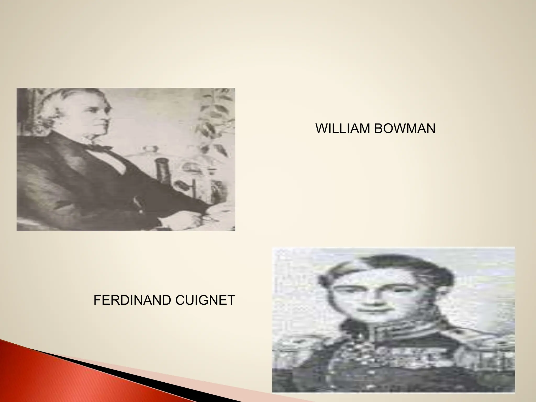
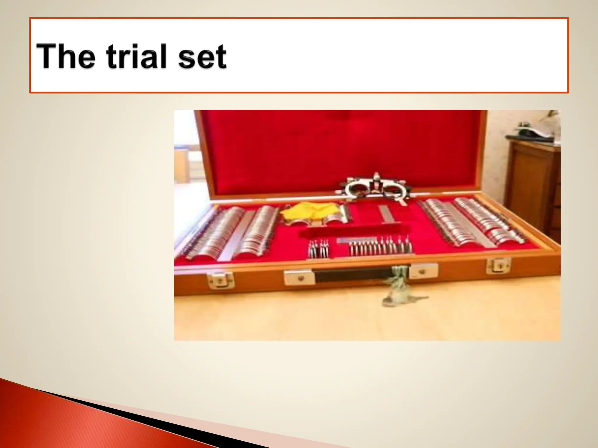
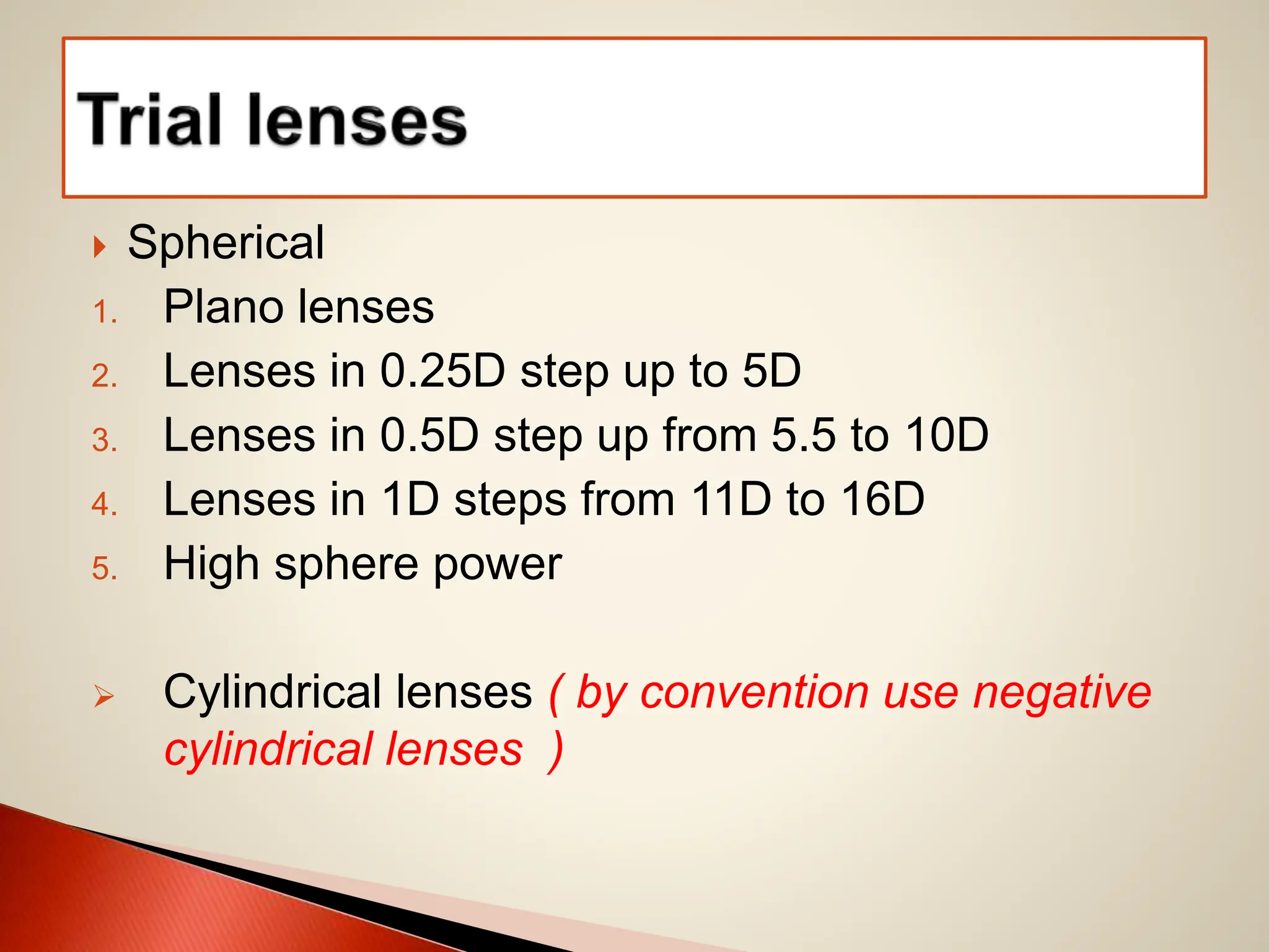
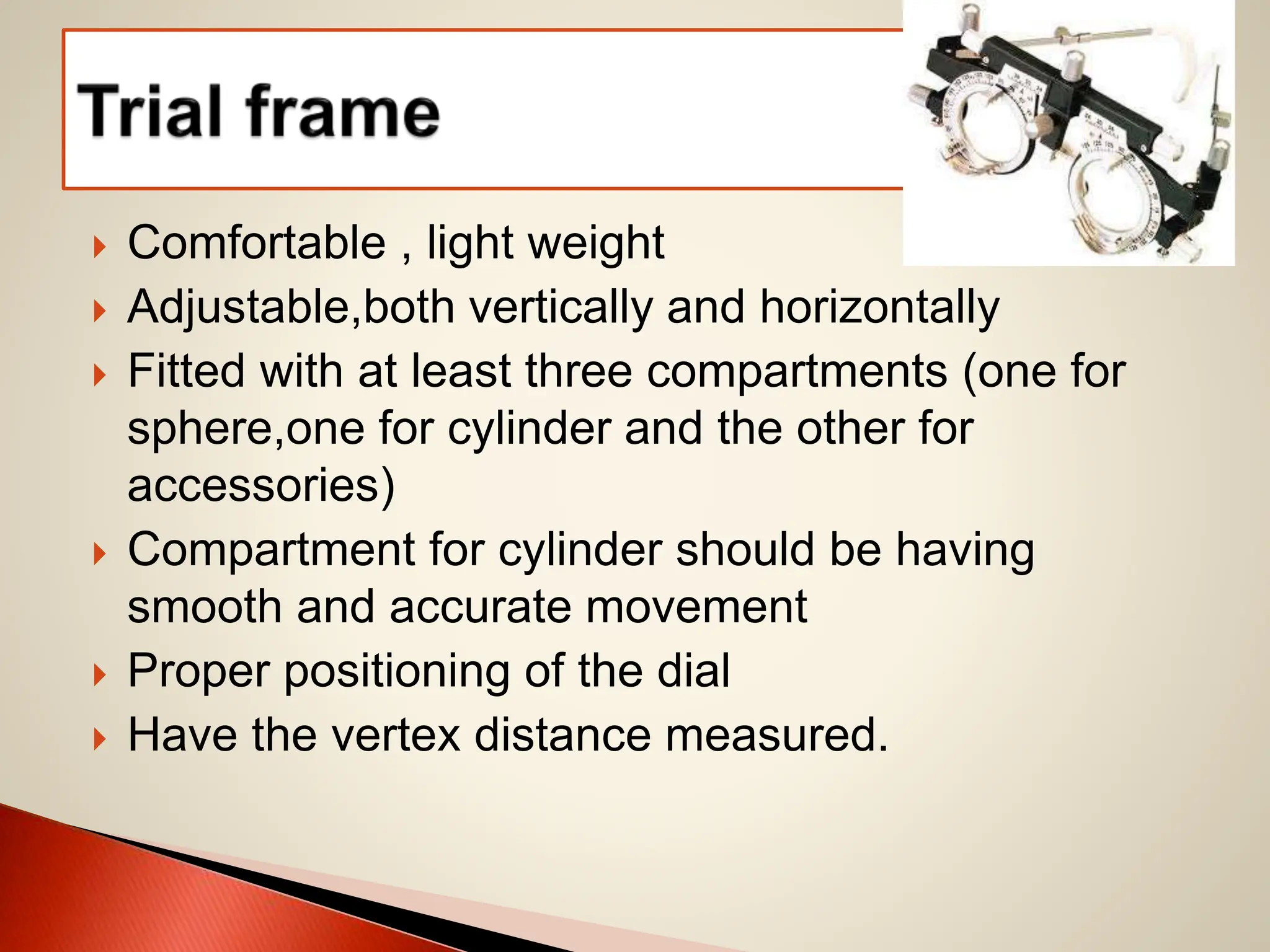
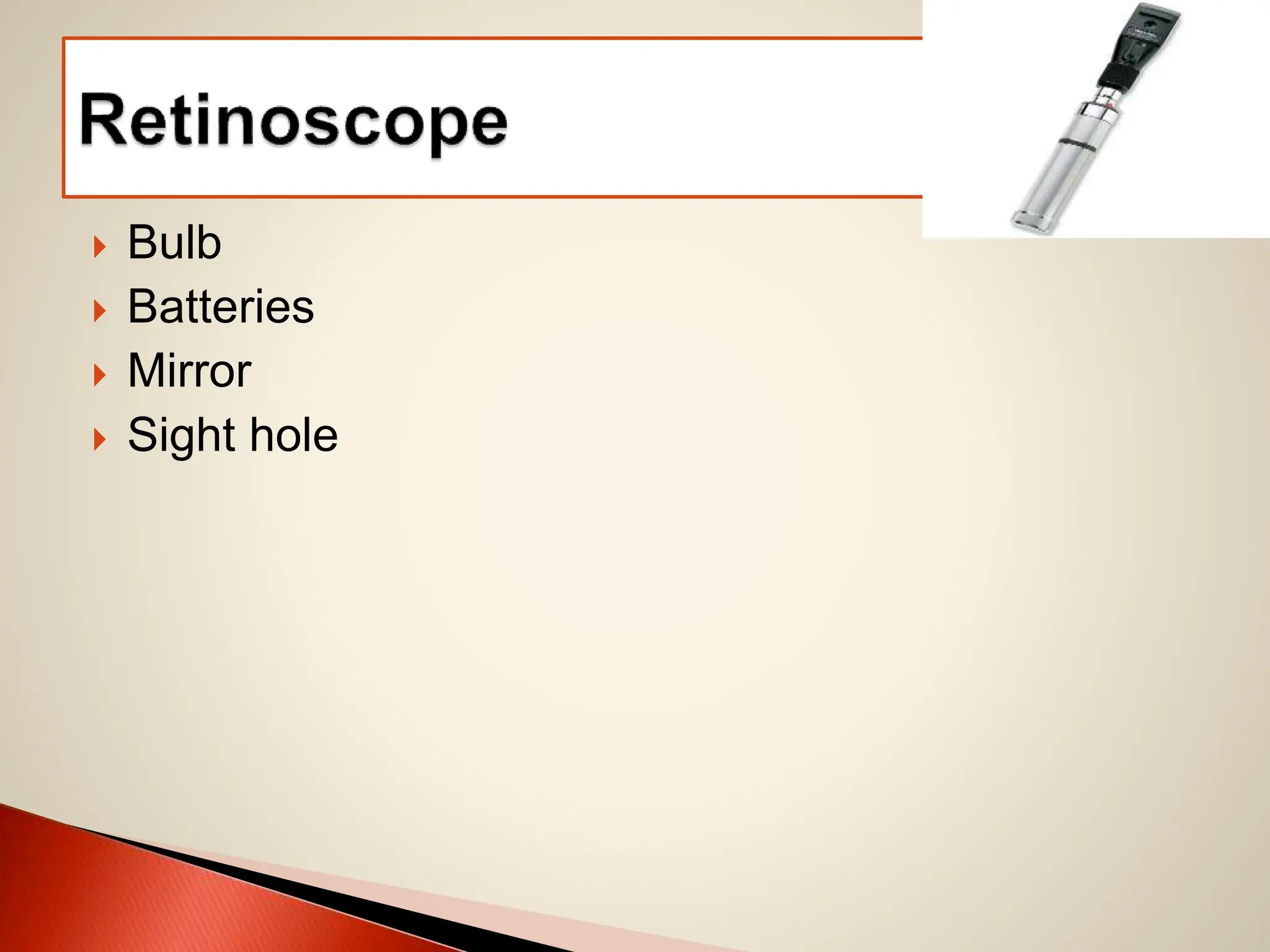
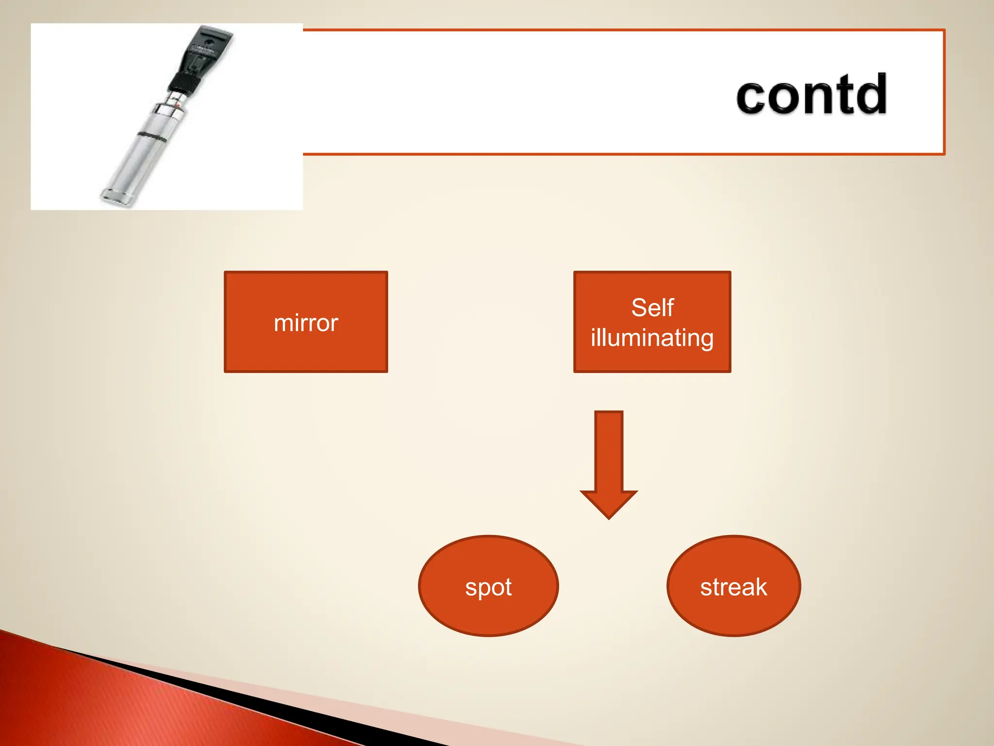
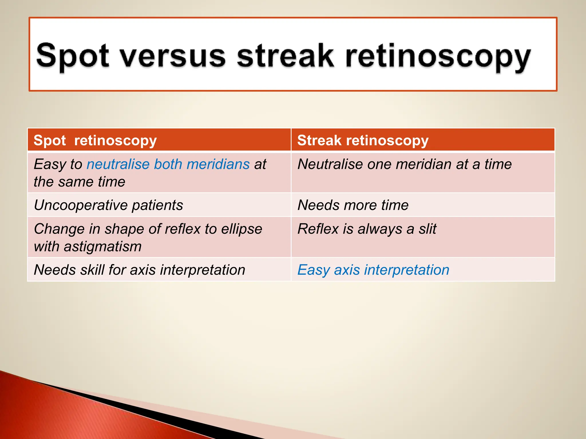
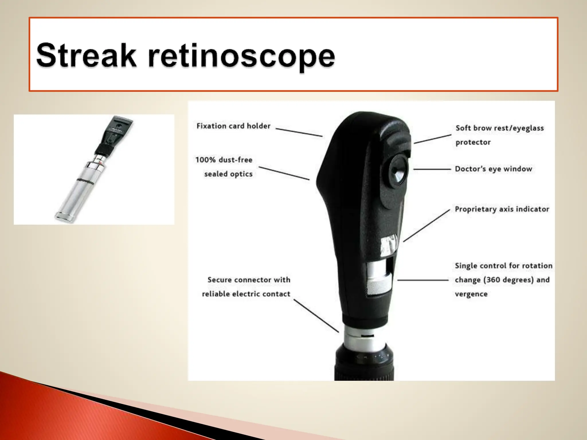
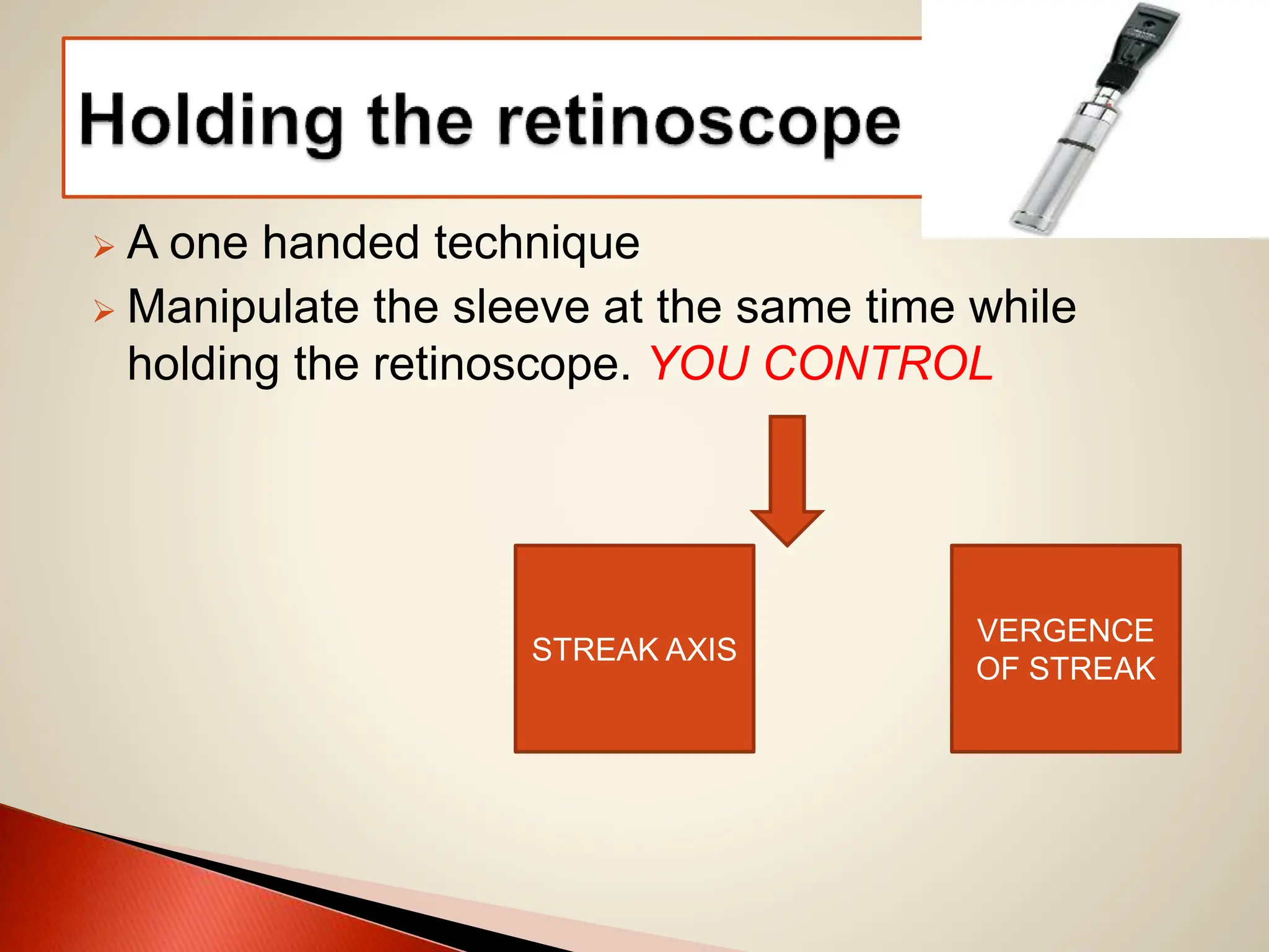
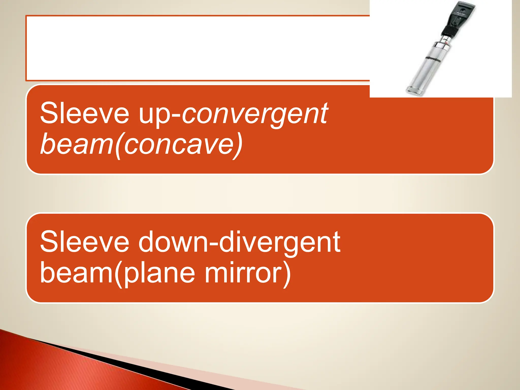
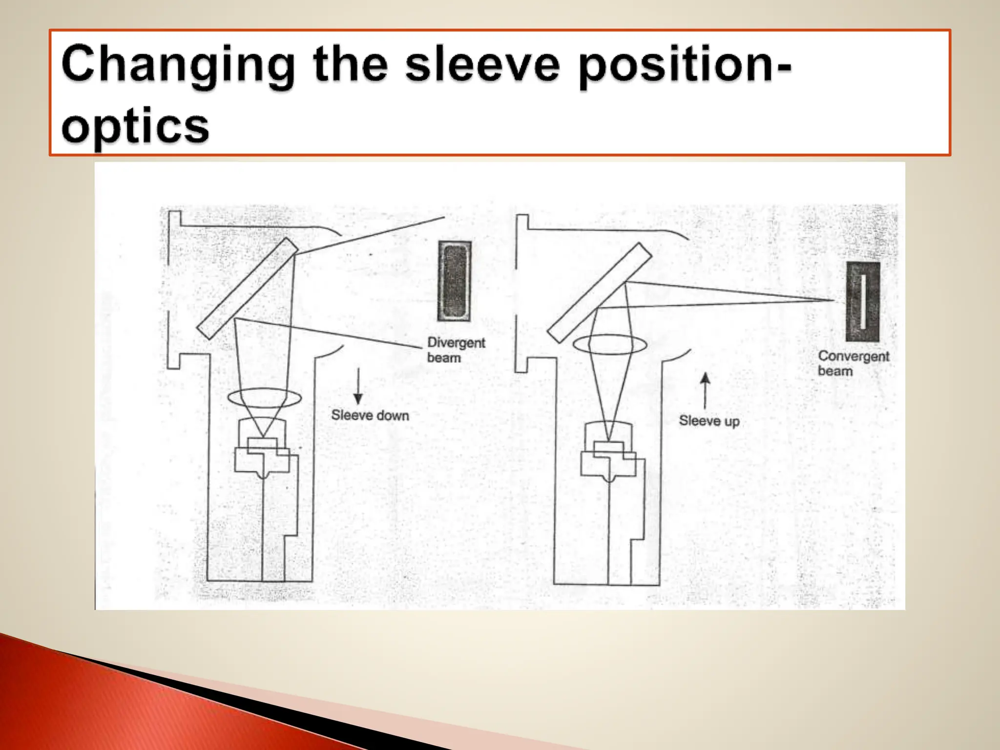
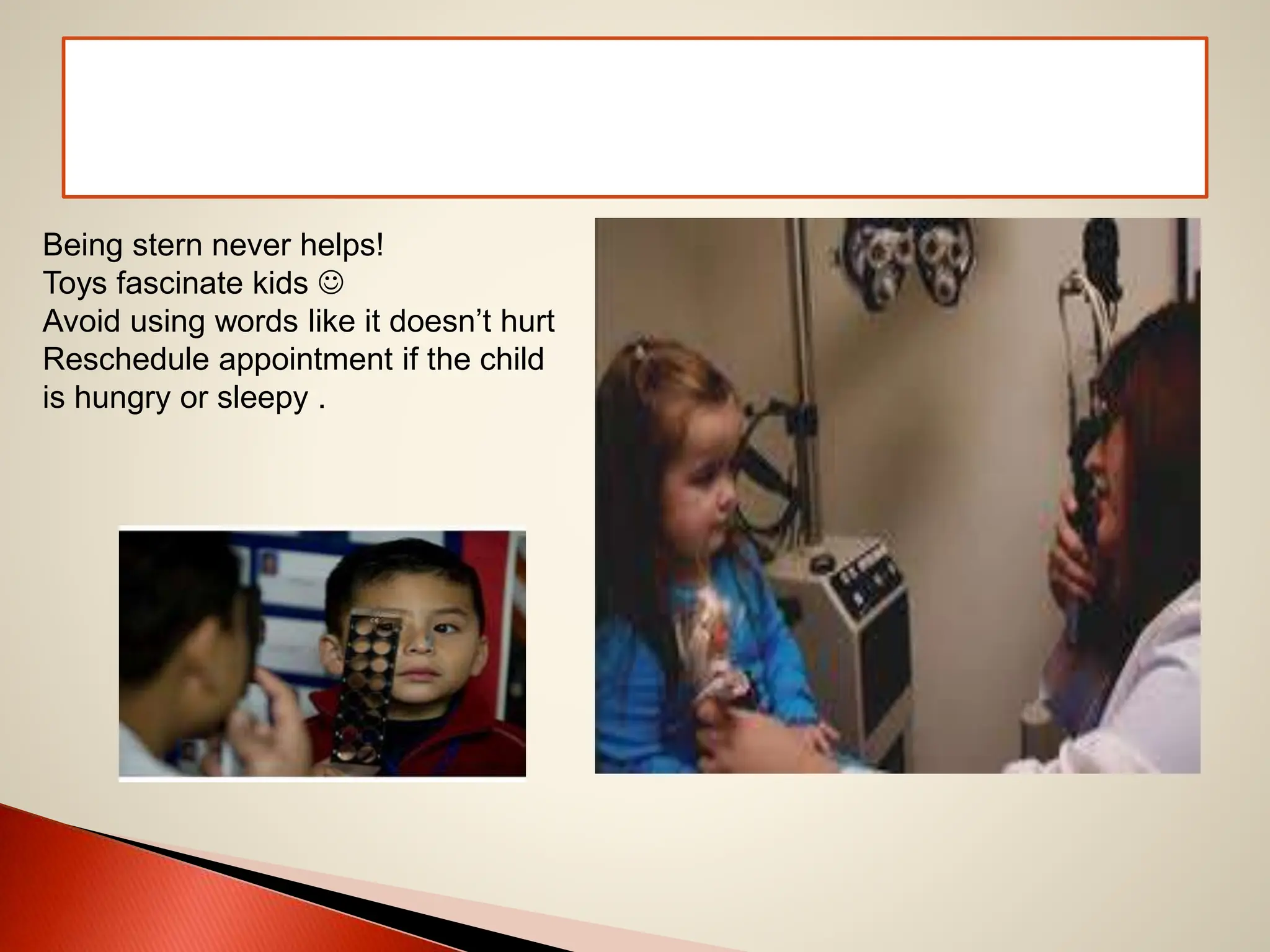
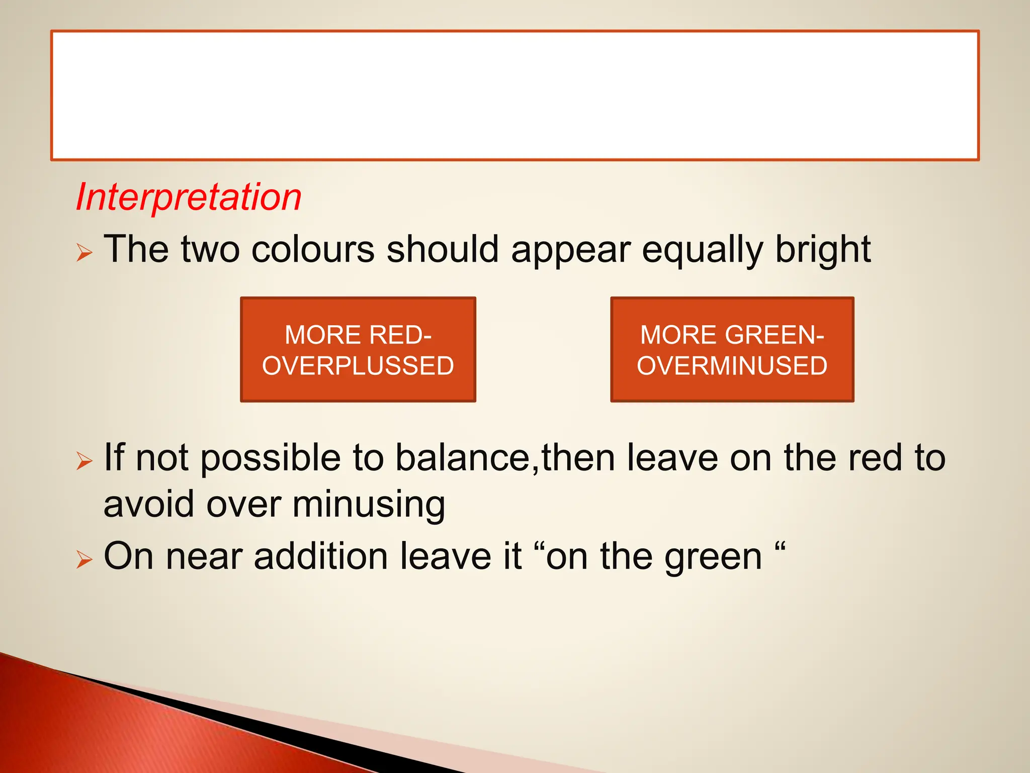
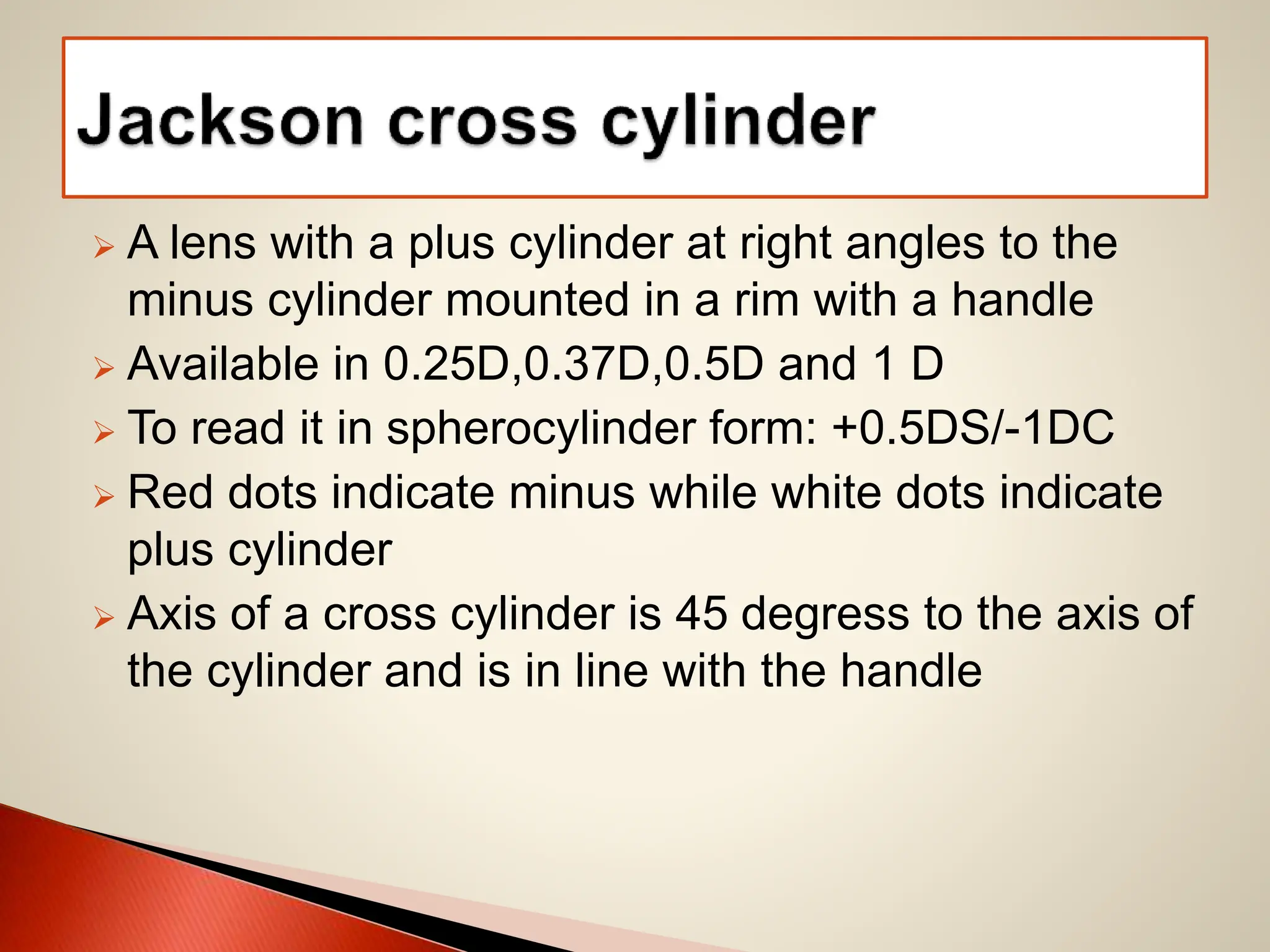
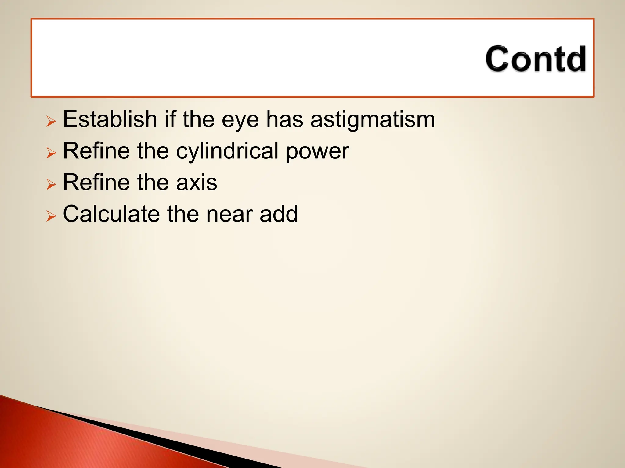
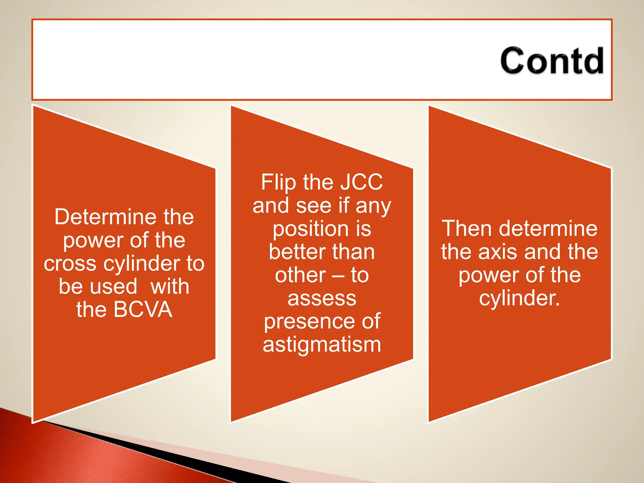
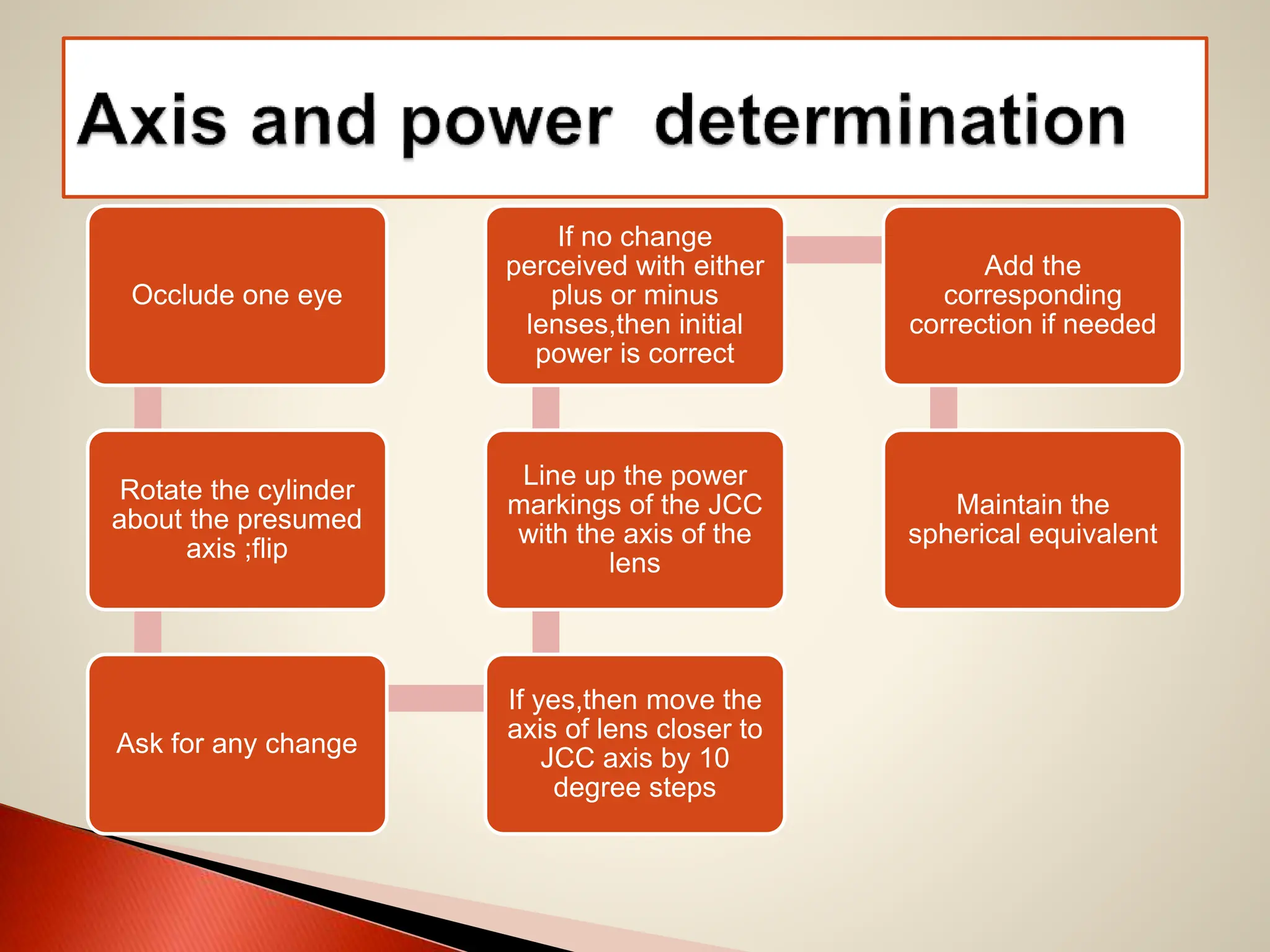
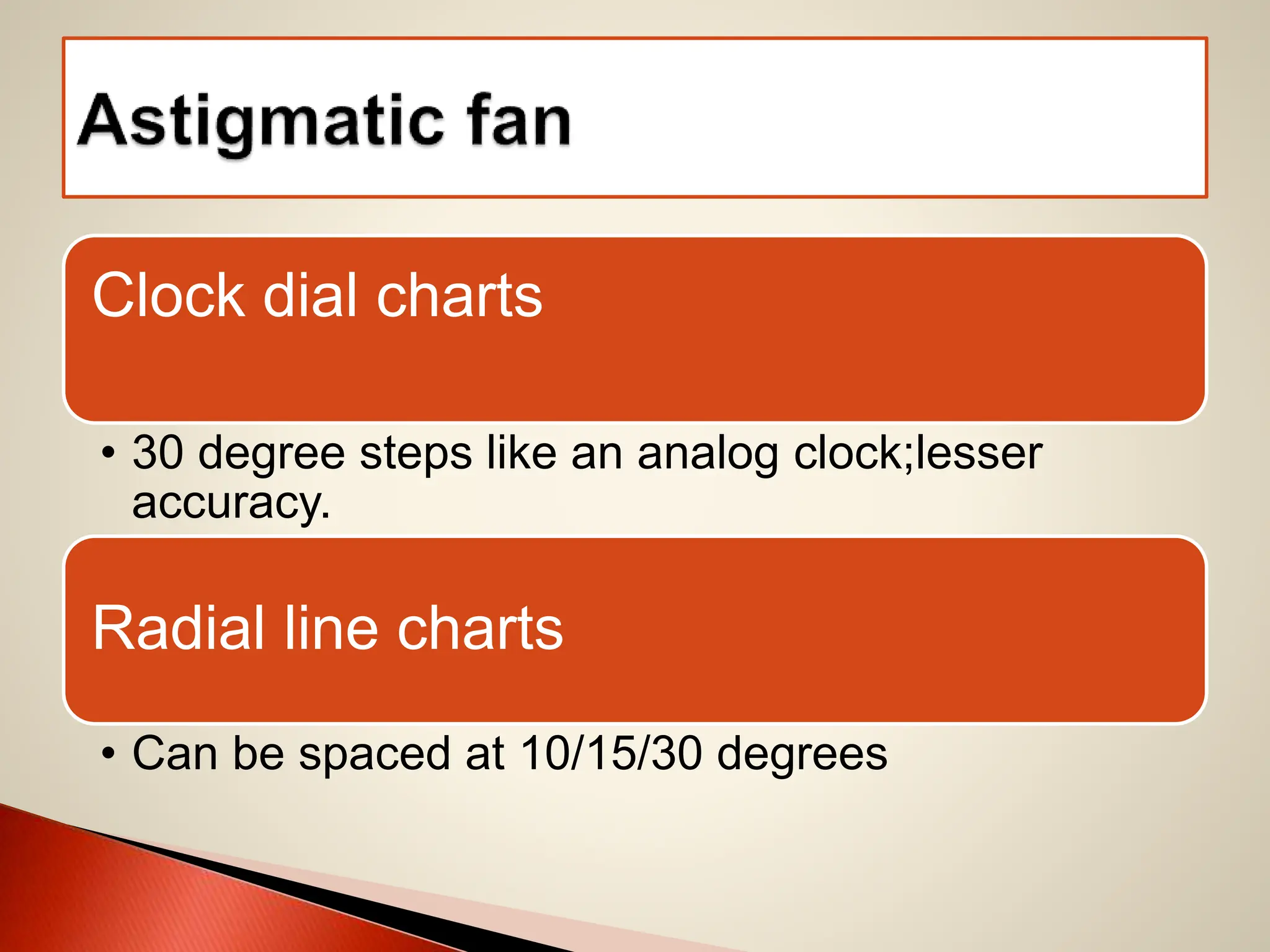
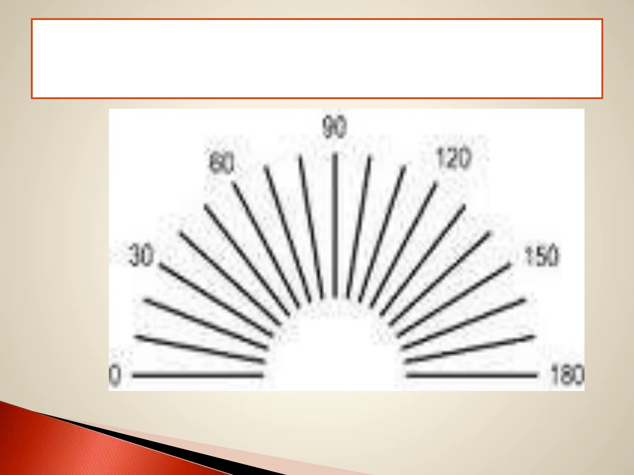
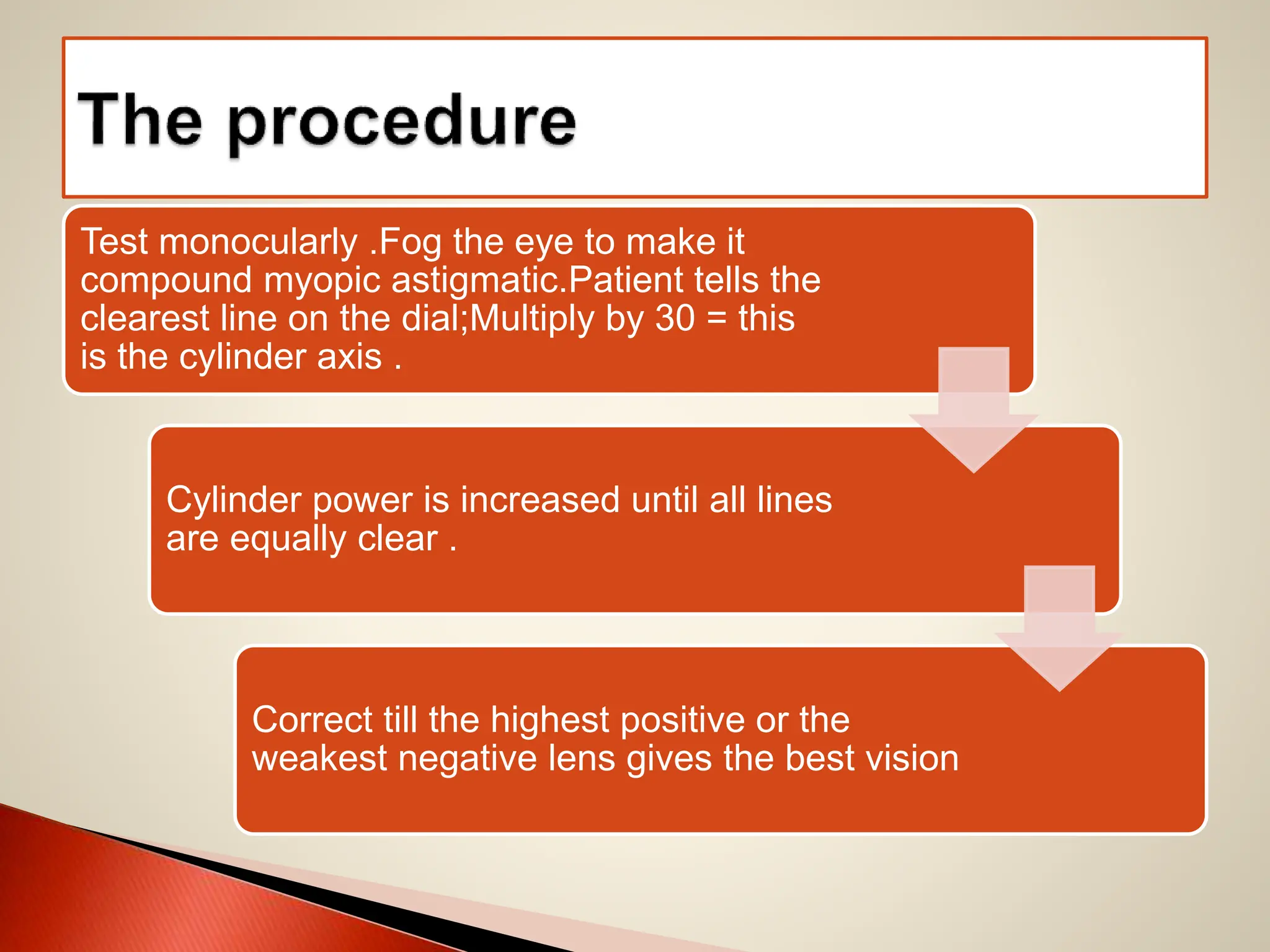
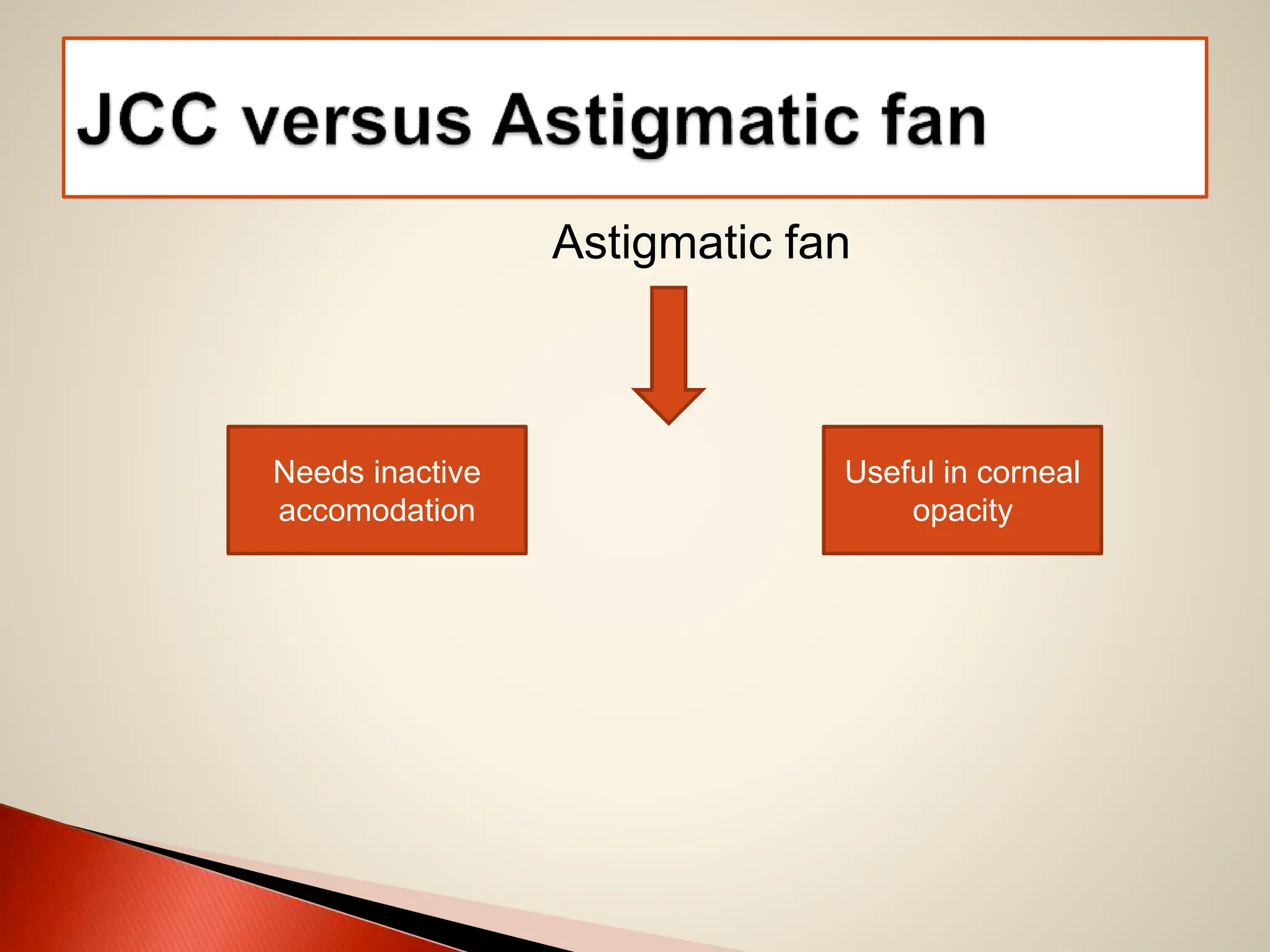
 History of refraction
 Definition of retinoscopy
 Types of retinoscopy
 Optics of retinoscopy
 Accessories needed to perform retinoscopy
 Objective retinoscopy
 Subjective retinoscopy-Duochrome test,Jackson Cross
Cylinder,Astigmatic Fan
 Near correction
 Binocular balancing
 Optics formulae for MCQs
 Dynamic retinoscopy
 Automated refractometers
 William Bowman (1859) saw the linear light reflex
with a Helmhotz Ophthalmoscope and used it in
diagnosis of corneal disorders like keratoconus
 French militiary ophthalmologist Ferdinand
Cuignet (1873 ) used the reflexes to measure the
errors of refraction.
 M.Mehgin proved the light reflex to be a fundal
reflex.
 H.Parent (1880) introduced the term retinoscopie
Shadow test
Skiascopy
Pupilloscopie
Koreskopie
WILLIAM BOWMAN
FERDINAND CUIGNET
The method of estimating the refractive state of the
eye is the technique of retinoscopy .
Duke-Elder’s Practice Of Refraction
Tenth Edition
Accomodation
Static Dynamic
Relaxed Active
Illumination stage : illumination of the subject’s
retina
Reflex stage : reflex imagery of this area onto the
observer
Projection stage : projection of the image by the
observer .
 History of the visual symptoms should be elicited
 Slit lamp examination should be carrried out
 Cover tests to determine any latent and manifest
deviations should be done
 Visual acuity should be tested both uniocularly
and binocularly and for distance and for near.
retinoscopy
Subjective
refraction
Refinement
of
refraction
Binocular
balancing
Near add
Test
muscle
balance
 A dark room
 A retinoscope
 A trial set
 A trial frame
 Spherical
1. Plano lenses
2. Lenses in 0.25D step up to 5D
3. Lenses in 0.5D step up from 5.5 to 10D
4. Lenses in 1D steps from 11D to 16D
5. High sphere power
 Cylindrical lenses ( by convention use negative
cylindrical lenses )
 The test lenses should ideally conform to in terms
of form and thickness to the spectacle lenses
being prescribed.
 Use reduced aperture lenses(thin lenses of
diameter 25mm)
 Preferably be planoconvex or planoconcave
 Prisms
 Occluder
 Pin hole disc
 Stenopic slit
 Red and green filters
 Maddox rod
 Comfortable , light weight
 Adjustable,both vertically and horizontally
 Fitted with at least three compartments (one for
sphere,one for cylinder and the other for
accessories)
 Compartment for cylinder should be having
smooth and accurate movement
 Proper positioning of the dial
 Have the vertex distance measured.
 Bulb
 Batteries
 Mirror
 Sight hole
mirror
Self
illuminating
spot streak
Spot retinoscopy Streak retinoscopy
Easy to neutralise both meridians at
the same time
Neutralise one meridian at a time
Uncooperative patients Needs more time
Change in shape of reflex to ellipse
with astigmatism
Reflex is always a slit
Needs skill for axis interpretation Easy axis interpretation
 Halogen light source
 Battery
 Mirror
 The vergence of the light can be controlled by the
sleeve by changing the distance between the lens
and the light source
 A one handed technique
 Manipulate the sleeve at the same time while
holding the retinoscope. YOU CONTROL
STREAK AXIS
VERGENCE
OF STREAK
Sleeve up-convergent
beam(concave)
Sleeve down-divergent
beam(plane mirror)
 More light enters the eye
 Small pupils/media opacities
 Remember the reflex movements are reversed in
comparison with the sleeve down position
To find the correct position of the sleeve to get a
plane mirror effect
 Hold the scope 33 cm from a flat surface
 The position of the sleeve that produces the
widest beam of light
 Parastop
 Distance from the retinoscope to the patient’s eye
 Ideal would be infinity;for practical purposes it is
6m or 20 feet
 Arm’s length
 Change the working distance in cases of very
small pupils or media opacities
 Choose a fixation target larger than 6/60
 A plain spot of light can also be used
 Eye levels of the subject and the examiner should
be same
 The examiner should not obstruct the patient’s
view of the fixation target
 Stay as close to the patient’s visual axis
A 10 degree off axis will produce a false astigmatic
judgement of 0.5D
 Ask the patient to keep both eyes open
 Not to look at the retinoscope light but at the
fixation target
 Tell him he can blink as he likes!
 Uniquely designed lens holder
 Ease of changing the lens during retinoscopy
 Typically, the patient sits behind the phoropter, and
looks through it at the distance vision chart and then at
near for individuals needing reading glasses.
Sometimes a retinoscope
maybe
used to provide the intial
setting in the phoropter .
They also measure phorias,
accomodative amplitudes and
Vergernces.
The major components of a
phoropter are
JCC,prisms and lenses.
 Pinhole optics allow the eye to focus on smaller
bundles of light entering the eye, improving focus.
 The stenopic slit found in all the trial sets is 1-2
mm by 15-35 mm in size. It splits an opaque disc
into two halves. It is useful in finding out the axis
of the cylinder.
Stenopic slit
 Check the direction of the reflex with the direction
of the movement of the retinoscope
 The reflex
Direction
Brightness
Width and speed
Axis
 With motion
 Against motion
 A neutral reflex
 An indeterminate reflex (scissor type/too dim)
Emmetropia,hyperopia,myopia < the dioptric
value of the working distance
Myopia more than the dioptric value of the
working distance
Myopia equal to the dioptric value of the
working distance
Is the motion of the reflex
parallel to the movement
of your retinoscope? No !
Reorient
 The relative brightness of the reflex is an indicator
of the degree of ametropia
 Dim reflex :
High refractive errors
Small pupil
Media opacity
 Increase the luminosity of the light source
 Dilate the pupil
 Reduce the working distance
 Sleeve up !
 Tells how far we are from the point of neutrality
 A narrower and a speedier streak indicates we are
nearing neutrality
 At neutrality the streak widens again and speeds
up more
 If the movement is “ with” add a plus lens
 If the movement is “against” add a minus lens
 If the reflex is dull to begin with start with a higher
power
 Neutralise one meridian with spherical
lenses.Then rotate 90 degress and assess the
reflex.
At neutralisation the patient’s far point is at the
plane of the retinoscope and no movement occurs
in other words the retinoscope is conjugate with
the patient’s retina.
 To confirm neutralisation,add an extra 0.25D lens
and look for reversal
 Move closer to the patient and a with movement
should appear
Agent Conc Maximu
m
cyclople
gia
Duration
of
cyclople
gia
Mydriasi
s
Recovery of
mydriasis
Tropicam
ide
1,2 20-25 4-6 20-30 6 hours
Cyclopen
tolate
0.5,1 20-45 24 15-45 1 day
Homatro
pine
2,5 30-90 72 10-30 1-3 days
Scopola
mine
0.25 30-60 7 days 40 3-10 days
Atropine 0.25,0.5,
1
120 15 days 30 7-10 days
Problems
Poor
technique
accomodation
Scissoring
/poor reflex
aberrations
Mixed aberrations( irregular astigmatism,decentred
lens,corneal scarring) lead to different nature of
the two halves of the reflex ( one part is relatively
myopic and the other hyperopic )
Find a lens that makes the two portions to meet at
the centre of the pupil
Sweep the reflex in all directions
Is there a change in the speed/brightness/direction
of the reflex ?
If yes then there is an
astigmatism
Immature cataracts can lead to confusing reflexes
An experienced examiner can get a rough guide as
to the refractive error and give an appropritae
subjective correction
 Vision maynot be a true indication of the degree of
hyperopia
 Do a cycloplegic refraction in hyperopia
 In a myope perform a dry retinoscopy as far as
possible
 A subjective refraction after objective evaluation is
the best method to prescribe in myopes
 Rule out pseudomyopia due to ciliary spasm
 Prescribe the correction as accepted under the
post mydriatic test
 Instruction about visual hygiene
 When treating larger amounts of astigmatism
prefer to undercorrect for the first time and
gradually increase.
Strive for patient comfort rather than theoretical
optical correction.
 In irregular astigmatism a compromise should be
arrived at by subjective refraction
 Never overplus near correction !
 A short statured person may have lesser working
distance !
 Infant’s eyes are hyperopic with a very strong
accomodation
 A nuclear cataract will have a myopic refraction
with various zones of refraction while a cortical
cataract will have a good central glow( do an
undilated examination)
Being stern never helps!
Toys fascinate kids 
Avoid using words like it doesn’t hurt
Reschedule appointment if the child
is hungry or sleepy .
Lens power formula
100 cm / focal length
Lens effectivity formula
D2 = d1 /1-s*d1
Spherical equivalent is sum of the (sphere + cylinder/2)
Amplitude of accomodation = 100/ near point of
accomodation
To tranpose:add the sphere and cylinder,change the
sign of the cylinder and add 90 to the axis of the
cylinder
Prentice rule: amount of deviation= decentration of
visual axis with respect to lens center * power of lens
 Infant , child
 Stroke patient
 Malingerers
 Mentally challenged
Determination of the refractive error by asking
and relying on the subject
When objective findings are determined-
retinoscopy/AR/power of glasses/keratometry
No data-takes visual acuity in assessment
 Determine the sphere
 Determine the cylinder- power and axis
 Binocular balancing
IDENTIFY THE BEST VISION SPHERE
MAXIMUM PLUS OR MINIMUM MINUS
TOLERATED
DUOCHROME TEST
ESTIMATE CORRECTION BASED ON ACUITY
DIVIDE BY 18 FOR SPHERE
(VALID FOR MANIFEST HYPEROPIA ONLY )
DIVIDE BY 9 FOR CYLINDER
Determine visual acuity
Snellen ETDRS
Visual acuity Spherical error Cylindrical error
6/6 - -
6/9 0.75 1.5
6/12 1 2
6/18 1.5 3
6/24 1.75 3.5
6/36 2.25 4.5
6/60 3 High
Cylinder error has a better acuity because there is a circle of least diffusion
 Objective
refraction
present
Blur
below
6/18
Reduce plus lenses and
refine
Remove the old
lens only after
Putting a newer
lens in the hyperopic correction
Control accomodation
DUOCHROME TEST
Described by Brown and Freeman
Principle : Chromatic aberration wherein green light
(535nm) is focussed 0.25D in front of retina and
red light ( 620 nm ) is focussed 0.25D behind it
with yellow light being taken as a reference.
• Red background
clearer and darker
Myope
• Green background
clearer and darker
hyperope
Procedure :
Determine the best sphere
Do this test monocularly
Ask which of the numbers or letters on the red or
green appear darker,sharper and clearer.
Interpretation
 The two colours should appear equally bright
 If not possible to balance,then leave on the red to
avoid over minusing
 On near addition leave it “on the green “
MORE RED-
OVERPLUSSED
MORE GREEN-
OVERMINUSED
 Ametropia should be corrected to 6/12 before
going ahead with the test
 The test may over minus in elderly patients
JACKSON CROSS CYLINDER
ASTIGMATIC FAN
 A lens with a plus cylinder at right angles to the
minus cylinder mounted in a rim with a handle
 Available in 0.25D,0.37D,0.5D and 1 D
 To read it in spherocylinder form: +0.5DS/-1DC
 Red dots indicate minus while white dots indicate
plus cylinder
 Axis of a cross cylinder is 45 degress to the axis of
the cylinder and is in line with the handle
 Establish if the eye has astigmatism
 Refine the cylindrical power
 Refine the axis
 Calculate the near add
Determine the
power of the
cross cylinder to
be used with
the BCVA
Flip the JCC
and see if any
position is
better than
other – to
assess
presence of
astigmatism
Then determine
the axis and the
power of the
cylinder.
Occlude one eye
Rotate the cylinder
about the presumed
axis ;flip
Ask for any change
If yes,then move the
axis of lens closer to
JCC axis by 10
degree steps
Line up the power
markings of the JCC
with the axis of the
lens
If no change
perceived with either
plus or minus
lenses,then initial
power is correct
Add the
corresponding
correction if needed
Maintain the
spherical equivalent
Clock dial charts
• 30 degree steps like an analog clock;lesser
accuracy.
Radial line charts
• Can be spaced at 10/15/30 degrees
Test monocularly .Fog the eye to make it
compound myopic astigmatic.Patient tells the
clearest line on the dial;Multiply by 30 = this
is the cylinder axis .
Cylinder power is increased until all lines
are equally clear .
Correct till the highest positive or the
weakest negative lens gives the best vision
Astigmatic fan
Needs inactive
accomodation
Useful in corneal
opacity
 To measure the refractive state when the patient
fixates for the near
 Test monocularly
 Aim is to use the reading addition to substitute for
about one third to half of the existing amplitude of
accomodation
 The reading correction is added to the power for
the distance
 No correction is made for the cylinder
Eg : to a distance prescription of +2.5DS/-1D CY at
180 the near prescription would read + 4DS/-
1DCY at 180
Do not over correct for near !
Dynamic
retinoscopy
Refining of
previous add
Cross
cylinder for
near
Amplitude of
accomodation
Expected age
value
 Simulate the patient’s habitual viewing
circumstances such as lighting and working
distance
 If tested binocularly may lead to a false low near
add due to convergent accomodation
 Give the distance prescription and measure the
amplitude of accomodation.
Amplitude of accomodation = 1 / near point
Measure the near point by a near point ruler with a
target carrier
“Push up” or “push away” method
Age 10 20 35 45 50 55 60 65 70
Dond
ers
14 10 5.5 3.5 2.5 1.75 1 0.5 0.25
Hofst
etter
15.5 12.5 8 5 3.5 2 0.5 0.5 0
 To balance the accomodation between the two
eyes
 Assess the best corrected acuity uniocularly
Alternate
cover
method
Humphriss
immediate
contrast
method
Borish
technique
Rule out
 Phorias
 Tropias
 Convergence insufficiency
 Fusional reserves
Infrared source
Fixation target
Badal optometer ( position of the lens is linearly
propotional to the refractive error with a constant
magnification )
COLLIMATION OF IR RAYS
BEAM
SPLITTER
REMOVES
REFLECTED
LIGHT FROM
CORNEA
LATERAL MOVEMENT OF THE SYSTEM TO FIND
OPTIMAL FOCUS OF SLIT ON RETINA
 Measures at least three meridians of the eye
 Uses sine squared function to measure the
refractive power
Power = sphere + ( cylinder sine 2@ )
 Autorefractors measure the refractive error of the
patient.
 Not dependent on patient or operator judgement
 Not reliable in pathological corneas like post
graft,keratoconus and post refractive surgery
 Pseudomyopia due to accomodation(can use
cycloplegics or auto-fogging )
 Anomalies in vitreous cause errors
 Patient fixates with both eyes on a near object
 Magnetic fixation cards incorporated in the
retinoscopes
 A small “ with “ movement is seen for near in
emmetropes with a normal accomodation
 Add lenses to achieve the neutral point – this
represent the accomodative power
 To check for accomodative disorders
 To determine the adequacy of cycloplegia
 Alert the practitioner to the presence of
uncorrected hypermetropia or anisometropia
 Useful in amblyopia therapy
Techniques of refraction is the process of calculation of glass power.

Techniques of refraction is the process of calculation of glass power.

  • 2.
     History ofrefraction  Definition of retinoscopy  Types of retinoscopy  Optics of retinoscopy  Accessories needed to perform retinoscopy  Objective retinoscopy  Subjective retinoscopy-Duochrome test,Jackson Cross Cylinder,Astigmatic Fan  Near correction  Binocular balancing  Optics formulae for MCQs  Dynamic retinoscopy  Automated refractometers
  • 3.
     William Bowman(1859) saw the linear light reflex with a Helmhotz Ophthalmoscope and used it in diagnosis of corneal disorders like keratoconus  French militiary ophthalmologist Ferdinand Cuignet (1873 ) used the reflexes to measure the errors of refraction.  M.Mehgin proved the light reflex to be a fundal reflex.  H.Parent (1880) introduced the term retinoscopie
  • 4.
  • 5.
  • 6.
    The method ofestimating the refractive state of the eye is the technique of retinoscopy . Duke-Elder’s Practice Of Refraction Tenth Edition
  • 7.
  • 8.
    Illumination stage :illumination of the subject’s retina Reflex stage : reflex imagery of this area onto the observer Projection stage : projection of the image by the observer .
  • 12.
     History ofthe visual symptoms should be elicited  Slit lamp examination should be carrried out  Cover tests to determine any latent and manifest deviations should be done  Visual acuity should be tested both uniocularly and binocularly and for distance and for near.
  • 13.
  • 14.
     A darkroom  A retinoscope  A trial set  A trial frame
  • 16.
     Spherical 1. Planolenses 2. Lenses in 0.25D step up to 5D 3. Lenses in 0.5D step up from 5.5 to 10D 4. Lenses in 1D steps from 11D to 16D 5. High sphere power  Cylindrical lenses ( by convention use negative cylindrical lenses )
  • 17.
     The testlenses should ideally conform to in terms of form and thickness to the spectacle lenses being prescribed.  Use reduced aperture lenses(thin lenses of diameter 25mm)  Preferably be planoconvex or planoconcave
  • 18.
     Prisms  Occluder Pin hole disc  Stenopic slit  Red and green filters  Maddox rod
  • 19.
     Comfortable ,light weight  Adjustable,both vertically and horizontally  Fitted with at least three compartments (one for sphere,one for cylinder and the other for accessories)  Compartment for cylinder should be having smooth and accurate movement  Proper positioning of the dial  Have the vertex distance measured.
  • 20.
     Bulb  Batteries Mirror  Sight hole
  • 21.
  • 22.
    Spot retinoscopy Streakretinoscopy Easy to neutralise both meridians at the same time Neutralise one meridian at a time Uncooperative patients Needs more time Change in shape of reflex to ellipse with astigmatism Reflex is always a slit Needs skill for axis interpretation Easy axis interpretation
  • 26.
     Halogen lightsource  Battery  Mirror  The vergence of the light can be controlled by the sleeve by changing the distance between the lens and the light source
  • 28.
     A onehanded technique  Manipulate the sleeve at the same time while holding the retinoscope. YOU CONTROL STREAK AXIS VERGENCE OF STREAK
  • 29.
  • 31.
     More lightenters the eye  Small pupils/media opacities  Remember the reflex movements are reversed in comparison with the sleeve down position
  • 32.
    To find thecorrect position of the sleeve to get a plane mirror effect  Hold the scope 33 cm from a flat surface  The position of the sleeve that produces the widest beam of light  Parastop
  • 33.
     Distance fromthe retinoscope to the patient’s eye  Ideal would be infinity;for practical purposes it is 6m or 20 feet  Arm’s length  Change the working distance in cases of very small pupils or media opacities
  • 34.
     Choose afixation target larger than 6/60  A plain spot of light can also be used  Eye levels of the subject and the examiner should be same  The examiner should not obstruct the patient’s view of the fixation target  Stay as close to the patient’s visual axis A 10 degree off axis will produce a false astigmatic judgement of 0.5D
  • 35.
     Ask thepatient to keep both eyes open  Not to look at the retinoscope light but at the fixation target  Tell him he can blink as he likes!
  • 36.
     Uniquely designedlens holder  Ease of changing the lens during retinoscopy  Typically, the patient sits behind the phoropter, and looks through it at the distance vision chart and then at near for individuals needing reading glasses.
  • 37.
    Sometimes a retinoscope maybe usedto provide the intial setting in the phoropter . They also measure phorias, accomodative amplitudes and Vergernces.
  • 38.
    The major componentsof a phoropter are JCC,prisms and lenses.
  • 40.
     Pinhole opticsallow the eye to focus on smaller bundles of light entering the eye, improving focus.  The stenopic slit found in all the trial sets is 1-2 mm by 15-35 mm in size. It splits an opaque disc into two halves. It is useful in finding out the axis of the cylinder.
  • 41.
  • 43.
     Check thedirection of the reflex with the direction of the movement of the retinoscope  The reflex Direction Brightness Width and speed Axis
  • 44.
     With motion Against motion  A neutral reflex  An indeterminate reflex (scissor type/too dim) Emmetropia,hyperopia,myopia < the dioptric value of the working distance Myopia more than the dioptric value of the working distance Myopia equal to the dioptric value of the working distance
  • 46.
    Is the motionof the reflex parallel to the movement of your retinoscope? No ! Reorient
  • 47.
     The relativebrightness of the reflex is an indicator of the degree of ametropia  Dim reflex : High refractive errors Small pupil Media opacity
  • 48.
     Increase theluminosity of the light source  Dilate the pupil  Reduce the working distance  Sleeve up !
  • 49.
     Tells howfar we are from the point of neutrality  A narrower and a speedier streak indicates we are nearing neutrality  At neutrality the streak widens again and speeds up more
  • 50.
     If themovement is “ with” add a plus lens  If the movement is “against” add a minus lens  If the reflex is dull to begin with start with a higher power  Neutralise one meridian with spherical lenses.Then rotate 90 degress and assess the reflex. At neutralisation the patient’s far point is at the plane of the retinoscope and no movement occurs in other words the retinoscope is conjugate with the patient’s retina.
  • 51.
     To confirmneutralisation,add an extra 0.25D lens and look for reversal  Move closer to the patient and a with movement should appear
  • 52.
    Agent Conc Maximu m cyclople gia Duration of cyclople gia Mydriasi s Recoveryof mydriasis Tropicam ide 1,2 20-25 4-6 20-30 6 hours Cyclopen tolate 0.5,1 20-45 24 15-45 1 day Homatro pine 2,5 30-90 72 10-30 1-3 days Scopola mine 0.25 30-60 7 days 40 3-10 days Atropine 0.25,0.5, 1 120 15 days 30 7-10 days
  • 54.
  • 55.
    Mixed aberrations( irregularastigmatism,decentred lens,corneal scarring) lead to different nature of the two halves of the reflex ( one part is relatively myopic and the other hyperopic ) Find a lens that makes the two portions to meet at the centre of the pupil
  • 56.
    Sweep the reflexin all directions Is there a change in the speed/brightness/direction of the reflex ? If yes then there is an astigmatism
  • 57.
    Immature cataracts canlead to confusing reflexes An experienced examiner can get a rough guide as to the refractive error and give an appropritae subjective correction
  • 58.
     Vision maynotbe a true indication of the degree of hyperopia  Do a cycloplegic refraction in hyperopia  In a myope perform a dry retinoscopy as far as possible  A subjective refraction after objective evaluation is the best method to prescribe in myopes  Rule out pseudomyopia due to ciliary spasm  Prescribe the correction as accepted under the post mydriatic test
  • 59.
     Instruction aboutvisual hygiene  When treating larger amounts of astigmatism prefer to undercorrect for the first time and gradually increase. Strive for patient comfort rather than theoretical optical correction.  In irregular astigmatism a compromise should be arrived at by subjective refraction  Never overplus near correction !  A short statured person may have lesser working distance !
  • 60.
     Infant’s eyesare hyperopic with a very strong accomodation  A nuclear cataract will have a myopic refraction with various zones of refraction while a cortical cataract will have a good central glow( do an undilated examination)
  • 61.
    Being stern neverhelps! Toys fascinate kids  Avoid using words like it doesn’t hurt Reschedule appointment if the child is hungry or sleepy .
  • 62.
    Lens power formula 100cm / focal length Lens effectivity formula D2 = d1 /1-s*d1 Spherical equivalent is sum of the (sphere + cylinder/2) Amplitude of accomodation = 100/ near point of accomodation To tranpose:add the sphere and cylinder,change the sign of the cylinder and add 90 to the axis of the cylinder Prentice rule: amount of deviation= decentration of visual axis with respect to lens center * power of lens
  • 63.
     Infant ,child  Stroke patient  Malingerers  Mentally challenged
  • 64.
    Determination of therefractive error by asking and relying on the subject When objective findings are determined- retinoscopy/AR/power of glasses/keratometry No data-takes visual acuity in assessment
  • 65.
     Determine thesphere  Determine the cylinder- power and axis  Binocular balancing
  • 66.
    IDENTIFY THE BESTVISION SPHERE MAXIMUM PLUS OR MINIMUM MINUS TOLERATED DUOCHROME TEST ESTIMATE CORRECTION BASED ON ACUITY DIVIDE BY 18 FOR SPHERE (VALID FOR MANIFEST HYPEROPIA ONLY ) DIVIDE BY 9 FOR CYLINDER Determine visual acuity Snellen ETDRS
  • 67.
    Visual acuity Sphericalerror Cylindrical error 6/6 - - 6/9 0.75 1.5 6/12 1 2 6/18 1.5 3 6/24 1.75 3.5 6/36 2.25 4.5 6/60 3 High Cylinder error has a better acuity because there is a circle of least diffusion
  • 68.
     Objective refraction present Blur below 6/18 Reduce pluslenses and refine Remove the old lens only after Putting a newer lens in the hyperopic correction Control accomodation
  • 69.
    DUOCHROME TEST Described byBrown and Freeman Principle : Chromatic aberration wherein green light (535nm) is focussed 0.25D in front of retina and red light ( 620 nm ) is focussed 0.25D behind it with yellow light being taken as a reference.
  • 70.
    • Red background clearerand darker Myope • Green background clearer and darker hyperope
  • 72.
    Procedure : Determine thebest sphere Do this test monocularly Ask which of the numbers or letters on the red or green appear darker,sharper and clearer.
  • 73.
    Interpretation  The twocolours should appear equally bright  If not possible to balance,then leave on the red to avoid over minusing  On near addition leave it “on the green “ MORE RED- OVERPLUSSED MORE GREEN- OVERMINUSED
  • 74.
     Ametropia shouldbe corrected to 6/12 before going ahead with the test  The test may over minus in elderly patients
  • 75.
  • 76.
     A lenswith a plus cylinder at right angles to the minus cylinder mounted in a rim with a handle  Available in 0.25D,0.37D,0.5D and 1 D  To read it in spherocylinder form: +0.5DS/-1DC  Red dots indicate minus while white dots indicate plus cylinder  Axis of a cross cylinder is 45 degress to the axis of the cylinder and is in line with the handle
  • 78.
     Establish ifthe eye has astigmatism  Refine the cylindrical power  Refine the axis  Calculate the near add
  • 79.
    Determine the power ofthe cross cylinder to be used with the BCVA Flip the JCC and see if any position is better than other – to assess presence of astigmatism Then determine the axis and the power of the cylinder.
  • 80.
    Occlude one eye Rotatethe cylinder about the presumed axis ;flip Ask for any change If yes,then move the axis of lens closer to JCC axis by 10 degree steps Line up the power markings of the JCC with the axis of the lens If no change perceived with either plus or minus lenses,then initial power is correct Add the corresponding correction if needed Maintain the spherical equivalent
  • 81.
    Clock dial charts •30 degree steps like an analog clock;lesser accuracy. Radial line charts • Can be spaced at 10/15/30 degrees
  • 83.
    Test monocularly .Fogthe eye to make it compound myopic astigmatic.Patient tells the clearest line on the dial;Multiply by 30 = this is the cylinder axis . Cylinder power is increased until all lines are equally clear . Correct till the highest positive or the weakest negative lens gives the best vision
  • 84.
  • 85.
     To measurethe refractive state when the patient fixates for the near  Test monocularly  Aim is to use the reading addition to substitute for about one third to half of the existing amplitude of accomodation
  • 86.
     The readingcorrection is added to the power for the distance  No correction is made for the cylinder Eg : to a distance prescription of +2.5DS/-1D CY at 180 the near prescription would read + 4DS/- 1DCY at 180
  • 87.
    Do not overcorrect for near !
  • 88.
    Dynamic retinoscopy Refining of previous add Cross cylinderfor near Amplitude of accomodation Expected age value
  • 89.
     Simulate thepatient’s habitual viewing circumstances such as lighting and working distance  If tested binocularly may lead to a false low near add due to convergent accomodation  Give the distance prescription and measure the amplitude of accomodation.
  • 90.
    Amplitude of accomodation= 1 / near point Measure the near point by a near point ruler with a target carrier “Push up” or “push away” method
  • 92.
    Age 10 2035 45 50 55 60 65 70 Dond ers 14 10 5.5 3.5 2.5 1.75 1 0.5 0.25 Hofst etter 15.5 12.5 8 5 3.5 2 0.5 0.5 0
  • 93.
     To balancethe accomodation between the two eyes  Assess the best corrected acuity uniocularly
  • 94.
  • 95.
    Rule out  Phorias Tropias  Convergence insufficiency  Fusional reserves
  • 96.
    Infrared source Fixation target Badaloptometer ( position of the lens is linearly propotional to the refractive error with a constant magnification )
  • 97.
    COLLIMATION OF IRRAYS BEAM SPLITTER REMOVES REFLECTED LIGHT FROM CORNEA LATERAL MOVEMENT OF THE SYSTEM TO FIND OPTIMAL FOCUS OF SLIT ON RETINA
  • 98.
     Measures atleast three meridians of the eye  Uses sine squared function to measure the refractive power Power = sphere + ( cylinder sine 2@ )
  • 99.
     Autorefractors measurethe refractive error of the patient.  Not dependent on patient or operator judgement  Not reliable in pathological corneas like post graft,keratoconus and post refractive surgery  Pseudomyopia due to accomodation(can use cycloplegics or auto-fogging )  Anomalies in vitreous cause errors
  • 100.
     Patient fixateswith both eyes on a near object  Magnetic fixation cards incorporated in the retinoscopes  A small “ with “ movement is seen for near in emmetropes with a normal accomodation  Add lenses to achieve the neutral point – this represent the accomodative power
  • 102.
     To checkfor accomodative disorders  To determine the adequacy of cycloplegia  Alert the practitioner to the presence of uncorrected hypermetropia or anisometropia  Useful in amblyopia therapy