these slides explain the objective refraction in optometry , and describes its types and its measurement , and it gives you in details the types of Retinoscopy.
2. Principle of retinoscopy
analysis of the light reflex created in the pupil by light reflected back
from the retina for the purpose of objectively determining the refractive
error
This reflected light starts with a light shining off a plane mirror of the
retinoscope and then passing through the pupil to eventually shine on
the retina
The light reflected back off the retina is viewed in the pupil by the
examiner “peeking” means *the examiner sees * through a small
aperture in the center of the retinoscope mirror.
Some modern retinoscopes produce a “spot” rather than a streak. These
spot retinoscopes are more difficult to use.
3. The term “objective refraction” is used when the refractive error of
an eye is determined without input by the patient .
Retinoscopy is a form of objective in which the judgment of a
human operator is required to determine the refractive error.
Retinscope is done with help of instrument called RETINOSCOPY.
refractive error can be estimated objectively by a process called
photorefraction.
Photorefraction is especially usueful when patient cooperation
cannot be well maintained
4.
5. Mildolt and O’leary (1978) concluded that the vitreoretinal reflex
contributes significantly to the fundus reflexes of young patients when
visible light is used in retinoscopy .
Common used synonyms of retinoscpy are “Skiascopy” and “skiametry”
and other synonyms occasionally seen in literature are “umbrascopy”
“pupilloscpy” “retinoskiascopy”
A restinoscpy is a small , handheld device that emits visible white light
towards the pupil of the eye being analyzed and allows the operator to
view the red reflex of light reflected back through the pupil from the
ocular fundus .
6.
7. When the R is on the eye
Right eye: the normal red reflex. Left eye: the absence of a red reflex is abnormal
and could indicate a serious condition
8. Retinoscpic can be performed on
who ?
infants
Uncooperative
(malingering)
patients
Low vision
patients
Mentally
infirm
9. Techniques of retinoscopy :
Static retinoscopy
Needs for the acc to be relaxed
Patient is asked to fixate at distance(6m) target
Dynamic retinoscopy
No needs to make the acc relaxed
Patient is asked to fixate at 30 or 50 cm target
11. Instrumentation ( structure) of R
Head which has
- Light bulb
- Peephole
- Mirror
Neck
• Sleeve for rotating either down or up
Handle (electrical supply )
12. Static streak retinoscopy
The technique of retinoscopy is used to objectively determine the
refractive status of the eye relative to the point of fixation .
Retinoscopy is usually the first technique performed in the ocular
examination that determines the patient’s refractive status , and it is
immediately followed by the subjective refraction .
The retinoscopic findings , therefore , usually serve as the starting
point for the subjective refraction and are independent confirmation of
the subjective results .
13.
14. The objective of static R is to find the position of the paraxial far point
]punctum remotum (PR) [ of the eye , an optical theory initially
advocated by landolt in 1878.
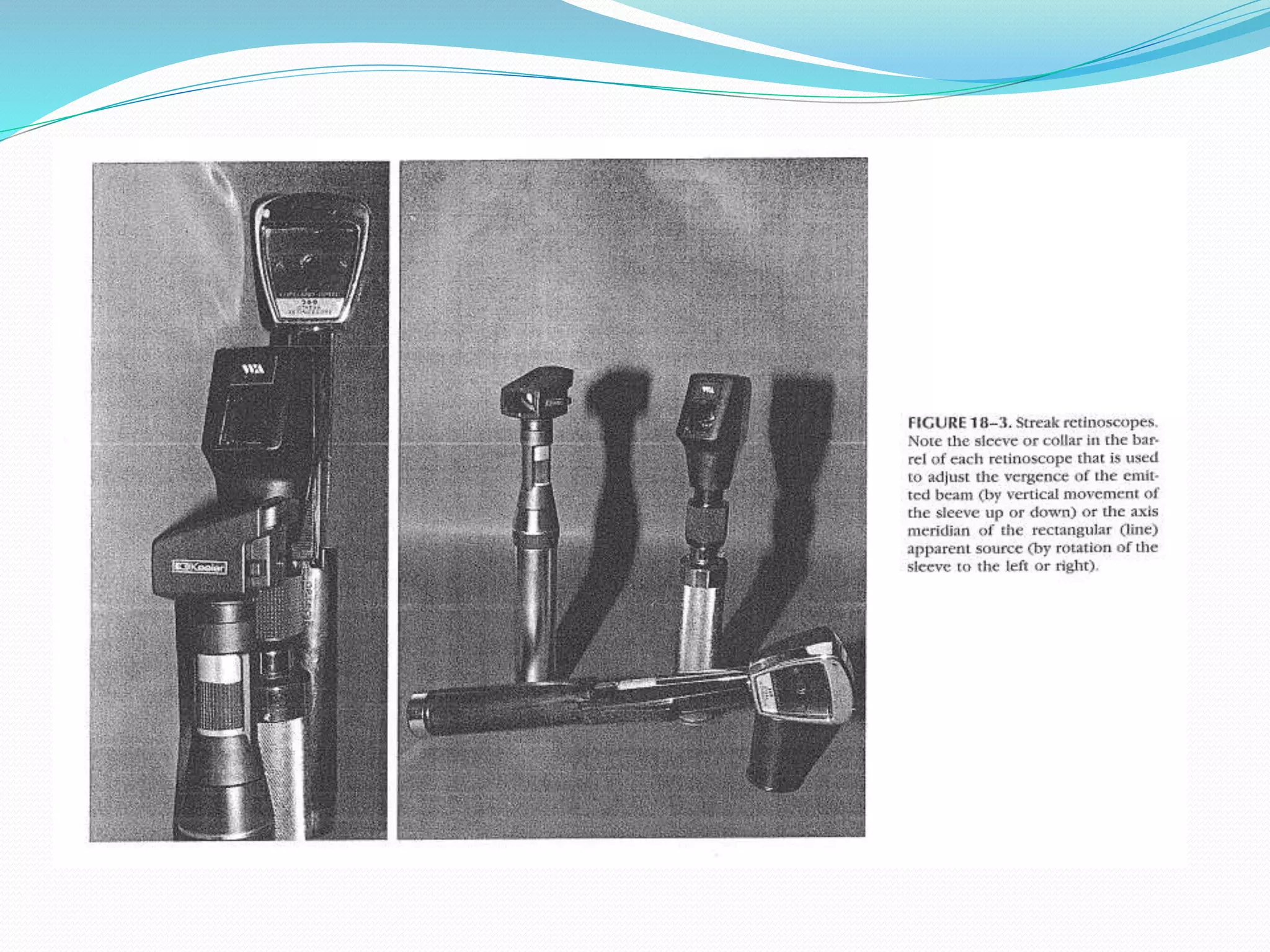
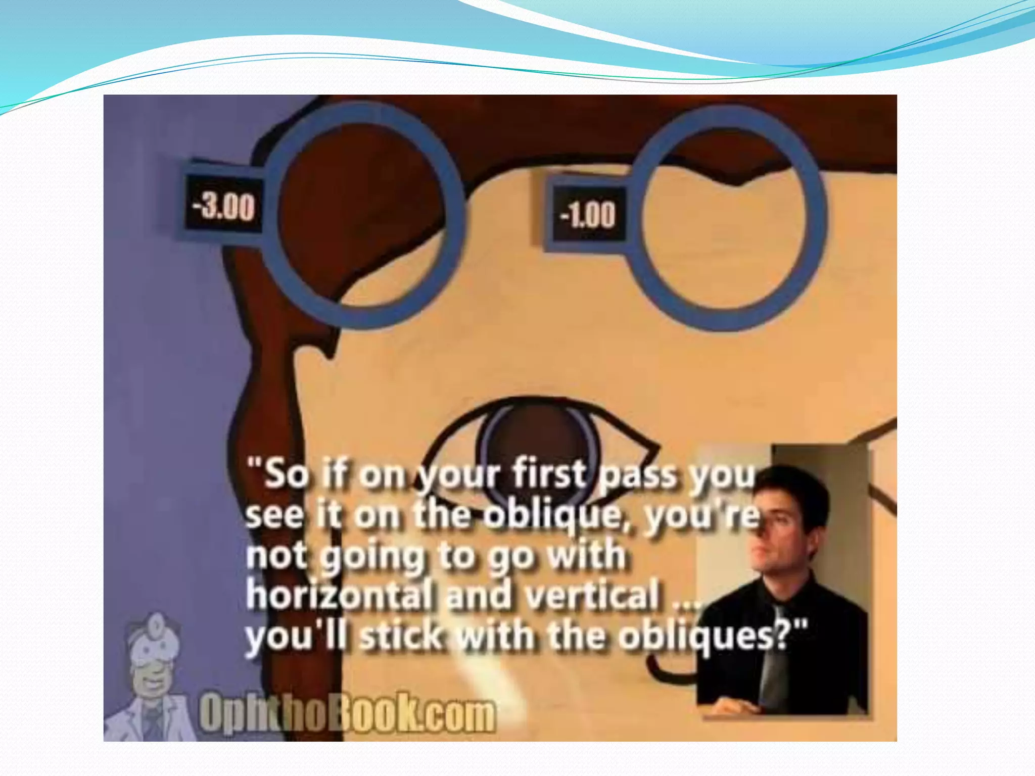
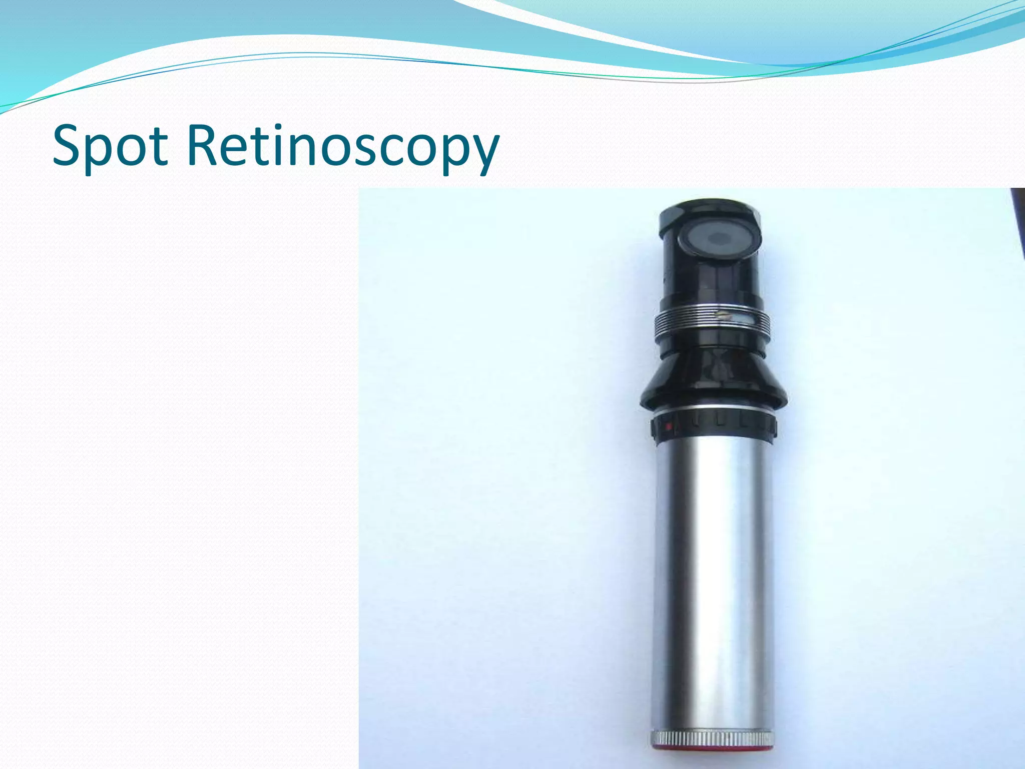
The “spot R” reflects a beam of light from a circular source whereas the
“streak retinoscpe” emits a beam from a line source .
Between these two forms of retinoscpes , the streak R is more useful
clinically because it can be more readily applied to the determination
of astigmatic corrections by assessment of the axis of cylinder and
refractive powers in the two primary amertopic meridians.
The use of streak retinoscpy has generally replaced the use of spot R in
ophthalmic practice .
15.
16.
17.
18. Basically the retinoscopist views through the aperture
of the R at a distance of 40 to 10 cm from the patient’s
eye and shines the beam of the retinoscope into the
pupil of patient's eye while the patient fixates a distant
target.
19. The attributes of the pupillary reflex that signify the relative position of
the far point are :
WITH motion :compared with movement of the incident divergent
rectangular beam ,indicates a far point location behind the retinoscope
aperture (between the operator and infinity)
AGAINST MOTION of the streak indicates a far point location between
the retinoscope aperture and the patient’s eye (moderately to highly
myopic eyes)
IF the retinoscopic reflex is at NEUTRALITY, showing neither “with”
nor “against” motion , the far point is located at the aperture of
retinoscope
20. Hyperopic patients
Light focuses behind the retina
Streak movement in same direction as the retinoscope
light (with motion )
Add plus lenses in this case to get neutralized ( to
bring the focus point at the retina )
21. Myopic patients
Light focuses at the point before the retina
Streak movement is in opposite of the retinoscope
light (against movement )
Add Minus (-) lens to move the focus point on the
retina
22. Emmetropic patients
No motion of the reflex observed in the pupil
Known as ( neutral motion)
23.
24. Scissors reflex
When two band appear which move towards and away
from each other like blades of scissors
Most of time occurs in only one meridian
Usually seen in kertoconus and irregular astigmatism
cases
28. Procedure
Patient sits at distance of 60 or 50 cm from the examiner
Patient is asked to fix his eyes at specific target to relax his accommodation
Now scope the R on patient’s eye and observe the light comes out of his eye .
Then determine if the motion is against , with or no motion
Make sure that you scope both meridians vertically and horizontally
If the motion in V and H is same then put + or – Sphere lenses
If the motion in V and H is NOT same then put + or – Cylindrical in case of
astigmatism
Repeat the procedure on left eye with same steps
29. Working distance ( WD)
Is the distance between the R and the patient’s eye
It can be calculated by this formula : D= 1/F
Which F is = the power of the lens .
E.g. F= 1.50D so the working distance is 0.66mm
1.50 /66cm (arm’s length ) so = 0.66 X 100=66 cm
• The length of an average’s persons arm is 66 cm
• The power of the lens that focuses at best parallel light rays
at 66 cm is +1.50 D
30. Neutralization
In case of myopia
Add – lenses and then put + lenses till it get neutralizes
In case of hyperopia
Add + lens till it get neutralizes
31. Recording R
degrees. Also record the monocular visual acuity
with the retinoscopy result. For example:
RE: 2.00 DS/-0.50 105 6/4.5
LE: 2.25 DS 6/4.52
OD: 2.00/1.00 105 20/203
OS: 1.75/0.75 70 20/25
32.
33.
34.
35. References
Primary care of optometry , page 183
Clinical procedure in primary eye care
Clinical refraction ,Chapter 18
Tom root ( youtube ) link :
https://www.youtube.com/watch?v=ezOoPKZwNDk
Editor's Notes
Photorefraction is a photograph or videograph of the pupils is interpreted currently by a trained operator or clinician
But there are now attempts to automated the interpretations of photographic and videographic refractors
Here if u divide 1/1.5 u will get 0.66 but in Meter unit so multiply by 100 to get it in cm so u will get 66 cm