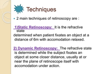
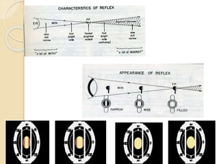
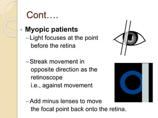
Retinoscopy is the primary objective method for determining a patient's refractive error. It involves using a retinoscope to illuminate the retina and observe the movement of the reflected light. For myopic patients, the light moves in the opposite direction of the retinoscope's movement, while for hyperopic patients it moves in the same direction. The goal is to find the neutralization point where no movement is seen, indicating the proper refractive correction. Factors like the working distance, type of mirror used, and patient's fixation can impact results. Retinoscopy is useful for initial refractive estimates and screening for ocular conditions.
different techniques of retinoscopy by which we can find amplitude of accomodation,and correct amount of hyperopia. we can also determine lead and lag of accomodation.
Detailed instumentaion and use of manual Lensometer and just a outline of automated lensometer.
I have used the picture of manual lensometer with out the parts describtion because i have explained orally by showing the picture..
Hope u all like it and may help you in learning better. :)
Contact lens for congenital aphakia and other eye conditions for infants and toddlers. The slide presentation encompasses indications for CL fitting in paediatric, contact lens options, fitting techniques, challenges and contact lens as myopia control.
DIRECT DOWNLOAD LINK ❤❤https://healthkura.com/retinoscopy/❤❤
Dear viewers Check Out my other piece of works at ❤❤❤ https://healthkura.com ❤❤❤
Retinoscopy and Objective Refraction and Subjective Refraction in spherical ametropia and astigmatism
Retinoscopy (Principle & Techniques of Retinoscopy) and objective refraction, Subjective Refracition
Best presentation about retinoscopy and objective refraction techniques, and basis of subjective refraction. If you want to master the technique of retinoscopy, this presentation can be your guidance and partner in your journey to retinoscopy, objective refraction and subjective refraction.
Presentation Layout:
Retinoscope, types of retinoscope and uses of retinoscope
-Introduction to retinoscopy and objective refraction
-Retinoscopy
- In spherical ametropia
- In astigmatism
- Others: strabismus, amblyopia, pediatric pt.,
cycloplegic refraction
-Static and Dynamic Retinoscopy
-Problems seeing reflex during retinoscopy
-Errors in retinoscopy
Objective of retinoscopy and objective refraction
-To locate the far point of the eye conjugate to the retina
- Myopia or hyperopia
-Bring far point to the infinity by using appropriate lenses
- Determines amount of ametropia by retinoscopy and objective refraction
References:
-Clinical Procedures in Optometry by Eskridge, Amos and Bartlett ,
-Primary Care Optometry by Grosvenor T.,
-Borish’s Clinical Refraction by Benjamin W. J.,
-Theory And Practice Of Optics And Refraction by AK Khurana
-Retinoscopy-Student Manual by ICEE Refractive Error Training Package (2009)
-Clinical Optics and Refraction By Andrew Keirl, Caroline Christie
-Clinical Refraction Guide - A Kumar Bhootra
-Clinical Procedures in Primary Eye Care by David B. Elliott
-Internet
Follow me to get in touch with optometric and ophthalmic updates.
these slides explain the objective refraction in optometry , and describes its types and its measurement , and it gives you in details the types of Retinoscopy.
Retinoscope is an objective refraction instrument used to
determine the spherocylindrical refractive error, as well as
observe optical aberrations, irregularities, and opacities.
The technique is called Retinoscopy/Skiascopy/Shadow Test
This presentation explain about retinoscope, the instrument, its history, its types, the procedure and different cases also the advantages and disadvantages of the instrument and the working lens
Techniques of refraction is the process of calculation of glass power.drbrijeshbhu
Refractive errors are most common cause of ocular morbidity. It affects all age groups, and ethnic profiles. There is no g nder discrimination. Most common symptoms are blur vission along with pain in eye ,headache and tiredness. Refraction is process of determination of eye and currect it with power glass power or contact lens power. It can subjective or objective.
Retinoscopy for undergraduates and post-graduates.
salient points covering examinations and PGMEE.
Detailed discussion of the technique of retinoscopy and its utility in deducing refractive errors.
Use of cycloplegic refraction and subjective refraction has been discussed.
This include a brief explanation of the clinical refraction methods in the eye examination procedure. In order to get the full video download the ppt. it includes a lot of important things
Dark Room Procedures for undergraduates(MB,BS) in the field of Ophthalmology are explained in simple terms in this presentation. Series of lectures taken at Central Park Medical College Lahore Pakistan.
Best Ayurvedic medicine for Gas and IndigestionSwastikAyurveda
Here is the updated list of Top Best Ayurvedic medicine for Gas and Indigestion and those are Gas-O-Go Syp for Dyspepsia | Lavizyme Syrup for Acidity | Yumzyme Hepatoprotective Capsules etc
Knee anatomy and clinical tests 2024.pdfvimalpl1234
This includes all relevant anatomy and clinical tests compiled from standard textbooks, Campbell,netter etc..It is comprehensive and best suited for orthopaedicians and orthopaedic residents.
Flu Vaccine Alert in Bangalore Karnatakaaddon Scans
As flu season approaches, health officials in Bangalore, Karnataka, are urging residents to get their flu vaccinations. The seasonal flu, while common, can lead to severe health complications, particularly for vulnerable populations such as young children, the elderly, and those with underlying health conditions.
Dr. Vidisha Kumari, a leading epidemiologist in Bangalore, emphasizes the importance of getting vaccinated. "The flu vaccine is our best defense against the influenza virus. It not only protects individuals but also helps prevent the spread of the virus in our communities," he says.
This year, the flu season is expected to coincide with a potential increase in other respiratory illnesses. The Karnataka Health Department has launched an awareness campaign highlighting the significance of flu vaccinations. They have set up multiple vaccination centers across Bangalore, making it convenient for residents to receive their shots.
To encourage widespread vaccination, the government is also collaborating with local schools, workplaces, and community centers to facilitate vaccination drives. Special attention is being given to ensuring that the vaccine is accessible to all, including marginalized communities who may have limited access to healthcare.
Residents are reminded that the flu vaccine is safe and effective. Common side effects are mild and may include soreness at the injection site, mild fever, or muscle aches. These side effects are generally short-lived and far less severe than the flu itself.
Healthcare providers are also stressing the importance of continuing COVID-19 precautions. Wearing masks, practicing good hand hygiene, and maintaining social distancing are still crucial, especially in crowded places.
Protect yourself and your loved ones by getting vaccinated. Together, we can help keep Bangalore healthy and safe this flu season. For more information on vaccination centers and schedules, residents can visit the Karnataka Health Department’s official website or follow their social media pages.
Stay informed, stay safe, and get your flu shot today!
- Video recording of this lecture in English language: https://youtu.be/lK81BzxMqdo
- Video recording of this lecture in Arabic language: https://youtu.be/Ve4P0COk9OI
- Link to download the book free: https://nephrotube.blogspot.com/p/nephrotube-nephrology-books.html
- Link to NephroTube website: www.NephroTube.com
- Link to NephroTube social media accounts: https://nephrotube.blogspot.com/p/join-nephrotube-on-social-media.html
Tom Selleck Health: A Comprehensive Look at the Iconic Actor’s Wellness Journeygreendigital
Tom Selleck, an enduring figure in Hollywood. has captivated audiences for decades with his rugged charm, iconic moustache. and memorable roles in television and film. From his breakout role as Thomas Magnum in Magnum P.I. to his current portrayal of Frank Reagan in Blue Bloods. Selleck's career has spanned over 50 years. But beyond his professional achievements. fans have often been curious about Tom Selleck Health. especially as he has aged in the public eye.
Follow us on: Pinterest
Introduction
Many have been interested in Tom Selleck health. not only because of his enduring presence on screen but also because of the challenges. and lifestyle choices he has faced and made over the years. This article delves into the various aspects of Tom Selleck health. exploring his fitness regimen, diet, mental health. and the challenges he has encountered as he ages. We'll look at how he maintains his well-being. the health issues he has faced, and his approach to ageing .
Early Life and Career
Childhood and Athletic Beginnings
Tom Selleck was born on January 29, 1945, in Detroit, Michigan, and grew up in Sherman Oaks, California. From an early age, he was involved in sports, particularly basketball. which played a significant role in his physical development. His athletic pursuits continued into college. where he attended the University of Southern California (USC) on a basketball scholarship. This early involvement in sports laid a strong foundation for his physical health and disciplined lifestyle.
Transition to Acting
Selleck's transition from an athlete to an actor came with its physical demands. His first significant role in "Magnum P.I." required him to perform various stunts and maintain a fit appearance. This role, which he played from 1980 to 1988. necessitated a rigorous fitness routine to meet the show's demands. setting the stage for his long-term commitment to health and wellness.
Fitness Regimen
Workout Routine
Tom Selleck health and fitness regimen has evolved. adapting to his changing roles and age. During his "Magnum, P.I." days. Selleck's workouts were intense and focused on building and maintaining muscle mass. His routine included weightlifting, cardiovascular exercises. and specific training for the stunts he performed on the show.
Selleck adjusted his fitness routine as he aged to suit his body's needs. Today, his workouts focus on maintaining flexibility, strength, and cardiovascular health. He incorporates low-impact exercises such as swimming, walking, and light weightlifting. This balanced approach helps him stay fit without putting undue strain on his joints and muscles.
Importance of Flexibility and Mobility
In recent years, Selleck has emphasized the importance of flexibility and mobility in his fitness regimen. Understanding the natural decline in muscle mass and joint flexibility with age. he includes stretching and yoga in his routine. These practices help prevent injuries, improve posture, and maintain mobilit
Basavarajeeyam is a Sreshta Sangraha grantha (Compiled book ), written by Neelkanta kotturu Basavaraja Virachita. It contains 25 Prakaranas, First 24 Chapters related to Rogas& 25th to Rasadravyas.
New Drug Discovery and Development .....NEHA GUPTA
The "New Drug Discovery and Development" process involves the identification, design, testing, and manufacturing of novel pharmaceutical compounds with the aim of introducing new and improved treatments for various medical conditions. This comprehensive endeavor encompasses various stages, including target identification, preclinical studies, clinical trials, regulatory approval, and post-market surveillance. It involves multidisciplinary collaboration among scientists, researchers, clinicians, regulatory experts, and pharmaceutical companies to bring innovative therapies to market and address unmet medical needs.
Title: Sense of Taste
Presenter: Dr. Faiza, Assistant Professor of Physiology
Qualifications:
MBBS (Best Graduate, AIMC Lahore)
FCPS Physiology
ICMT, CHPE, DHPE (STMU)
MPH (GC University, Faisalabad)
MBA (Virtual University of Pakistan)
Learning Objectives:
Describe the structure and function of taste buds.
Describe the relationship between the taste threshold and taste index of common substances.
Explain the chemical basis and signal transduction of taste perception for each type of primary taste sensation.
Recognize different abnormalities of taste perception and their causes.
Key Topics:
Significance of Taste Sensation:
Differentiation between pleasant and harmful food
Influence on behavior
Selection of food based on metabolic needs
Receptors of Taste:
Taste buds on the tongue
Influence of sense of smell, texture of food, and pain stimulation (e.g., by pepper)
Primary and Secondary Taste Sensations:
Primary taste sensations: Sweet, Sour, Salty, Bitter, Umami
Chemical basis and signal transduction mechanisms for each taste
Taste Threshold and Index:
Taste threshold values for Sweet (sucrose), Salty (NaCl), Sour (HCl), and Bitter (Quinine)
Taste index relationship: Inversely proportional to taste threshold
Taste Blindness:
Inability to taste certain substances, particularly thiourea compounds
Example: Phenylthiocarbamide
Structure and Function of Taste Buds:
Composition: Epithelial cells, Sustentacular/Supporting cells, Taste cells, Basal cells
Features: Taste pores, Taste hairs/microvilli, and Taste nerve fibers
Location of Taste Buds:
Found in papillae of the tongue (Fungiform, Circumvallate, Foliate)
Also present on the palate, tonsillar pillars, epiglottis, and proximal esophagus
Mechanism of Taste Stimulation:
Interaction of taste substances with receptors on microvilli
Signal transduction pathways for Umami, Sweet, Bitter, Sour, and Salty tastes
Taste Sensitivity and Adaptation:
Decrease in sensitivity with age
Rapid adaptation of taste sensation
Role of Saliva in Taste:
Dissolution of tastants to reach receptors
Washing away the stimulus
Taste Preferences and Aversions:
Mechanisms behind taste preference and aversion
Influence of receptors and neural pathways
Impact of Sensory Nerve Damage:
Degeneration of taste buds if the sensory nerve fiber is cut
Abnormalities of Taste Detection:
Conditions: Ageusia, Hypogeusia, Dysgeusia (parageusia)
Causes: Nerve damage, neurological disorders, infections, poor oral hygiene, adverse drug effects, deficiencies, aging, tobacco use, altered neurotransmitter levels
Neurotransmitters and Taste Threshold:
Effects of serotonin (5-HT) and norepinephrine (NE) on taste sensitivity
Supertasters:
25% of the population with heightened sensitivity to taste, especially bitterness
Increased number of fungiform papillae
2. WHAT IS
OBJECTIVE REFRACTION ???
Where the result depends purely
on the examiners judgement to
determine the optimum optical
correction.
3. Methods for objective
refraction
Keratometry
Ophthalmoscopy
Optometers
Auto refraction
Photorefraction
&
Retinoscopy [Most important & common
method ]
4. What is
Retinoscopy & Retinoscope?
Retinoscopy or skiascopy is the primary method
for objective determination of the total refractive
status of the eye.
Retinoscopy is done with the help of an instrument
called Retinoscope.
It illuminates the inside of the eye,to observe the
light that is reflected from retina.By examining how
emerging rays change,refractive power of eye can
be determined.
5. History~
Sir William Bowman in 1859,reported the
movement of light and shadow effect.
Used since 1873 – reflecting mirror spot
retinoscopes, externally illuminated.
Modern streak design that brought
significant change in 1927, by Jack C.
Copeland.
7. Techniques
2 main techniques of retinoscopy are :
1)Static Retinoscopy: It is the refractive
state
determined when patient fixates an object at a
distance of 6m with accomodation relaxed.
2) Dynamic Retinoscopy: The refractive state
is determined while the subject fixates an
object at some closer distance, usually at or
near the plane of retinoscope itself with
accomodation under action.
8. Cont…
Static Retinoscopy include
Spot retinoscope: Light source is spot
of light.
Streak retinoscope: The bulb is
constructed so that is provides a beam
in the form of a streak rather than a
spot.
10. Static Vs
Dynamic
Accomodation fully
relaxed
Working distance
lens added or
subtracted from
the objective finding
Fixates letters at 6m
Only ametropia or
emmetropia can be
determined
-Accomodation fully
in play
-No influence of
working distance
-Fixates at the bulb of
retinoscope
-Accomodative lag
can be determined
11. Significance of
spot & streak
Round filament
Scoped in any
meridian
Assessment of
the contact lens
fitting
Dealing with
pediatric patients
Vision screening
programs
-Linear filament
-Quickly change
from plano mirror
to concave mirror
-Narrowing the
width makes it
easy to pin down
the principal
meridians
12. Principle of retinoscopy
To locate the far point of the eye/ plane
conjugate to the retina
Bring far point to the infinity by using
appropriate lenses
Accommodation at a minimum.
13. Continue......
Mirror with central hole
Subject
Observer
Outgoing light
Incoming light
Variable condensing lens
14. Origin of Retinoscopic Reflex
Interface between the vitreous and
retina.
Pigment epithelium of retina or
Bruch’s membrane
16. Illumination Stage
Depends on ~
concept of the immediate source of light
the movement of the illuminated area of
the fundus ,with the movement of
reflecting mirror.
Plane mirror : immediate source of light
moves with the movement of the mirror.
Concave mirror : immediate source of
light moves against the movement of the
mirror.
18. Reflex Stage
Depending upon the refractive status
of
the eye:~
19. Characteristics of reflex
1.Speed: large refractive errors have a
slow-moving reflex, small errors have a fast
reflex.
2.Brilliance: large errors have dull reflex,
small errors have a bright reflex. Becomes
brighter when neutrality approaches.
3)Width: Narrow in high degree error &
widen in low degree error.
20.
21. Mirror effect…
Plane mirror effect:
◦ Effective source lies behind the plane of
mirror (most commonly used)
◦ The rays of light form the source goes
parallel or slightly diverging
◦ Does not cross between the source and the
patient’s eye-
with movement – hyperopia
Against movement - myopia
22. Concave mirror effect:
◦ Generally not used
• keep the effective source in front of the
plane
of the mirror, so that the rays emitted from
source are more converging and cross at
a
certain distance between patient and the
source
with movement – myopia
Against movement - hyperopia
24. Working distance & lens
selection
Beginning retinoscopy, the examiner must
choose a WD.
Depends upon the length of the examiner’s
arm. If arm permits-
1. 66cm-WD=+1.50D
2. 50cm -WD=+2.00D
25. Prerequisite for
objective retinoscopy
Dark room
Retinoscope
A trial set
A trial frame
Distance fixation target
26. PROCEDURE
~Patient sits at a distance of
66cm/50cm from the examiner.
~Patient is asked to fix at a distance
target to relax accommodation.
~Light is thrown on the patient’s
eye from retinoscope.
~By rocking the light slowly the
characteristics of the reflex
are observed.
~Then neutralizing the reflex.
~Examiner must be examined the
patient’s Rt eye by his/her Rt eye
& vice versa.
27. Nature of reflexes in ametropia
(plane mirror)
Myopic far point of accommodation
located at a finite distance
infront of the eye
Hyperopes far point of
accommodation is located at
some point behind the
primary focal plane of the eye
• Emmetropic eyes far point
of accommodation is located
at infinity
28. Observation System
• When we view the reflex in patient’s eye, it seems to
move in the direction
• If the retinoscope is tilted upward, reflex will move
to the opposite direction ;in case of myope;
• same direction of retinoscopic light & reflex ; in
case of hyperope and emmetrope;
• no movement (with working lens) at all in case of
emmetrope.
29. Streak motion
Hyperopic patients
◦ Light focuses behind the retina
◦ Streak movement in
same direction as the
retinoscope . i.e.,
displays with motion
◦ Add plus lenses to bring
the focusing point up to the retina
30. Cont….
• Myopic patients
– Light focuses at the point
before the retina
–Streak movement in
opposite direction as the
retinoscope
i.e., against movement
–Add minus lenses to move
the focal point back onto the retina.
31. Emmetropic patients
◦ No motion of the reflex observed in the
pupil
◦ Also known as neutral motion or complete
flashing
32. Spherical or Cylindrical ???
Streak both the
horizontal and vertical
meridian to determine
the astigmatism
33. Finding cylinder axis
1.Break: seen when the streak
is not parallel to the principal
meridian and disappears when
the streak is rotated to
the correct axis.
2. Width: reflex appears
narrowest when the streak
aligns with the axis.
34. 3.Intensity: Line is brighter when the streak
is on the correct axis.
4. Skew: When the streak
is off-axis,it will move in a
slightly different dirrection
from the pupillary reflex
and move in the same
dirrection when the streak
is aligned with the principal
meridian.
35. Straddling:
Finding axis can be confirmed by this
technique.
Performed with the estimated correcting
cylinder in place.
Streak is turned 45⁰ off-axis in both
dirrections.
If the axis is :
Correct – widths should be equal in
both position.
Incorrect – widths will be unequal.
37. Finding the cylinder power
With 2 sphere : After the 2 principal meridians are
identified, spherical techniques are applied to each
axis.
With a sphere and a cylinder :
1st neutralize one axis by a spherical lens
↓
Over the lens,neutralize the other axis 90⁰
away by a cyliderical lens
38. Scissors reflex
When 2 band reflexes appear which move
towards & away from each other like the
blades of scissors….
Most of the time occurs in only one meridian
Seen in Keratoconus & irregular astigmatism
pt’s
.
◦ Neutralization~ ?
39. Neutralization point
Point at which the peephole becomes
conjugate with the patient’s retina.
Point at which the reversal of the
reflex is observed.
40. Neutralization . . .
With motion – Plus lenses are
added until neutrality occur.
Against motion – Minus lenses
are added…
The width of reflex widens progressively as
the neutralization is approached & at the
end point ~streak disappears
~pupil completely
illuminated
41. End point of neutrality
1.Over correction of ±0.25D
2.On altering the WD.
3.Changing the mirror.
42. Clinical use…
Objective determination of refractive error
Starting point of subjective refraction
To find out regular & irregular astigmatism
Helpful for non-communicative or non-verbal
pt’s
Screening for ocular disorders [keratoconus,
media opacities]
Some special assessments can be
determined [Accommodation
stability,Accommodative lag]
43. Errors of retinoscopy
1. Incorrect WD.
2. Failure of the patient to fixate the
distant target.
3. Scoping of the patient’s visual axis.
4. Failure to obtain a reversal.
5. Failure to locate the principal
meridians.
6. Failure to recognize scissors motion.
44. References…
Primary Care Optometry~
TheodoreGrosvenor
Clinical Procedures in Optometry~
Theory & Practice of Optics &
Refraction~A.K.Khurana
Internet