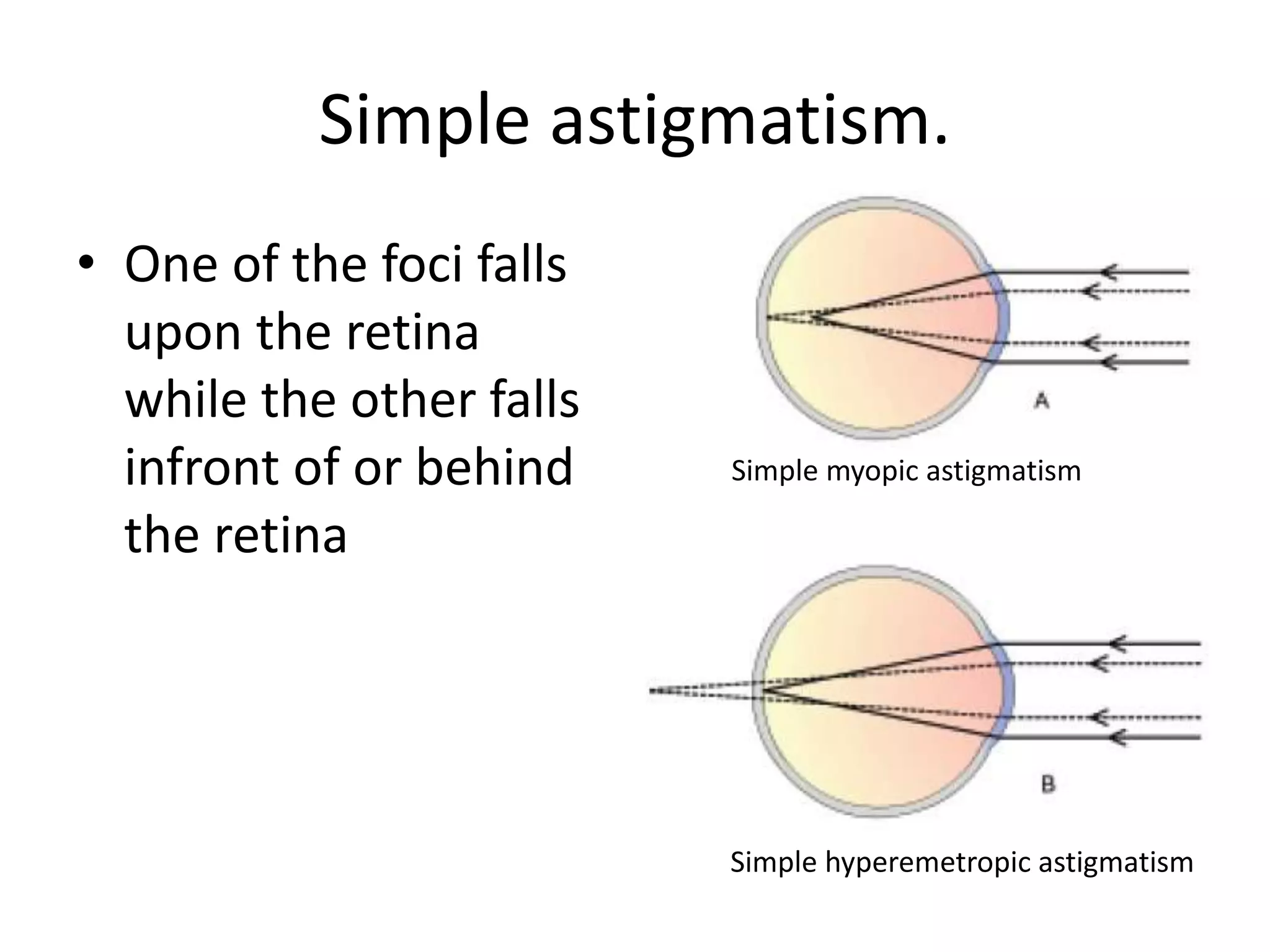
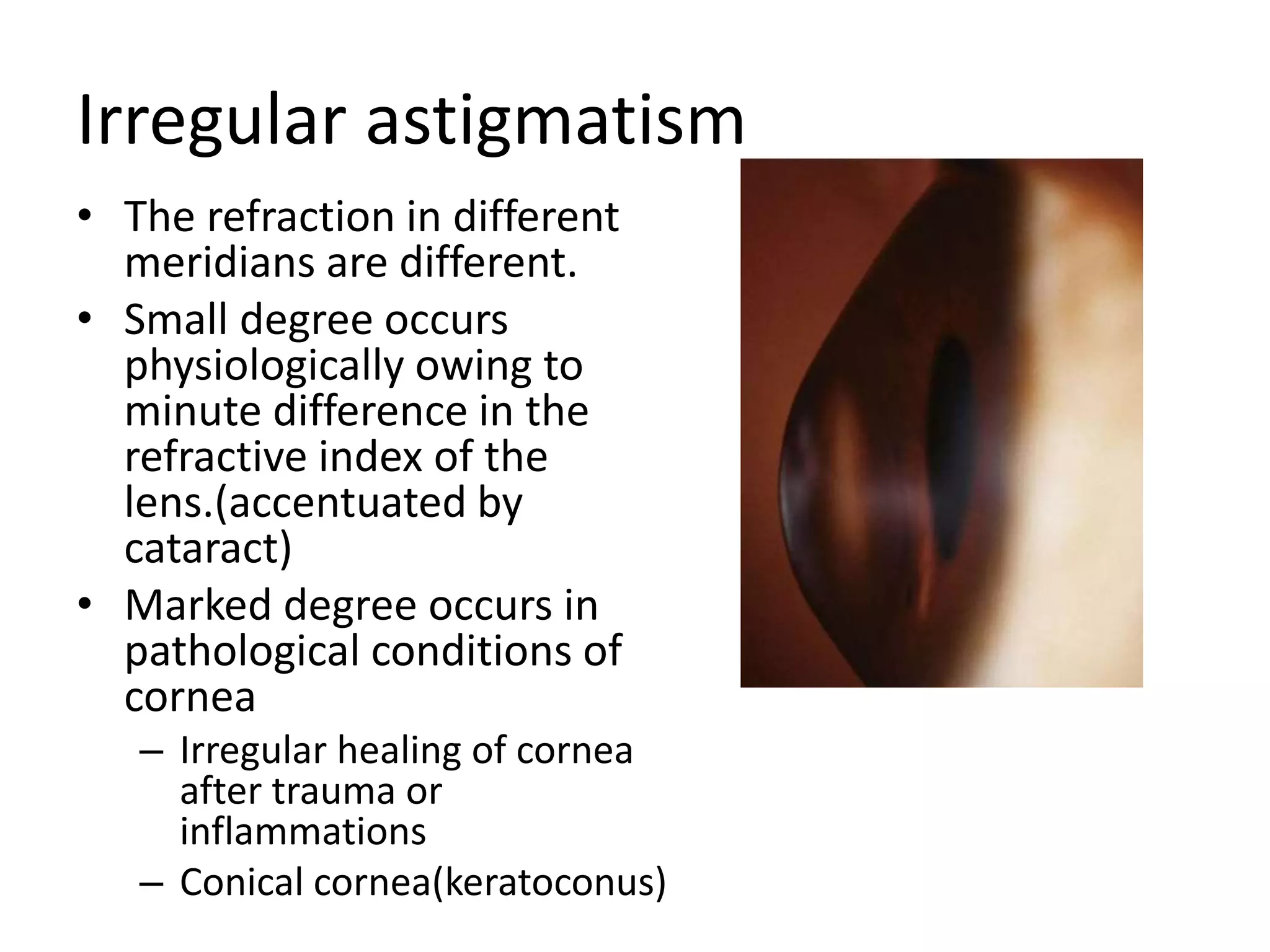
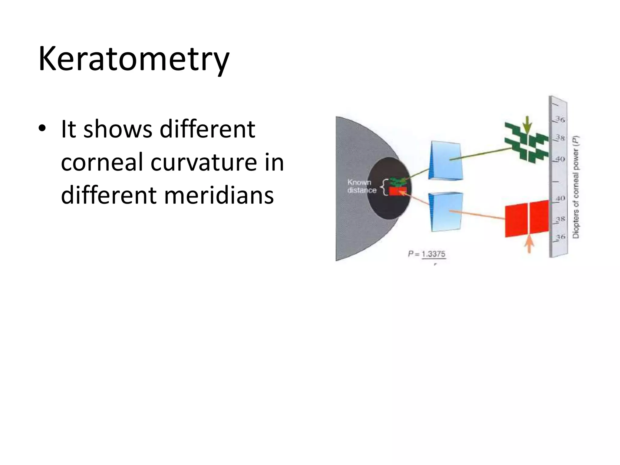
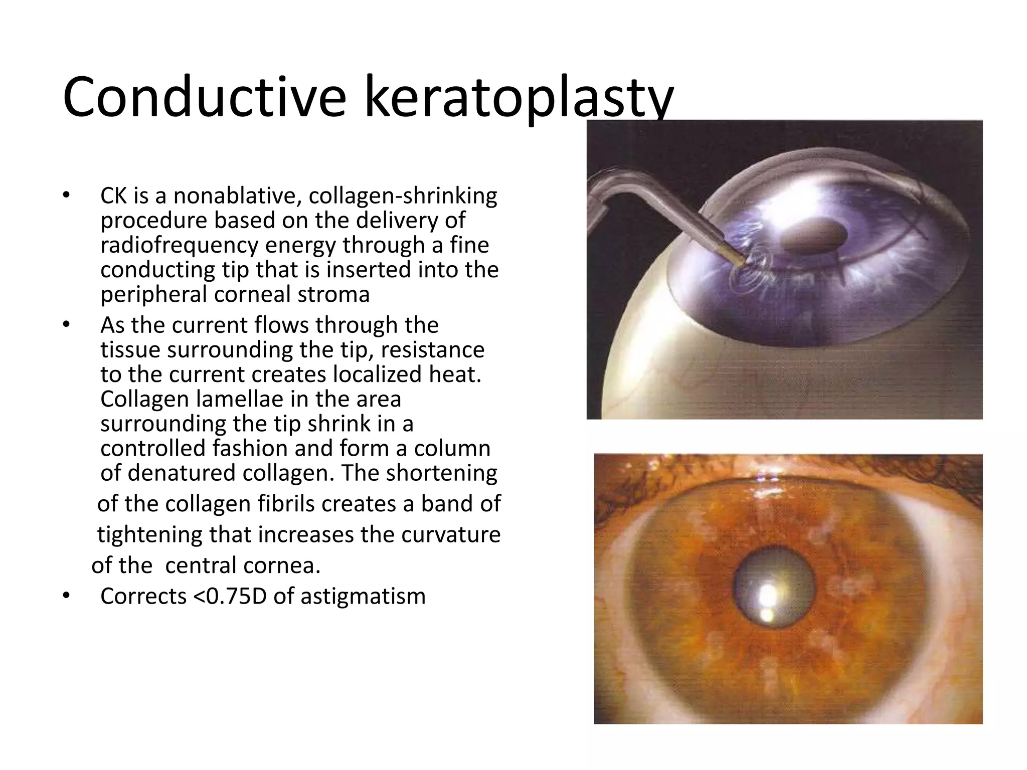
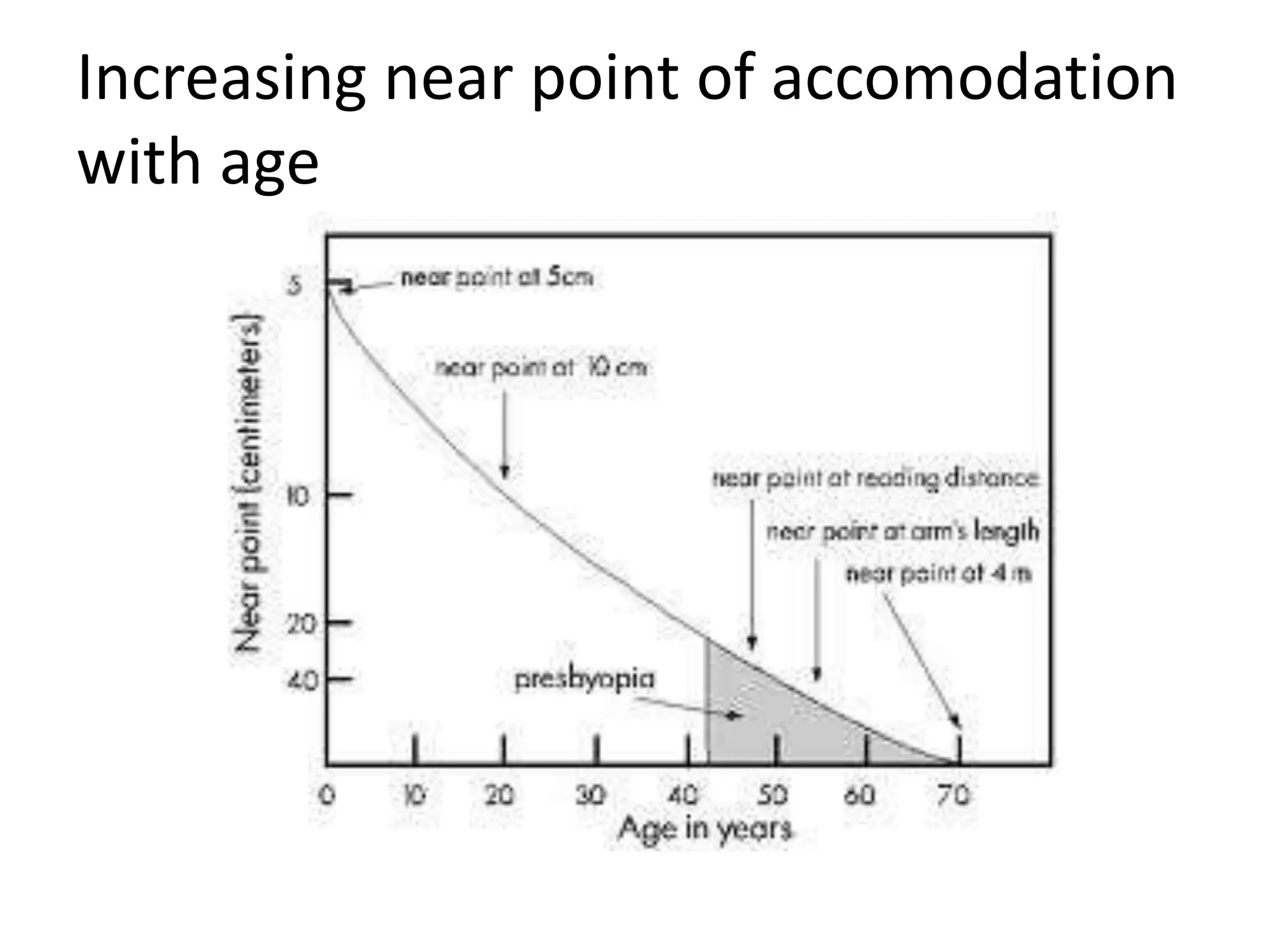
ASTIGMATISM, PRESBYOPIA
AND APHAKIA
Resident(1st year)
National Academy of Medical Sciences
ASTIGMATISM
ASTIGMATISM
Greek; A-without, Stigmos-
a point/a spot (Coined by
William Whewell;1849)
Definition
It is that condition of
refraction wherein a point
focus of light cannot be
formed upon the retina.
It occurs due to unequal
refraction of light in
different meridians.
Etiology of astigmatism
Corneal Astigmatism
Congenital
-occurrence is almost invariable in small degrees
-most commonly the vertical curve is greater than
the horizontal curve( about 0.5D). This is called
direct astigmatism.
-physiological
Acquired astigmatism
• Trauma to the cornea
including surgeries
• Pressure of swellings
on the lid( chalazion or
tumor)
• Pterygium
Corneal astigmatism contd…
Acquired astigmatism
• Conical cornea/
keratoconus
• Corneal inflammation
and ulceration and
scars
Lenticular astigmatism
• Lenticonus
• Oblique placement of
the lens
Lenticular astigmatism
• Traumatic subluxation
of the lens
• Decentration of IOL
• Index astigmatism
Retinal astigmatism
• The posterior pole of the eye may be placed
obliquely as when it bulges backwards in a
staphyloma in high myopia.
Types of astigmatism
Regular astigmatism
• The two principle meridians are at right angles
and are therefore susceptible to correction
– With the rule astigmatism
– Against the rule astigmatism
– Oblique astigmatism
Irregular astigmatism
• There are irregularities in the curvature of the
meridians so that no geometric figure is adhered
to
• Cannot be corrected adequately by spectacles
Optics of regular astigmatism
Sturm’s conoid
With-The-Rule (WTR) Astigmatism
• The two principal meridians
are at right angle to each
other with the vertical
meridian being more curved
than the horizontal i.e. the
greatest refractive power is
at 90˚(+/-20˚)
• Corrected by convex
cylinder at 90˚(+/-20˚) or
concave cylinder at 180 (+/-
20˚)
Against-The-Rule (ATR) Astigmatism
• The two principal meridians
are at right angle to each
other with the horizontal
meridian being more
curved than the vertical i.e.
the greatest refractive
power is at 180˚(+/-20˚)
• Corrected by convex
cylinder at 180 ˚(+/-20˚) or
concave cylinder at 90 ˚(+/-
20˚)
Oblique (OBL) Astigmatism
• The two principal
meridia are not
horizontal or vertical
though they are at
right angle to each
other.
Bi-oblique Astigmatism
• The two priciple
meridians are not at
right angle to each
other but are
crossed obliquely.
30˚
100˚
Regular astigmatism can further be
classified as
• Simple astigmatism
• Compound astigmatism
• Mixed astigmatism
Simple astigmatism.
• One of the foci falls
upon the retina
while the other falls
infront of or behind
the retina
Simple myopic astigmatism
Simple hyperemetropic astigmatism
Compound astigmatism
• Neither of the foci
fall upon the retina
but both are placed
in-front of or behind
the retina Compound myopic astigmatism
Compound hypermetropic astigmatism
Mixed astigmatism
• One focus is infront
of the retina while
the other is behind
so that the refraction
is hypermetropic in
one direction and
myopic in the other
direction
Mixed astigmatism
Irregular astigmatism
• The refraction in different
meridians are different.
• Small degree occurs
physiologically owing to
minute difference in the
refractive index of the
lens.(accentuated by
cataract)
• Marked degree occurs in
pathological conditions of
cornea
– Irregular healing of cornea
after trauma or
inflammations
– Conical cornea(keratoconus)
Symptoms of astigmatism
• Reduced Visual acuity
• Peculiar vision
• Asthenopia( more with hypermetropic
astigmatism)
• Head tilt in oblique astigmatism in children
• Half closure of the eyelids
Clinical Tests
• Visual acuity tests – distance and near
• Autorefraction
• Keratometry
• Retinoscopy
– Most reliable source of information for cylinder power
and axis
• Monocular subjective refraction
– Astigmatic fan test
– Jackson cross cylinder
Keratometry
• It shows different
corneal curvature in
different meridians
Retinoscopy
• Shows different power in different
meridians.
• If the streak passes exactly through
the axis of the astigmatism, a sharply
defined reflex band is seen which
moves parallel to the band of the
light outside the pupil.
• If it doesn’t pass exactly through the
astigmatic axis, the reflex becomes
poorly defined and tends to remain
fixed in the axis producing a break in
the alignment between the reflex in
the pupil and the band outside it
tending to lie intermediate between
the latter and the true astigmatic axis
Cross-cylinders
• Used to refine the axis and
the power of the cylinder.
• Done after the cylindrical
correction has been made.
• Combination of a negative
and positive cylinder of
equal strength (usually
0.5D) mounted at an angle
of 90 degrees
• In practice combined into
one piece of glass
Refinement of the axis
• The cross-cylinder is placed with its
axis at 45˚ to the axis of the cylinder in
the trial frame( first with +0.5 cylinder
and then with -0.5 cylinder or vive-
versa) and the patient is asked to tell
any change in the visual acuity.
• If the patient notices no difference
then the correcting cylinder in the
trial frame is correct.
• If visual improvement is attained by
one or the other the correcting
cylinder is turned slightly in the
direction of the axis of the cylinder of
the same denomination in the cross
cylinder.
• It is repeated until the position of the
trial cylinder is found at which
rotation of the cross cylinder gives no
alteration in distinctness.
Refinement of the power of the cylinder
• The cross-cylinder is placed
in the same direction as the
axis of the cylinder in the
trial frame and then
perpendicular to it.
• If the visual acuity is
unimproved in either
position the power of
cylinder in the trial frame is
correct.
• If visual acuity is improved
in any of the positions then
change should be made.
Astigmatic fan test(Maddox V test)
• The patient is asked to look
at the figure and if any of
the line is more distinct
than the other then
astigmatism is present.
• Rotating the V slightly to the
direction of the blacker limb
an intermediate position is
reached when the two
limbs of V are equally
distinct. This gives the
direction at right angles to
the exact axis of the
correcting cylinder.
Management
Optical correction
• Spectacles or contact
lenses
– Appropriate cylindrical
lens
– Toric soft contact lenses
or toric rigid gas
permeable contact
lenses
Guidelines for optical correction
• Small astigmatism(0.5D or less) should be treated
only if there is visual deterioration or asthenopic
symptoms.
• High astigmatism should be fully corrected
• Change in the axis of the lenses in patients used
to the previous axis should be done cautiously
• New astigmatism correction in adults is not
tolerated. In such cases it is better to undercorrect
and give full correction gradually.
• Bi-oblique astigmatism, mixed and high
astigmatism are better treated by contact lenses
• Spherical component should also be corrected.
Management
Refractive Surgery
Incisional correction of
Astigmatism
• Transverse keratotomy
• Arcuate keratotomy incisions
are placed in the cornea at
7mm optical zone) to the
steepest corneal meridian.
• Can correct upto 4-6D of
astigmatism.
Limbal relaxing incisions
• Incision is given at the
limbus
• Correct -1 to -2D of
astigmatism.
Conductive keratoplasty
• CK is a nonablative, collagen-shrinking
procedure based on the delivery of
radiofrequency energy through a fine
conducting tip that is inserted into the
peripheral corneal stroma
• As the current flows through the
tissue surrounding the tip, resistance
to the current creates localized heat.
Collagen lamellae in the area
surrounding the tip shrink in a
controlled fashion and form a column
of denatured collagen. The shortening
of the collagen fibrils creates a band of
tightening that increases the curvature
of the central cornea.
• Corrects <0.75D of astigmatism
Laser based corneal refractive procedure
• Photo refractive
keratotomy(uses cylindrical
photoablation pattern.)—can
correct upto 3D of astigmatism
• Astigmatic LASIK can correct
upto 5D
• Wavefront-guided and
wavefront-optimised LASIK can
correct
– -10 to -12D of Myopic astigmatism
– +6D of hyperopic astigmatism and
– +5D of mixed astigmatism
Intraocular lens for correcting
astigmatism
• Angle supported anterior chamber phakic IOL
• Toric intraocular implant
Treatment of irregular astigmatism
• Spectacles though may not provide full
correction some amount of correction can be
attempted
• Contact lenses
• Surgical correction by keratoplasty
PRESBYOPIA
Definition (Latin; old man’s eye/aged eye)
Presbyopia refers to the slow, normal,
naturally occurring, age-related, irreversible
reduction in maximal accommodative
amplitude (i.e., recession of the near point)
sufficient to cause symptoms of blur and
ocular discomfort or asthenopia at the
customary near working distance.
• first reported clinically between 40 and 45
years of age, with its peak onset between 42
and 44 years
• Its onset may occur any time from 38 to 48
years of age, depending on a variety of
factors.
• From approximately age 52 years on, the
prevalence of presbyopia is considered to be
essentially 100%
Risk factors for development of presbyopia
Refractive error
• Hyperopes have their near point considerably
further away than emmetropes(exhibit apparent
relatively reduced accommodative amplitudes)
and thus effectively become presbyopic a few
years earlier than either myopes or emmetropes.
• In the myopes develop presbyopia later in life and
if he has the error of -4D, presbyopia will never
develop
Ambient Temperature
• With the eyeball being peripheral to the body
core, it may exhibit considerable surface
temperature variations because of the
influence of ambient temperature.
• There is an inverse relation between ambient
temperature and age of onset of presbyopia
Factors contributing to the age related
decrease in accomodation
• Decrease in modulus of elasticity of lens
capsule—cannot effectively mold the
underlying lens substance
• Increase in stiffness of the lens substance—
energy required to deform the lens substance
increases
• Increase in size/volume of lens—decreases
the effectivity of lens capsule function
• Anterior shift of the equatorial zonular fibres
due to inrease in the size of the lens
• Decrease in equatorial zonular fibres
• Increase in the number of disulfide bond in the
lens substance—stabilises the collagen
molecules within the capsule and the lens by
the process of cross-linking—lens substance
becomes rigid and capsule becomes less elastic
• Mechanical and anatomical changes in the
ciliary muscle
• Decrease in choroidal elasticity
Amplitude of accomodation and age
• The amplitude of accommodation represents the
maximal accommodative level, or closest near
focusing response, that can be produced with
maximal voluntary effort in the fully corrected
eye.
• It is calculated as
Accomodative amplitude(A)= diopteric power
needed to see clearly at near point-diopteric
power needed to see clearly at far point
At the age of 10 yrs
Near point=7cm
Far point=∞
Hence,
A= 100/7-1/∞
=14-0
=14D
At the age of 40 yrs
Near point=25cm
Far point=∞
Hence,
A=100/25-1/∞
=4-0
=4D
Increasing near point of accomodation
with age
Amplitude of accomodation with age
Variation of amplitude of accomodation with age
Symptoms
• Reduced vision at customary near-work distance.
• Drowsiness after short period of reading or near work
• Reading materials must be held farther away
• Asthenopia related to attempts at excessive
accommodative effort is reported. It may even lead to
an accommodative spasm and pseudomyopia.
• Transient diplopia and variable esophoria may be
experienced as a result of the increased
accommodative response/effort and the consequent
synkinetically overdriven accommodative convergence
that may be difficult to control consistently using
compensatory negative fusional vergence.":"
Near Acuity Tests
• Reading distance at 16 inches(40 cm)
Treatment
• The treatment of presbyopia is to
provide the patient with convex
lenses so that his accomodation
is reinforced and his near point
brought within a useful working
distance.
• To do this
– know the working distance of the
individual
– estimate his refraction
– determine the amplitude of
accomodation
– supplement this by appropriate
strength of lens allowing him
sufficient reserve of accomodation
• One-third of the amplitude of accomodation
should be kept for reserve if the patient is to
work comfortably
• The near correction should never be
overcorrected. It should be such that the
patient should be able to read the near vision
chart satisfactory not only at his reading
distance but also some 12-15cm further away.
Example
• Emmetropic patient, whose working distance is
25cm has his near point receded to 50cm
• His amplitude of accomodation is 2D
• With 1/3rd kept for reserve, remaining amplitude
is 2/3rd i.e. 1.3D
• To see at the distance of 25cm, he will require
amplitude of 4D
• Hence the prebyopic lens he requires is
4-1.3=2.7D
Treatment
Optical correction
• Spectacles
– Single vision reading glasses
– Multifocal lenses containing near Add
• Bifocal lenses
• Trifocal lenses
• Progressive addition lenses
• Contact lenses
– Single vision contact lenses with glasses
– Monovision contact lenses
– Bifocal and multifocal contact lenses
– Modified monovision contact lenses
Correcting Presbyopia: Contact Lens
Monovision
Dominant eye:
mainly
corrected
for distance
Non-dominant eye:
mainly corrected
for
near
59-67%
Patients Tolerate
Brain merges two images
to see near and far
without
glasses
Refractive surgery for presbyopia
Non-Accomodative treatment of presbyopia
• Monovision
• Conductive keratoplasty
• Multifocal IOL implants
• Custom or Multifocal Ablations
• Corneal Inlays
Refractive surgery for presbyopia
Accomodative treatment of Presbyopia
• Scleral surgery
• Accomodating IOLs
Monovision by refractive surgery
In this mild myopia is created in the near eye in
the presbyopic or peripresbyopic population.
Also called modified monovision.
The best candidates for modified monovision
are myopic patients over the age of 40
Conductive keratoplasty
• conductive keratoplasty (CK) is a
nonablative, collagen-shrinking
procedure approved for the
correction of low levels of
hyperopia(+0.75 to +3.25 D). In
CK, radiofrequency (RF) energy
is delivered through a fine
conducting tip inserted into the
peripheral corneal stroma in a
circular pattern. The application
of RF energy shrinks the
collagen in the periphery, which
steepens the central cornea and
induces a myopic shift.
• Treatment of presbyopia inn
hyperopic and emmetropic
individuals
Multifocal IOLs
• They are good options
for patients
undergoing cataract
surgery.
ReZoom Multifocal IOL showing 5
concentric refractive zones
ReSTOR multifocal IOL with apodized
diffractive changes in lens surface
Custom or multifocal
ablations
• Uses eximer laser to create a
multifocal cornea.
Corneal Inlays
• Placing a biocompatible polymer
lens in the central cornea either
beneath a LASIK flap or via a
stromal tunnel.
• Inlays create near vision through
different methods like change in
corneal curvature, multifocality,
pin-hole effect.
Accomodative treatment of
presbyopia
Scleral surgery
• Increase zonular tension
by weakening or altering
the sclera over the ciliary
body to allow for its
passive expansion
• Consists of
– Anterior ciliary
sclerotomy
– Placement of scleral
expansion bands
Accomodating IOLs
• During ciliary muscle
contraction, forward
displacement of IOL
led to the increase in
effective IOL power
and increase in near
vision
Crystalens has flexible hinge in the
haptic at the proximal end and a
polyamide footplate at the distal end
APHAKIA
Definition :
• A=without , phakia = lens
• Absence of crystalline lens from the patellar
fossa
CAUSES
• Congenital absence of crystalline lens
• Surgical aphakia after cataract extraction
• Post-traumatic absorption of lens
• Traumatic extrusion of lens
• Posterior dislocation of lens
Optics of an aphakic eye
• High hypermetropia
• Total power of the eye reduced to +44 D
• Aphakic eye consists of a curved surface, ie.
cornea , separating two media of different
refractive indices, air (1) and aqueous and
vitreous humor (1.33)
• Anterior focal point becomes 23.2 mm in front
of the anterior surface of the cornea
• Posterior focal point lies 31 mm behind the
anterior surface of the cornea
• Total loss of accomodation
CLINICAL FEATURES
• Symptoms: defective vision for near and far
• Signs:
- Limbal scar may be seen in case of surgical aphakia
- Deep anterior chamber
- Jet black pupil
- Iridodonesis
- Purkinje image test: 3rd and 4th images are absent
- Fundus examination: small hypermetropic disc
TREATMENT
Optical treatment:
• Spectacles:
- Standard aphakic glass ~+12.5 D
Advantages: safe, easy and inexpensive
method of correcting aphakia
Difficulties encountered in correcting
aphakia with the use of spectacles:
- Image magnification of
~30% (cannot be used to
correct unilateral aphakia
as it will result in diplopia)
- Distance misjudgement
leading to accidents
- Spherical aberrations
resulting in “pin-cushion”
effect
Jack-in-the-box
phenomenon
• Prismatic deviation
occurring at the periphery
of a strong lens gives rise to
a ring of blindness around
the central field.( about
15˚extending from 50˚-65˚
from central fixation)
• When the eyes move, the
circle of blindness also
moves(in opposite
direction) so that a person
or and object appear and
disappear like Jack in the
box.
• Restricted field vision
• Coloured vision due to
chromatic abberation
• Cosmetic blemish
• Cumbersome to use
• Problem of near vision
Contact lenses
Advantages:
- Less image magnification
(6-8%)
- Elimination of
aberrations and prismatic
effects of thick glasses
- Better field of vision
- Cosmetically more
acceptable
- Suitable for unilateral
aphakia
• Disadvantages of contact lenses:
- Costly
- Cumbersome to use esp in children and
elderly
- Associated corneal complications
IOL implantation
• Best available method of
correcting aphakia
• The commonest
modality being
employed nowadays.
Refractive surgery for aphakia
• Keratophakia: plus power lens is placed
intrastromally to increase the curvature of
anterior cornea.
– Homoplastic(prepared from donor cornea)
– Alloplastic( prepared from synthetic material)
• Epikeratophakia: a lenticule prepared from the
donor cornea is sutured to the surface of
patient’s cornea after removing the
epithelium
References
• Duke-Elder’s Practice of Refraction(10th edition)
• AK Khurana Theory and Practice of Optics and
Refraction
• American Academy of Ophthalmology, Basic and
Clinical Science Course,Clinical Optics(2011-2012)
• American Academy of Ophthalmology, Basic and
Clinical Science Course, Refractive Surgery (2012-
2013)
• Borish’s Clinical Refraction
• Ophthalmology -Myron Yanoff & Jay S.Duker
WE WILL
RISE AGAIN

Astigmatism, presbyopia and aphakia

  • 1.
    ASTIGMATISM, PRESBYOPIA AND APHAKIA Resident(1styear) National Academy of Medical Sciences
  • 2.
  • 3.
    ASTIGMATISM Greek; A-without, Stigmos- apoint/a spot (Coined by William Whewell;1849) Definition It is that condition of refraction wherein a point focus of light cannot be formed upon the retina. It occurs due to unequal refraction of light in different meridians.
  • 4.
    Etiology of astigmatism CornealAstigmatism Congenital -occurrence is almost invariable in small degrees -most commonly the vertical curve is greater than the horizontal curve( about 0.5D). This is called direct astigmatism. -physiological
  • 5.
    Acquired astigmatism • Traumato the cornea including surgeries • Pressure of swellings on the lid( chalazion or tumor) • Pterygium
  • 6.
    Corneal astigmatism contd… Acquiredastigmatism • Conical cornea/ keratoconus • Corneal inflammation and ulceration and scars
  • 7.
    Lenticular astigmatism • Lenticonus •Oblique placement of the lens
  • 8.
    Lenticular astigmatism • Traumaticsubluxation of the lens • Decentration of IOL • Index astigmatism
  • 9.
    Retinal astigmatism • Theposterior pole of the eye may be placed obliquely as when it bulges backwards in a staphyloma in high myopia.
  • 10.
    Types of astigmatism Regularastigmatism • The two principle meridians are at right angles and are therefore susceptible to correction – With the rule astigmatism – Against the rule astigmatism – Oblique astigmatism Irregular astigmatism • There are irregularities in the curvature of the meridians so that no geometric figure is adhered to • Cannot be corrected adequately by spectacles
  • 11.
    Optics of regularastigmatism Sturm’s conoid
  • 12.
    With-The-Rule (WTR) Astigmatism •The two principal meridians are at right angle to each other with the vertical meridian being more curved than the horizontal i.e. the greatest refractive power is at 90˚(+/-20˚) • Corrected by convex cylinder at 90˚(+/-20˚) or concave cylinder at 180 (+/- 20˚)
  • 13.
    Against-The-Rule (ATR) Astigmatism •The two principal meridians are at right angle to each other with the horizontal meridian being more curved than the vertical i.e. the greatest refractive power is at 180˚(+/-20˚) • Corrected by convex cylinder at 180 ˚(+/-20˚) or concave cylinder at 90 ˚(+/- 20˚)
  • 14.
    Oblique (OBL) Astigmatism •The two principal meridia are not horizontal or vertical though they are at right angle to each other.
  • 15.
    Bi-oblique Astigmatism • Thetwo priciple meridians are not at right angle to each other but are crossed obliquely. 30˚ 100˚
  • 16.
    Regular astigmatism canfurther be classified as • Simple astigmatism • Compound astigmatism • Mixed astigmatism
  • 17.
    Simple astigmatism. • Oneof the foci falls upon the retina while the other falls infront of or behind the retina Simple myopic astigmatism Simple hyperemetropic astigmatism
  • 18.
    Compound astigmatism • Neitherof the foci fall upon the retina but both are placed in-front of or behind the retina Compound myopic astigmatism Compound hypermetropic astigmatism
  • 19.
    Mixed astigmatism • Onefocus is infront of the retina while the other is behind so that the refraction is hypermetropic in one direction and myopic in the other direction Mixed astigmatism
  • 20.
    Irregular astigmatism • Therefraction in different meridians are different. • Small degree occurs physiologically owing to minute difference in the refractive index of the lens.(accentuated by cataract) • Marked degree occurs in pathological conditions of cornea – Irregular healing of cornea after trauma or inflammations – Conical cornea(keratoconus)
  • 21.
    Symptoms of astigmatism •Reduced Visual acuity • Peculiar vision • Asthenopia( more with hypermetropic astigmatism) • Head tilt in oblique astigmatism in children • Half closure of the eyelids
  • 22.
    Clinical Tests • Visualacuity tests – distance and near • Autorefraction • Keratometry • Retinoscopy – Most reliable source of information for cylinder power and axis • Monocular subjective refraction – Astigmatic fan test – Jackson cross cylinder
  • 23.
    Keratometry • It showsdifferent corneal curvature in different meridians
  • 24.
    Retinoscopy • Shows differentpower in different meridians. • If the streak passes exactly through the axis of the astigmatism, a sharply defined reflex band is seen which moves parallel to the band of the light outside the pupil. • If it doesn’t pass exactly through the astigmatic axis, the reflex becomes poorly defined and tends to remain fixed in the axis producing a break in the alignment between the reflex in the pupil and the band outside it tending to lie intermediate between the latter and the true astigmatic axis
  • 25.
    Cross-cylinders • Used torefine the axis and the power of the cylinder. • Done after the cylindrical correction has been made. • Combination of a negative and positive cylinder of equal strength (usually 0.5D) mounted at an angle of 90 degrees • In practice combined into one piece of glass
  • 26.
    Refinement of theaxis • The cross-cylinder is placed with its axis at 45˚ to the axis of the cylinder in the trial frame( first with +0.5 cylinder and then with -0.5 cylinder or vive- versa) and the patient is asked to tell any change in the visual acuity. • If the patient notices no difference then the correcting cylinder in the trial frame is correct. • If visual improvement is attained by one or the other the correcting cylinder is turned slightly in the direction of the axis of the cylinder of the same denomination in the cross cylinder. • It is repeated until the position of the trial cylinder is found at which rotation of the cross cylinder gives no alteration in distinctness.
  • 27.
    Refinement of thepower of the cylinder • The cross-cylinder is placed in the same direction as the axis of the cylinder in the trial frame and then perpendicular to it. • If the visual acuity is unimproved in either position the power of cylinder in the trial frame is correct. • If visual acuity is improved in any of the positions then change should be made.
  • 28.
    Astigmatic fan test(MaddoxV test) • The patient is asked to look at the figure and if any of the line is more distinct than the other then astigmatism is present. • Rotating the V slightly to the direction of the blacker limb an intermediate position is reached when the two limbs of V are equally distinct. This gives the direction at right angles to the exact axis of the correcting cylinder.
  • 29.
    Management Optical correction • Spectaclesor contact lenses – Appropriate cylindrical lens – Toric soft contact lenses or toric rigid gas permeable contact lenses
  • 30.
    Guidelines for opticalcorrection • Small astigmatism(0.5D or less) should be treated only if there is visual deterioration or asthenopic symptoms. • High astigmatism should be fully corrected • Change in the axis of the lenses in patients used to the previous axis should be done cautiously • New astigmatism correction in adults is not tolerated. In such cases it is better to undercorrect and give full correction gradually. • Bi-oblique astigmatism, mixed and high astigmatism are better treated by contact lenses • Spherical component should also be corrected.
  • 31.
    Management Refractive Surgery Incisional correctionof Astigmatism • Transverse keratotomy • Arcuate keratotomy incisions are placed in the cornea at 7mm optical zone) to the steepest corneal meridian. • Can correct upto 4-6D of astigmatism.
  • 32.
    Limbal relaxing incisions •Incision is given at the limbus • Correct -1 to -2D of astigmatism.
  • 33.
    Conductive keratoplasty • CKis a nonablative, collagen-shrinking procedure based on the delivery of radiofrequency energy through a fine conducting tip that is inserted into the peripheral corneal stroma • As the current flows through the tissue surrounding the tip, resistance to the current creates localized heat. Collagen lamellae in the area surrounding the tip shrink in a controlled fashion and form a column of denatured collagen. The shortening of the collagen fibrils creates a band of tightening that increases the curvature of the central cornea. • Corrects <0.75D of astigmatism
  • 34.
    Laser based cornealrefractive procedure • Photo refractive keratotomy(uses cylindrical photoablation pattern.)—can correct upto 3D of astigmatism • Astigmatic LASIK can correct upto 5D • Wavefront-guided and wavefront-optimised LASIK can correct – -10 to -12D of Myopic astigmatism – +6D of hyperopic astigmatism and – +5D of mixed astigmatism
  • 35.
    Intraocular lens forcorrecting astigmatism • Angle supported anterior chamber phakic IOL • Toric intraocular implant
  • 36.
    Treatment of irregularastigmatism • Spectacles though may not provide full correction some amount of correction can be attempted • Contact lenses • Surgical correction by keratoplasty
  • 37.
  • 38.
    Definition (Latin; oldman’s eye/aged eye) Presbyopia refers to the slow, normal, naturally occurring, age-related, irreversible reduction in maximal accommodative amplitude (i.e., recession of the near point) sufficient to cause symptoms of blur and ocular discomfort or asthenopia at the customary near working distance.
  • 39.
    • first reportedclinically between 40 and 45 years of age, with its peak onset between 42 and 44 years • Its onset may occur any time from 38 to 48 years of age, depending on a variety of factors. • From approximately age 52 years on, the prevalence of presbyopia is considered to be essentially 100%
  • 40.
    Risk factors fordevelopment of presbyopia Refractive error • Hyperopes have their near point considerably further away than emmetropes(exhibit apparent relatively reduced accommodative amplitudes) and thus effectively become presbyopic a few years earlier than either myopes or emmetropes. • In the myopes develop presbyopia later in life and if he has the error of -4D, presbyopia will never develop
  • 41.
    Ambient Temperature • Withthe eyeball being peripheral to the body core, it may exhibit considerable surface temperature variations because of the influence of ambient temperature. • There is an inverse relation between ambient temperature and age of onset of presbyopia
  • 42.
    Factors contributing tothe age related decrease in accomodation • Decrease in modulus of elasticity of lens capsule—cannot effectively mold the underlying lens substance • Increase in stiffness of the lens substance— energy required to deform the lens substance increases • Increase in size/volume of lens—decreases the effectivity of lens capsule function
  • 43.
    • Anterior shiftof the equatorial zonular fibres due to inrease in the size of the lens • Decrease in equatorial zonular fibres • Increase in the number of disulfide bond in the lens substance—stabilises the collagen molecules within the capsule and the lens by the process of cross-linking—lens substance becomes rigid and capsule becomes less elastic • Mechanical and anatomical changes in the ciliary muscle • Decrease in choroidal elasticity
  • 44.
    Amplitude of accomodationand age • The amplitude of accommodation represents the maximal accommodative level, or closest near focusing response, that can be produced with maximal voluntary effort in the fully corrected eye. • It is calculated as Accomodative amplitude(A)= diopteric power needed to see clearly at near point-diopteric power needed to see clearly at far point
  • 45.
    At the ageof 10 yrs Near point=7cm Far point=∞ Hence, A= 100/7-1/∞ =14-0 =14D
  • 46.
    At the ageof 40 yrs Near point=25cm Far point=∞ Hence, A=100/25-1/∞ =4-0 =4D
  • 47.
    Increasing near pointof accomodation with age
  • 48.
    Amplitude of accomodationwith age Variation of amplitude of accomodation with age
  • 49.
    Symptoms • Reduced visionat customary near-work distance. • Drowsiness after short period of reading or near work • Reading materials must be held farther away • Asthenopia related to attempts at excessive accommodative effort is reported. It may even lead to an accommodative spasm and pseudomyopia. • Transient diplopia and variable esophoria may be experienced as a result of the increased accommodative response/effort and the consequent synkinetically overdriven accommodative convergence that may be difficult to control consistently using compensatory negative fusional vergence.":"
  • 50.
    Near Acuity Tests •Reading distance at 16 inches(40 cm)
  • 51.
    Treatment • The treatmentof presbyopia is to provide the patient with convex lenses so that his accomodation is reinforced and his near point brought within a useful working distance. • To do this – know the working distance of the individual – estimate his refraction – determine the amplitude of accomodation – supplement this by appropriate strength of lens allowing him sufficient reserve of accomodation
  • 52.
    • One-third ofthe amplitude of accomodation should be kept for reserve if the patient is to work comfortably • The near correction should never be overcorrected. It should be such that the patient should be able to read the near vision chart satisfactory not only at his reading distance but also some 12-15cm further away.
  • 53.
    Example • Emmetropic patient,whose working distance is 25cm has his near point receded to 50cm • His amplitude of accomodation is 2D • With 1/3rd kept for reserve, remaining amplitude is 2/3rd i.e. 1.3D • To see at the distance of 25cm, he will require amplitude of 4D • Hence the prebyopic lens he requires is 4-1.3=2.7D
  • 54.
    Treatment Optical correction • Spectacles –Single vision reading glasses – Multifocal lenses containing near Add • Bifocal lenses • Trifocal lenses • Progressive addition lenses
  • 55.
    • Contact lenses –Single vision contact lenses with glasses – Monovision contact lenses – Bifocal and multifocal contact lenses – Modified monovision contact lenses
  • 56.
    Correcting Presbyopia: ContactLens Monovision Dominant eye: mainly corrected for distance Non-dominant eye: mainly corrected for near 59-67% Patients Tolerate Brain merges two images to see near and far without glasses
  • 57.
    Refractive surgery forpresbyopia Non-Accomodative treatment of presbyopia • Monovision • Conductive keratoplasty • Multifocal IOL implants • Custom or Multifocal Ablations • Corneal Inlays
  • 58.
    Refractive surgery forpresbyopia Accomodative treatment of Presbyopia • Scleral surgery • Accomodating IOLs
  • 59.
    Monovision by refractivesurgery In this mild myopia is created in the near eye in the presbyopic or peripresbyopic population. Also called modified monovision. The best candidates for modified monovision are myopic patients over the age of 40
  • 60.
    Conductive keratoplasty • conductivekeratoplasty (CK) is a nonablative, collagen-shrinking procedure approved for the correction of low levels of hyperopia(+0.75 to +3.25 D). In CK, radiofrequency (RF) energy is delivered through a fine conducting tip inserted into the peripheral corneal stroma in a circular pattern. The application of RF energy shrinks the collagen in the periphery, which steepens the central cornea and induces a myopic shift. • Treatment of presbyopia inn hyperopic and emmetropic individuals
  • 61.
    Multifocal IOLs • Theyare good options for patients undergoing cataract surgery. ReZoom Multifocal IOL showing 5 concentric refractive zones ReSTOR multifocal IOL with apodized diffractive changes in lens surface
  • 62.
    Custom or multifocal ablations •Uses eximer laser to create a multifocal cornea. Corneal Inlays • Placing a biocompatible polymer lens in the central cornea either beneath a LASIK flap or via a stromal tunnel. • Inlays create near vision through different methods like change in corneal curvature, multifocality, pin-hole effect.
  • 63.
    Accomodative treatment of presbyopia Scleralsurgery • Increase zonular tension by weakening or altering the sclera over the ciliary body to allow for its passive expansion • Consists of – Anterior ciliary sclerotomy – Placement of scleral expansion bands
  • 64.
    Accomodating IOLs • Duringciliary muscle contraction, forward displacement of IOL led to the increase in effective IOL power and increase in near vision Crystalens has flexible hinge in the haptic at the proximal end and a polyamide footplate at the distal end
  • 65.
  • 66.
    Definition : • A=without, phakia = lens • Absence of crystalline lens from the patellar fossa
  • 67.
    CAUSES • Congenital absenceof crystalline lens • Surgical aphakia after cataract extraction • Post-traumatic absorption of lens • Traumatic extrusion of lens • Posterior dislocation of lens
  • 68.
    Optics of anaphakic eye • High hypermetropia • Total power of the eye reduced to +44 D • Aphakic eye consists of a curved surface, ie. cornea , separating two media of different refractive indices, air (1) and aqueous and vitreous humor (1.33) • Anterior focal point becomes 23.2 mm in front of the anterior surface of the cornea • Posterior focal point lies 31 mm behind the anterior surface of the cornea • Total loss of accomodation
  • 70.
    CLINICAL FEATURES • Symptoms:defective vision for near and far • Signs: - Limbal scar may be seen in case of surgical aphakia - Deep anterior chamber - Jet black pupil - Iridodonesis - Purkinje image test: 3rd and 4th images are absent - Fundus examination: small hypermetropic disc
  • 71.
    TREATMENT Optical treatment: • Spectacles: -Standard aphakic glass ~+12.5 D Advantages: safe, easy and inexpensive method of correcting aphakia
  • 72.
    Difficulties encountered incorrecting aphakia with the use of spectacles: - Image magnification of ~30% (cannot be used to correct unilateral aphakia as it will result in diplopia) - Distance misjudgement leading to accidents - Spherical aberrations resulting in “pin-cushion” effect
  • 73.
    Jack-in-the-box phenomenon • Prismatic deviation occurringat the periphery of a strong lens gives rise to a ring of blindness around the central field.( about 15˚extending from 50˚-65˚ from central fixation) • When the eyes move, the circle of blindness also moves(in opposite direction) so that a person or and object appear and disappear like Jack in the box.
  • 74.
    • Restricted fieldvision • Coloured vision due to chromatic abberation • Cosmetic blemish • Cumbersome to use • Problem of near vision
  • 76.
    Contact lenses Advantages: - Lessimage magnification (6-8%) - Elimination of aberrations and prismatic effects of thick glasses - Better field of vision - Cosmetically more acceptable - Suitable for unilateral aphakia
  • 77.
    • Disadvantages ofcontact lenses: - Costly - Cumbersome to use esp in children and elderly - Associated corneal complications
  • 78.
    IOL implantation • Bestavailable method of correcting aphakia • The commonest modality being employed nowadays.
  • 79.
    Refractive surgery foraphakia • Keratophakia: plus power lens is placed intrastromally to increase the curvature of anterior cornea. – Homoplastic(prepared from donor cornea) – Alloplastic( prepared from synthetic material) • Epikeratophakia: a lenticule prepared from the donor cornea is sutured to the surface of patient’s cornea after removing the epithelium
  • 80.
    References • Duke-Elder’s Practiceof Refraction(10th edition) • AK Khurana Theory and Practice of Optics and Refraction • American Academy of Ophthalmology, Basic and Clinical Science Course,Clinical Optics(2011-2012) • American Academy of Ophthalmology, Basic and Clinical Science Course, Refractive Surgery (2012- 2013) • Borish’s Clinical Refraction • Ophthalmology -Myron Yanoff & Jay S.Duker
  • 81.