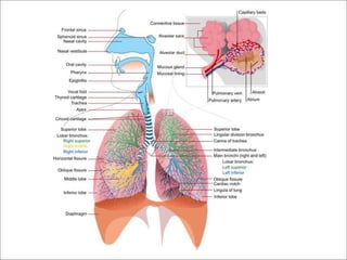
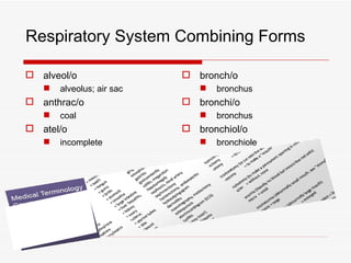
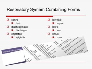
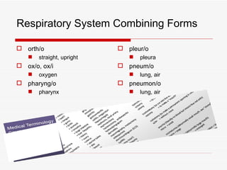
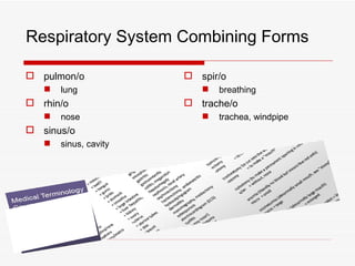
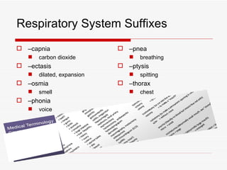
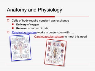
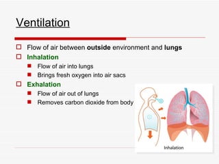
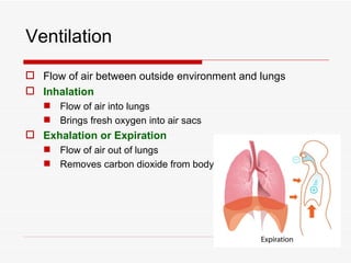
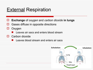
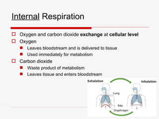
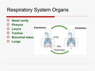
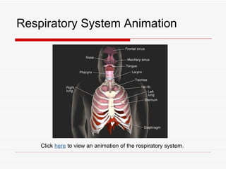
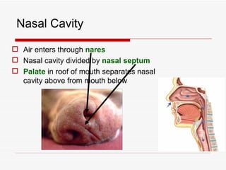
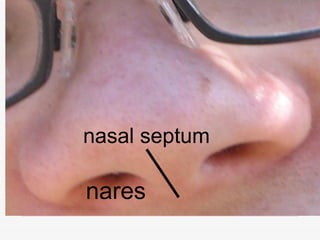
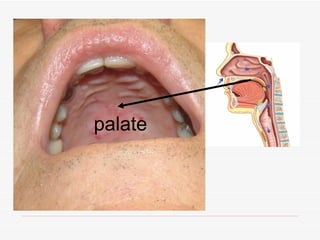
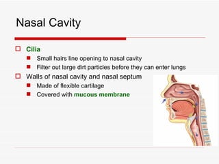
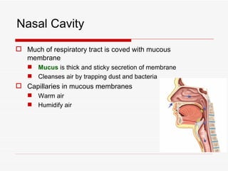
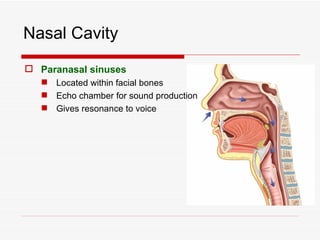
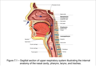
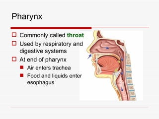
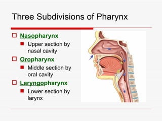
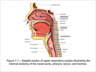
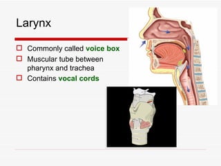
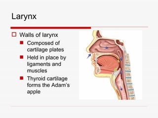
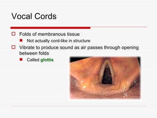
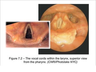
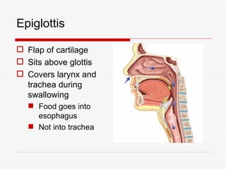
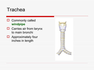
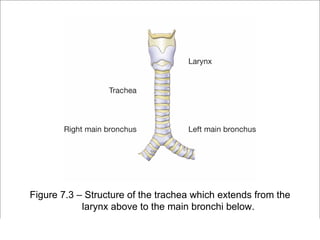
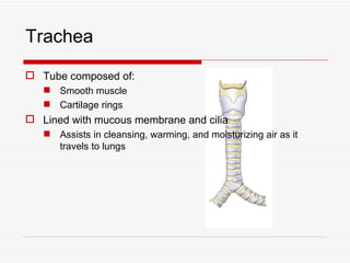
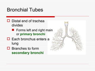
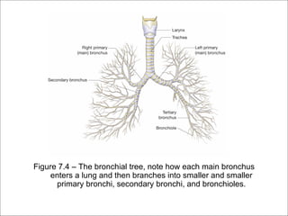
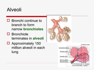
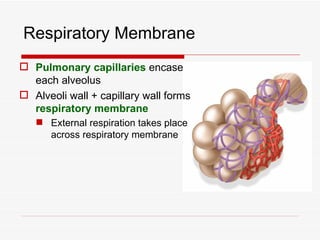
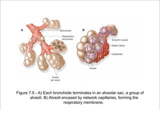
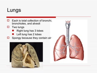
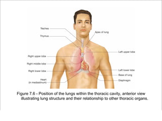
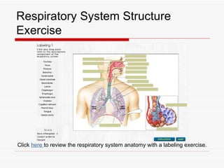
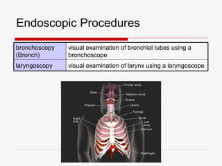
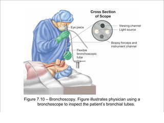
The respiratory system has several functions including inhaling oxygen and exhaling carbon dioxide. It is comprised of organs like the nasal cavity, pharynx, larynx, trachea, bronchial tubes, and lungs. The nasal cavity warms and humidifies air before it reaches the lungs. The pharynx is shared by the respiratory and digestive systems. The larynx contains the vocal cords which produce sound. The trachea transports air to the bronchi and lungs. In the lungs, bronchioles divide into alveoli where gas exchange occurs across the respiratory membrane.
In humans, the respiratory tract is the part of the anatomy of the respiratory system involved with the process of respiration. Air is breathed in through the nose or the mouth. In the nasal cavity, a layer of mucous membrane acts as a filter and traps pollutants and other harmful substances found in the air.
The respiratory system is the network of organs and tissues that help you breathe. It includes your airways, lungs, and blood vessels. The muscles that power your lungs are also part of the respiratory system. These parts work together to move oxygen throughout the body and clean out waste gases like carbon dioxide.
This PPT covers Anatomy and Physiology of respiratory system. Anatomy of respiratory organs, Mechanism of respiration, Internal Respiration, external respiration, Transport of oxygen in blood, Transport of carbon dioxide in blood, Regulation of respiration, lung volume and lung capacities are explained.
Anatomically the respiratory system is divided into
Upper respiratory tract
From the nostril to the vocal cord
Lower respiratory tract
The lower respiratory tract is from bellow the vocal cord upto the alveoli
Unit-III, Chapter-1- Respiratory System.pptAudumbar Mali
B. Pharm. First Year, Sem:II,
Unit III
Respiratory system 10 hours
Anatomy of respiratory system with special reference to anatomy of lungs,
mechanism of respiration, regulation of respiration
Lung Volumes and capacities transport of respiratory gases, artificial respiration,
and resuscitation methods.
Respiratory system PowerPoint templates designed by skilled, experienced and professional graphic designers that are intended to solve the big issue of time consumption. These are fully editable slides and organized in a way to simplify the most complex topics and present it in an attractive manner. You can easily incorporate charts, diagrams and animations along with the content to these layouts in order to explore new technologies and trends of businesses in a unique way. All editable slides are digitally created to pinpoint and clarify the message, and summarize information regarding topic.
The content in the slide are solely depended upon the syllabus of Purbanchal University for third-semester students. This content of the respiratory system will be enough for B.Pharmacy students studying anatomy and physiology
Anatomy of respiratory system with special reference to anatomy of lungs,
mechanism of respiration, regulation of respiration
Lung Volumes and capacities transport of respiratory gases, artificial respiration,
and resuscitation methods.
a biological system consisting of specific organs and structures used for the process of respiration in an organism, intake and exchange of oxygen and carbon dioxide between an organism and the environment, explore anatomy of the upper and lower respiratory tracts, from nasal passages to the lungs
Title: Sense of Smell
Presenter: Dr. Faiza, Assistant Professor of Physiology
Qualifications:
MBBS (Best Graduate, AIMC Lahore)
FCPS Physiology
ICMT, CHPE, DHPE (STMU)
MPH (GC University, Faisalabad)
MBA (Virtual University of Pakistan)
Learning Objectives:
Describe the primary categories of smells and the concept of odor blindness.
Explain the structure and location of the olfactory membrane and mucosa, including the types and roles of cells involved in olfaction.
Describe the pathway and mechanisms of olfactory signal transmission from the olfactory receptors to the brain.
Illustrate the biochemical cascade triggered by odorant binding to olfactory receptors, including the role of G-proteins and second messengers in generating an action potential.
Identify different types of olfactory disorders such as anosmia, hyposmia, hyperosmia, and dysosmia, including their potential causes.
Key Topics:
Olfactory Genes:
3% of the human genome accounts for olfactory genes.
400 genes for odorant receptors.
Olfactory Membrane:
Located in the superior part of the nasal cavity.
Medially: Folds downward along the superior septum.
Laterally: Folds over the superior turbinate and upper surface of the middle turbinate.
Total surface area: 5-10 square centimeters.
Olfactory Mucosa:
Olfactory Cells: Bipolar nerve cells derived from the CNS (100 million), with 4-25 olfactory cilia per cell.
Sustentacular Cells: Produce mucus and maintain ionic and molecular environment.
Basal Cells: Replace worn-out olfactory cells with an average lifespan of 1-2 months.
Bowman’s Gland: Secretes mucus.
Stimulation of Olfactory Cells:
Odorant dissolves in mucus and attaches to receptors on olfactory cilia.
Involves a cascade effect through G-proteins and second messengers, leading to depolarization and action potential generation in the olfactory nerve.
Quality of a Good Odorant:
Small (3-20 Carbon atoms), volatile, water-soluble, and lipid-soluble.
Facilitated by odorant-binding proteins in mucus.
Membrane Potential and Action Potential:
Resting membrane potential: -55mV.
Action potential frequency in the olfactory nerve increases with odorant strength.
Adaptation Towards the Sense of Smell:
Rapid adaptation within the first second, with further slow adaptation.
Psychological adaptation greater than receptor adaptation, involving feedback inhibition from the central nervous system.
Primary Sensations of Smell:
Camphoraceous, Musky, Floral, Pepperminty, Ethereal, Pungent, Putrid.
Odor Detection Threshold:
Examples: Hydrogen sulfide (0.0005 ppm), Methyl-mercaptan (0.002 ppm).
Some toxic substances are odorless at lethal concentrations.
Characteristics of Smell:
Odor blindness for single substances due to lack of appropriate receptor protein.
Behavioral and emotional influences of smell.
Transmission of Olfactory Signals:
From olfactory cells to glomeruli in the olfactory bulb, involving lateral inhibition.
Primitive, less old, and new olfactory systems with different path
Recomendações da OMS sobre cuidados maternos e neonatais para uma experiência pós-natal positiva.
Em consonância com os ODS – Objetivos do Desenvolvimento Sustentável e a Estratégia Global para a Saúde das Mulheres, Crianças e Adolescentes, e aplicando uma abordagem baseada nos direitos humanos, os esforços de cuidados pós-natais devem expandir-se para além da cobertura e da simples sobrevivência, de modo a incluir cuidados de qualidade.
Estas diretrizes visam melhorar a qualidade dos cuidados pós-natais essenciais e de rotina prestados às mulheres e aos recém-nascidos, com o objetivo final de melhorar a saúde e o bem-estar materno e neonatal.
Uma “experiência pós-natal positiva” é um resultado importante para todas as mulheres que dão à luz e para os seus recém-nascidos, estabelecendo as bases para a melhoria da saúde e do bem-estar a curto e longo prazo. Uma experiência pós-natal positiva é definida como aquela em que as mulheres, pessoas que gestam, os recém-nascidos, os casais, os pais, os cuidadores e as famílias recebem informação consistente, garantia e apoio de profissionais de saúde motivados; e onde um sistema de saúde flexível e com recursos reconheça as necessidades das mulheres e dos bebês e respeite o seu contexto cultural.
Estas diretrizes consolidadas apresentam algumas recomendações novas e já bem fundamentadas sobre cuidados pós-natais de rotina para mulheres e neonatos que recebem cuidados no pós-parto em unidades de saúde ou na comunidade, independentemente dos recursos disponíveis.
É fornecido um conjunto abrangente de recomendações para cuidados durante o período puerperal, com ênfase nos cuidados essenciais que todas as mulheres e recém-nascidos devem receber, e com a devida atenção à qualidade dos cuidados; isto é, a entrega e a experiência do cuidado recebido. Estas diretrizes atualizam e ampliam as recomendações da OMS de 2014 sobre cuidados pós-natais da mãe e do recém-nascido e complementam as atuais diretrizes da OMS sobre a gestão de complicações pós-natais.
O estabelecimento da amamentação e o manejo das principais intercorrências é contemplada.
Recomendamos muito.
Vamos discutir essas recomendações no nosso curso de pós-graduação em Aleitamento no Instituto Ciclos.
Esta publicação só está disponível em inglês até o momento.
Prof. Marcus Renato de Carvalho
www.agostodourado.com
Flu Vaccine Alert in Bangalore Karnatakaaddon Scans
As flu season approaches, health officials in Bangalore, Karnataka, are urging residents to get their flu vaccinations. The seasonal flu, while common, can lead to severe health complications, particularly for vulnerable populations such as young children, the elderly, and those with underlying health conditions.
Dr. Vidisha Kumari, a leading epidemiologist in Bangalore, emphasizes the importance of getting vaccinated. "The flu vaccine is our best defense against the influenza virus. It not only protects individuals but also helps prevent the spread of the virus in our communities," he says.
This year, the flu season is expected to coincide with a potential increase in other respiratory illnesses. The Karnataka Health Department has launched an awareness campaign highlighting the significance of flu vaccinations. They have set up multiple vaccination centers across Bangalore, making it convenient for residents to receive their shots.
To encourage widespread vaccination, the government is also collaborating with local schools, workplaces, and community centers to facilitate vaccination drives. Special attention is being given to ensuring that the vaccine is accessible to all, including marginalized communities who may have limited access to healthcare.
Residents are reminded that the flu vaccine is safe and effective. Common side effects are mild and may include soreness at the injection site, mild fever, or muscle aches. These side effects are generally short-lived and far less severe than the flu itself.
Healthcare providers are also stressing the importance of continuing COVID-19 precautions. Wearing masks, practicing good hand hygiene, and maintaining social distancing are still crucial, especially in crowded places.
Protect yourself and your loved ones by getting vaccinated. Together, we can help keep Bangalore healthy and safe this flu season. For more information on vaccination centers and schedules, residents can visit the Karnataka Health Department’s official website or follow their social media pages.
Stay informed, stay safe, and get your flu shot today!
These simplified slides by Dr. Sidra Arshad present an overview of the non-respiratory functions of the respiratory tract.
Learning objectives:
1. Enlist the non-respiratory functions of the respiratory tract
2. Briefly explain how these functions are carried out
3. Discuss the significance of dead space
4. Differentiate between minute ventilation and alveolar ventilation
5. Describe the cough and sneeze reflexes
Study Resources:
1. Chapter 39, Guyton and Hall Textbook of Medical Physiology, 14th edition
2. Chapter 34, Ganong’s Review of Medical Physiology, 26th edition
3. Chapter 17, Human Physiology by Lauralee Sherwood, 9th edition
4. Non-respiratory functions of the lungs https://academic.oup.com/bjaed/article/13/3/98/278874
The prostate is an exocrine gland of the male mammalian reproductive system
It is a walnut-sized gland that forms part of the male reproductive system and is located in front of the rectum and just below the urinary bladder
Function is to store and secrete a clear, slightly alkaline fluid that constitutes 10-30% of the volume of the seminal fluid that along with the spermatozoa, constitutes semen
A healthy human prostate measures (4cm-vertical, by 3cm-horizontal, 2cm ant-post ).
It surrounds the urethra just below the urinary bladder. It has anterior, median, posterior and two lateral lobes
It’s work is regulated by androgens which are responsible for male sex characteristics
Generalised disease of the prostate due to hormonal derangement which leads to non malignant enlargement of the gland (increase in the number of epithelial cells and stromal tissue)to cause compression of the urethra leading to symptoms (LUTS
Ethanol (CH3CH2OH), or beverage alcohol, is a two-carbon alcohol
that is rapidly distributed in the body and brain. Ethanol alters many
neurochemical systems and has rewarding and addictive properties. It
is the oldest recreational drug and likely contributes to more morbidity,
mortality, and public health costs than all illicit drugs combined. The
5th edition of the Diagnostic and Statistical Manual of Mental Disorders
(DSM-5) integrates alcohol abuse and alcohol dependence into a single
disorder called alcohol use disorder (AUD), with mild, moderate,
and severe subclassifications (American Psychiatric Association, 2013).
In the DSM-5, all types of substance abuse and dependence have been
combined into a single substance use disorder (SUD) on a continuum
from mild to severe. A diagnosis of AUD requires that at least two of
the 11 DSM-5 behaviors be present within a 12-month period (mild
AUD: 2–3 criteria; moderate AUD: 4–5 criteria; severe AUD: 6–11 criteria).
The four main behavioral effects of AUD are impaired control over
drinking, negative social consequences, risky use, and altered physiological
effects (tolerance, withdrawal). This chapter presents an overview
of the prevalence and harmful consequences of AUD in the U.S.,
the systemic nature of the disease, neurocircuitry and stages of AUD,
comorbidities, fetal alcohol spectrum disorders, genetic risk factors, and
pharmacotherapies for AUD.
Acute scrotum is a general term referring to an emergency condition affecting the contents or the wall of the scrotum.
There are a number of conditions that present acutely, predominantly with pain and/or swelling
A careful and detailed history and examination, and in some cases, investigations allow differentiation between these diagnoses. A prompt diagnosis is essential as the patient may require urgent surgical intervention
Testicular torsion refers to twisting of the spermatic cord, causing ischaemia of the testicle.
Testicular torsion results from inadequate fixation of the testis to the tunica vaginalis producing ischemia from reduced arterial inflow and venous outflow obstruction.
The prevalence of testicular torsion in adult patients hospitalized with acute scrotal pain is approximately 25 to 50 percent
TEST BANK for Operations Management, 14th Edition by William J. Stevenson, Ve...kevinkariuki227
TEST BANK for Operations Management, 14th Edition by William J. Stevenson, Verified Chapters 1 - 19, Complete Newest Version.pdf
Prix Galien International 2024 Forum ProgramLevi Shapiro
June 20, 2024, Prix Galien International and Jerusalem Ethics Forum in ROME. Detailed agenda including panels:
- ADVANCES IN CARDIOLOGY: A NEW PARADIGM IS COMING
- WOMEN’S HEALTH: FERTILITY PRESERVATION
- WHAT’S NEW IN THE TREATMENT OF INFECTIOUS,
ONCOLOGICAL AND INFLAMMATORY SKIN DISEASES?
- ARTIFICIAL INTELLIGENCE AND ETHICS
- GENE THERAPY
- BEYOND BORDERS: GLOBAL INITIATIVES FOR DEMOCRATIZING LIFE SCIENCE TECHNOLOGIES AND PROMOTING ACCESS TO HEALTHCARE
- ETHICAL CHALLENGES IN LIFE SCIENCES
- Prix Galien International Awards Ceremony
These lecture slides, by Dr Sidra Arshad, offer a quick overview of physiological basis of a normal electrocardiogram.
Learning objectives:
1. Define an electrocardiogram (ECG) and electrocardiography
2. Describe how dipoles generated by the heart produce the waveforms of the ECG
3. Describe the components of a normal electrocardiogram of a typical bipolar leads (limb II)
4. Differentiate between intervals and segments
5. Enlist some common indications for obtaining an ECG
Study Resources:
1. Chapter 11, Guyton and Hall Textbook of Medical Physiology, 14th edition
2. Chapter 9, Human Physiology - From Cells to Systems, Lauralee Sherwood, 9th edition
3. Chapter 29, Ganong’s Review of Medical Physiology, 26th edition
4. Electrocardiogram, StatPearls - https://www.ncbi.nlm.nih.gov/books/NBK549803/
5. ECG in Medical Practice by ABM Abdullah, 4th edition
6. ECG Basics, http://www.nataliescasebook.com/tag/e-c-g-basics
New Directions in Targeted Therapeutic Approaches for Older Adults With Mantl...i3 Health
i3 Health is pleased to make the speaker slides from this activity available for use as a non-accredited self-study or teaching resource.
This slide deck presented by Dr. Kami Maddocks, Professor-Clinical in the Division of Hematology and
Associate Division Director for Ambulatory Operations
The Ohio State University Comprehensive Cancer Center, will provide insight into new directions in targeted therapeutic approaches for older adults with mantle cell lymphoma.
STATEMENT OF NEED
Mantle cell lymphoma (MCL) is a rare, aggressive B-cell non-Hodgkin lymphoma (NHL) accounting for 5% to 7% of all lymphomas. Its prognosis ranges from indolent disease that does not require treatment for years to very aggressive disease, which is associated with poor survival (Silkenstedt et al, 2021). Typically, MCL is diagnosed at advanced stage and in older patients who cannot tolerate intensive therapy (NCCN, 2022). Although recent advances have slightly increased remission rates, recurrence and relapse remain very common, leading to a median overall survival between 3 and 6 years (LLS, 2021). Though there are several effective options, progress is still needed towards establishing an accepted frontline approach for MCL (Castellino et al, 2022). Treatment selection and management of MCL are complicated by the heterogeneity of prognosis, advanced age and comorbidities of patients, and lack of an established standard approach for treatment, making it vital that clinicians be familiar with the latest research and advances in this area. In this activity chaired by Michael Wang, MD, Professor in the Department of Lymphoma & Myeloma at MD Anderson Cancer Center, expert faculty will discuss prognostic factors informing treatment, the promising results of recent trials in new therapeutic approaches, and the implications of treatment resistance in therapeutic selection for MCL.
Target Audience
Hematology/oncology fellows, attending faculty, and other health care professionals involved in the treatment of patients with mantle cell lymphoma (MCL).
Learning Objectives
1.) Identify clinical and biological prognostic factors that can guide treatment decision making for older adults with MCL
2.) Evaluate emerging data on targeted therapeutic approaches for treatment-naive and relapsed/refractory MCL and their applicability to older adults
3.) Assess mechanisms of resistance to targeted therapies for MCL and their implications for treatment selection
2. Respiratory System at a Glance
Functions of the Respiratory System
Inhale fresh air into lungs
Exchange oxygen for carbon dioxide
Exhale stale air
3. Respiratory System at a Glance
Organs of the Respiratory System
Nasal cavity
Pharynx
Larynx
Trachea
Bronchial tubes
Lungs
10. Anatomy and Physiology
Cells of body require constant gas exchange
Delivery of oxygen
Removal of carbon dioxide
Respiratory system works in conjunction with ….
Cardiovascular system to meet this need
11. Respiration
Must be continuous to meet cells’ needs
Subdivided into three distinct parts:
Ventilation
Inhalation
Exhalation
12. Ventilation
Flow of air between outside environment and lungs
Inhalation
Flow of air into lungs
Brings fresh oxygen into air sacs
Exhalation
Flow of air out of lungs
Removes carbon dioxide from body
13. Ventilation
Flow of air between outside environment and lungs
Inhalation
Flow of air into lungs
Brings fresh oxygen into air sacs
Exhalation or Expiration
Flow of air out of lungs
Removes carbon dioxide from body
14. External Respiration
Exchange of oxygen and carbon dioxide in lungs
Gases diffuse in opposite directions
Oxygen
Leaves air sacs and enters blood stream
Carbon dioxide
Leaves blood stream and enters air sacs
15. Internal Respiration
Oxygen and carbon dioxide exchange at cellular level
Oxygen
Leaves bloodstream and is delivered to tissue
Used immediately for metabolism
Carbon dioxide
Waste product of metabolism
Leaves tissue and enters bloodstream
18. Nasal Cavity
Air enters through nares
Nasal cavity divided by nasal septum
Palate in roof of mouth separates nasal
cavity above from mouth below
21. Nasal Cavity
Cilia
Small hairs line opening to nasal cavity
Filter out large dirt particles before they can enter lungs
Walls of nasal cavity and nasal septum
Made of flexible cartilage
Covered with mucous membrane
22. Nasal Cavity
Much of respiratory tract is coved with mucous
membrane
Mucus is thick and sticky secretion of membrane
Cleanses air by trapping dust and bacteria
Capillaries in mucous membranes
Warm air
Humidify air
23. Nasal Cavity
Paranasal sinuses
Located within facial bones
Echo chamber for sound production
Gives resonance to voice
25. Pharynx
Commonly called throat
Used by respiratory and
digestive systems
At end of pharynx
Air enters trachea
Food and liquids enter
esophagus
26. Three Subdivisions of Pharynx
Nasopharynx
Upper section by
nasal cavity
Oropharynx
Middle section by
oral cavity
Laryngopharynx
Lower section by
larynx
27. Tonsils
Lymphatic tissue
Removes
pathogens in air
and food
Three pairs
Adenoids
Palatine
Lingual
28. Eustachian or Auditory Tube
Opening found in nasopharynx
Other end opens into middle ear
Tube opens with each swallow
Equalizes air pressure between
middle ear and outside
atmosphere
29. Figure 7.1 – Sagittal section of upper respiratory system illustrating the
internal anatomy of the nasal cavity, pharynx, larynx, and trachea.
30. Larynx
Commonly called voice box
Muscular tube between
pharynx and trachea
Contains vocal cords
31. Larynx
Walls of larynx
Composed of
cartilage plates
Held in place by
ligaments and
muscles
Thyroid cartilage
forms the Adam’s
apple
32. Vocal Cords
Folds of membranous tissue
Not actually cord-like in structure
Vibrate to produce sound as air passes through opening
between folds
Called glottis
38. Trachea
Tube composed of:
Smooth muscle
Cartilage rings
Lined with mucous membrane and cilia
Assists in cleansing, warming, and moisturizing air as it
travels to lungs
39. Bronchial Tubes
Distal end of trachea
divides
Forms left and right main
or primary bronchi
Each bronchus enters a
lung
Branches to form
secondary bronchi
40. Figure 7.4 – The bronchial tree, note how each main bronchus
enters a lung and then branches into smaller and smaller
primary bronchi, secondary bronchi, and bronchioles.
41. Alveoli
Bronchi continue to
branch to form
narrow bronchioles
Bronchiole
terminates in alveoli
Approximately 150
million alveoli in each
lung
42. Respiratory Membrane
Pulmonary capillaries encase
each alveolus
Alveoli wall + capillary wall forms
respiratory membrane
External respiration takes place
across respiratory membrane
44. Lungs
Each is total collection of bronchi,
bronchioles, and alveoli
Two lungs
Right lung has 3 lobes
Left lung has 2 lobes
Spongy because they contain air
48. Pleura
Parietal pleura
Outer membrane that lines wall of chest cavity
Visceral pleura
Inner membrane that adheres to surface of lungs
Pleura is folded to form a sac around each lung called
pleural cavity
Serous fluid between two pleural layers reduces friction
when two layers rub together during ventilation
52. Pulmonary Function Tests
It is important to measure actual volume of air flowing in
and out of lungs
Can then determine lung capacity
Respiratory therapist
Measures lung volumes
Pulmonary function tests
53. Lung Volumes
Tidal volume (TV) Volume of air moving in and out of lungs in
a single relaxed breath
Inspiratory reserve Volume of air that can be forcefully inhaled
volume (IRV) after a normal inhale
Expiratory reserve Volume of air that can be forcefully exhaled
volume (ERV) after a normal exhale
Residual volume (RV) Volume of air remaining in lungs after a
forced exhale
55. Respiratory Muscles - Inhalation
Diaphragm
Muscle separates abdomen from thoracic cavity
Contracts and moves down into abdominal cavity
Causes decrease of pressure, negative pressure, within
chest cavity
Air then enters lungs (inhalation) to equalize pressure
56. Respiratory Muscles - Inhalation
Intercostal muscles
Located between ribs
Raise rib cage to further enlarge thoracic cavity
Increases negative pressure
Assists with forceful inhalation
58. Respiratory Muscles - Exhalation
Unforced exhale results when:
Diaphragm and intercostal muscles relax
Thoracic cavity becomes smaller
Creates positive thoracic pressure
Air flows out of lungs to equalize pressure
For forceful exhale
Use additional chest and neck muscles to further decrease
size of thoracic cavity
Create greater positive pressure
59. Respiratory Rate
One of the vital signs (VS), along with heart rate,
temperature, and blood pressure
Respiratory rate dependent on level of CO2 in blood
When CO2 level is high, we breathe more rapidly to
expel excess
If CO2 levels drop, respiratory rate will also drop until
CO2 builds up in bloodstream
60. Respiratory Rates by Age Group
Age Respirations per Minute
Newborn 30–60
1-year-old 18–30
16-year-old 16–20
Adult 12–20
62. Word Building with bronch/o
–gram bronchogram record of bronchus
–itis bronchitis inflammation of bronchus
–plasty bronchoplasty surgical repair of bronchus
–genic bronchogenic produced by bronchus
–scope bronchoscope instrument to view bronchus
involuntary muscle contraction of
–spasm bronchospasm
bronchus
–ial bronchial pertaining to bronchus
63. Word Building with bronchi/o and
diaphragmat/o
–ectasis bronchiectasis dilated bronchus
–ic diaphragmatic pertaining to diaphragm
64. Word Building with laryng/o
–ectomy laryngectomy surgical removal of larynx
–itis laryngitis inflammation of larynx
–plasty laryngoplasty surgical repair of larynx
–scope laryngoscope instrument to view larynx
–eal laryngeal pertaining to larynx
–plegia laryngoplegia paralysis of larynx
65. Word Building with lob/o & pleur/o
–ectomy lobectomy surgical removal of lobe
puncture of pleura to withdraw
–centesis pleurocentesis
fluid
–ectomy pleurectomy surgical removal of pleura
–dynia pleurodynia pleura pain
66. Word Building with ox/o and ox/i
–meter oximeter instrument to measure oxygen
an– –ia anoxia condition of no oxygen
blood condition of insufficient
hypo– –emia hypoxemia
oxygen
hypo– –ia hypoxia condition of insufficient oxygen
67. Word Building with pharyng/o
and pulmon/o
–itis pharyngitis inflammation of pharynx
–eal pharyngeal pertaining to pharynx
inflammation of nose and
nas/o –itis nasopharyngitis
pharynx
–logist pulmonologist lung specialist
–ary pulmonary pertaining to lungs
68. Word Building with rhin/o
–itis rhinitis inflammation of nose
abnormal condition of fungus in
myc/o –osis rhinomycosis
nose
–plasty rhinoplasty surgical repair of nose
–rrhagia rhinorrhagia rapid flow (of blood) from nose
–rrhea rhinorrhea nose discharge
69. Word Building with sinus/o & thorac/o
pan– –itis pansinusitis inflammation of all sinuses
–algia thoracalgia chest pain
–ic thoracic pertaining to the chest
–otomy thoracotomy incision into chest
70. Word Building with trache/o
endo– –al endotracheal pertaining to within trachea
–otomy tracheotomy incision into trachea
–stenosis tracheostenosis narrowing of trachea
71. Word Building with –phonia & –capnia
a– aphonia no voice
dys– dysphonia abnormal voice
a– acapnia no carbon dioxide
hyper– hypercapnia excessive carbon dioxide
72. Word Building with –osmia & –thorax
an– anosmia no smell
hem/o hemothorax blood in the chest
py/o pyothorax pus in the chest
pneum/o pneumothorax air in the chest
73. Word Building with –pnea
a– apnea no breathing
brady– bradypnea slow breathing
dys– dyspnea difficult, labored breathing
eu– eupnea normal breathing
hyper– hyperpnea excessive (deep) breathing
hypo– hypopnea insufficient (shallow) breathing
ortho– orthopnea (sitting) straight breathing
tachy– tachypnea rapid breathing
74. Respiratory System Vocabulary
lack of oxygen; can lead to unconsciousness and
asphyxia
death
withdrawing fluid using suction; removing phlegm from
aspiration
patient’s airway; inhaling food or liquid into trachea
Cheyne-
abnormal breathing pattern with long periods of apnea
Stokes
followed by deep & rapid breathing
respiration
abnormal widening and thickening of fingers due to
clubbing
chronic oxygen deficiency
75. Respiratory System Vocabulary
cyanosis blue skin caused by low oxygen in blood
epistaxis a nosebleed
hemoptysis cough up blood or blood-stained sputum
hyperventilation breathing too fast and too deep
hypoventilation breathing too slow and too shallow
branch of medicine involving diagnosis and
internal medicine treatment of diseases of internal organs;
physician is an internist
76. Respiratory System Vocabulary
two-pronged plastic device to deliver
nasal cannula
oxygen into the nose
difficulty breathing made worse by lying
orthopnea
flat; patient breaths better sitting up
branch of medicine involving diagnosis and
otorhinolaryngology treatment of diseases of the ear, nose, and
throat
77. Respiratory System Vocabulary
patent open or unblocked
using fingers to tap on surface to determine
percussion
condition beneath surface
phlegm thick mucus secreted by respiratory tract
grating sound made when layers of pleura rub
pleural rub
together during respiration
branch of medicine involving diagnosis and
pulmonology treatment of diseases of respiratory system;
physician is a pulmonologist
78. Respiratory System Vocabulary
abnormal crackling sound during inspiration; indicates
rales
fluid or mucus in airway
musical sound during expiration; caused by bronchial
rhonchi
tube spasms
respiratory allied health specialty; assists with respiratory and
therapy cardiopulmonary disorders
shortness of indicates that a patient is having difficulty breathing;
breath (SOB) also called dyspnea
80. Respiratory System Vocabulary
sputum phlegm coughed up from respiratory tract
harsh, high-pitched breath sound; indicates obstruction
stridor
in the airway
thoracic branch of medicine involving diagnosis and treatment of
surgery diseases of respiratory system using surgical means
81. Upper Respiratory System Pathology
acute respiratory condition in children; characterized
croup
by barking type of cough
bacterial infection characterized by formation of thick
diphtheria
membranous film across throat; high mortality rate
bacterial infection of upper respiratory system;
pertussis
characterized by whooping cough
82. Bronchial Tube Pathology
difficulty breathing caused by bronchospasms,
asthma
dyspnea, coughing, and wheezing
enlarged bronchi due to destruction of bronchial
bronchiectasis
wall; result of infections
bronchogenic
cancerous tumor originating in bronchi
carcinoma
83. Asthma Video
Click here to view a video on asthma.
85. Lung Pathology
adult respiratory acute respiratory failure; characterized by
distress syndrome tachypnea, dyspnea, cyanosis, and
(ARDS) hypoxemia
type of pneumoconiosis; coal dust collecting in
anthracosis
lungs; also called black lung or miner’s lung
type of pneumoconiosis; asbestos fibers
asbestosis
collecting in lungs
86. Lung Pathology
condition in which alveoli in a
atelectasis portion of lung collapses; prevents
gas exchange in lung
chronic
progressive, chronic, and usually
obstructive
irreversible group of conditions; like
pulmonary
emphysema; lungs have decreased
disease
capacity to function
(COPD)
genetic condition; produces very
cystic fibrosis
thick mucus that causes severe
(CF)
congestion in lungs
87. COPD Video
Click here to view a video on chronic obstructive pulmonary diseases.
89. Lung Pathology
chronic lung condition characterized by
emphysema
destruction of alveolar walls
histoplasmosis fungal infection of the lungs
infant respiratory most common in premature infants;
distress syndrome characterized by tachypnea; previously
(IRDS) called hyaline membrane disease
influenza viral infection of respiratory system
90. Lung Pathology
severe bacterial infection causing
Legionnaire’s disease
pneumonia, liver, and kidney damage
less severe but longer lasting form of
Mycoplasma
bacterial pneumonia; also called walking
pneumonia
pneumonia
accumulation of foreign particles, such as
pneumoconiosis
coal dust, in the lungs
91. Lung Pathology
Pneumocystis carinii pneumonia caused by a fungus; an
pneumonia (PCP) opportunistic infection seen in AIDS patients
inflammatory condition of lungs; results in
pneumonia
alveoli filling with fluid
excessive amount of tissue fluid
pulmonary edema
accumulating in the lung tissues
floating blood clot obstructs pulmonary
pulmonary embolism
artery; causes infarct of lung tissue
92. Lung Pathology
formation of fibrous scar tissue in lung;
pulmonary fibrosis
reduced ability to expand lungs
severe acute
acute viral respiratory infection; begins like flu
respiratory
but quickly progresses; very high mortality rate
syndrome (SARS)
type of pneumoconiosis; accumulation of silica
silicosis
dust in lungs
93. Lung Pathology
breathing stops repeatedly during sleep;
sleep apnea
causes drop in oxygen levels
unexpected and unexplained death of
sudden infant death
apparently well infant; stops breathing for
syndrome (SIDS)
unknown reasons
bacterial lung infection; results in inflammation
tuberculosis (TB)
and calcification of lungs
95. Pleural Cavity Pathology
accumulation of pus in pleural
empyema
space; also called pyothorax
accumulation of fluid in pleural
pleural effusion cavity; prevents lungs from
fully expanding
inflammation of pleura;
pleurisy characterized by sharp pain
with each breath
collection of air in pleural
pneumothorax cavity; may result in collapsed
lung
97. Clinical Laboratory Tests
arterial blood blood test of oxygen and carbon dioxide levels in
gases (ABGs) the blood
sputum culture & cultures sputum for bacterial growth, if present,
sensitivity (C&S) then determines best antibiotic to use
sputum cytology examining sputum for malignant cells
98. Diagnostic Imaging
X-ray of lung after inhaling radiopaque
bronchography
substance
chest X-ray X-ray of the organs of the thoracic cavity
pulmonary X-ray of lungs after injecting dye into blood
angiography vessel
nuclear medicine test; radioactive air is inhaled
ventilation-
for ventilation portion; radioactive dye is injected
perfusion scan
for perfusion portion; looks for pulmonary emboli
99. Endoscopic Procedures
bronchoscopy visual examination of bronchial tubes using a
(Bronch) bronchoscope
laryngoscopy visual examination of larynx using a laryngoscope
101. Pulmonary Function Tests
measures oxygen level in
oximetry blood; uses oximeter on
patient’s finger tip
pulmonary group of tests to measure air
function test flow in and out of lungs, lung
(PFT) volumes, and gas exchange
measures lung capacity using
spirometry
a spirometer
102. Oximetry Video
Click here to view a video on performing oximetry.
104. Additional Diagnostic Procedures
monitoring patient
polysomnography sleeping to identify sleep
apnea
test for cystic fibrosis;
this disease causes
sweat test
large amount of salt in
sweat
introducing purified
protein derivative (PPD)
tuberculin skin
under the skin;
tests (TB test)
determines if person has
been exposed to TB
105. Respiratory Therapy
medication suspended in a
aerosol mist and inhaled; delivered by
therapy a nebulizer or metered dose
inhaler (single puff dose)
placing a tube through the
endotracheal
mouth and into the trachea to
intubation
keep airway open
intermittent
method for assisting patients in
positive
breathing with a machine that
pressure
produces an increase in
breathing
positive thoracic pressure
(IPPB)
110. Respiratory Therapy
drainage of bronchial secretions
by placing patient in positions
postural
using gravity to promote
drainage
drainage; cystic fibrosis
treatment
supplement providing additional oxygen
oxygen concentration to improve oxygen
therapy levels in bloodstream
machine that provides artificial
ventilator ventilation for a patient unable to
breathe alone
111. Nasal Cannula Video
Click here to view a video on applying a nasal cannula.
112. Surgical Procedures
surgical puncture of chest
thoracentesis wall to remove fluids; also
called thoracocentesis
insertion of tube (a chest
thoracostomy tube) into chest to drain off
fluid or air
emergency procedure to
create an opening directly
tracheostomy into trachea so person can
breathe easier; also called
tracheotomy
115. Additional Procedures
cardiopulmonary emergency treatment given to persons
resuscitation (CPR) when respiration and heart stop
technique for removing foreign body
Heimlich maneuver
obstructing trachea or pharynx
116. Respiratory System Pharmacology
antibiotic kills bacteria Amoxil, Cipro
blocks histamine released Allegra, Claritan,
antihistamine
during allergy attack Benadryl
Hycodan, Vicks
antitussive relieves urge to cough
Formula 44
117. Respiratory System Pharmacology
relaxes
Proventil,
bronchospas
bronchodilator Ventolin,
ms; treats
Theo-Dur
asthma
reduces
Flonase,
inflammation
corticosteroids Nasonex,
of respiratory
Azmacort
tract
reduces
congestion in Afrin, Drixoral,
decongestant
respiratory Sudafed
system
118. Respiratory System Pharmacology
improves ability to cough
expectorant Robitussin, Mucinex
up mucus
liquefies mucus so it is
mucolytic Mucomyst
easier to cough up
119. Respiratory System Abbreviations
ABGs arterial blood gases
ARDS adult respiratory distress syndrome
Bronch bronchoscopy
CO2 carbon dioxide
COPD chronic obstructive pulmonary disease
CPR cardiopulmonary resuscitation
C&S culture and sensitivity
120. Respiratory System Abbreviations
CTA clear to auscultation
CXR chest X-ray
DOE dyspnea on exertion
DPT diphtheria, pertussis, tetanus
ENT ear, nose, and throat
ERV expiratory reserve volume
FRC functional residual capacity
121. Respiratory System Abbreviations
HMD hyaline membrane disease
IC inspiratory capacity
IPPB intermittent positive pressure breathing
IRDS infant respiratory distress syndrome
IRV inspiratory reserve volume
LLL left lower lung
LUL left upper lung
122. Respiratory System Abbreviations
MDI metered dose inhaler
O2 oxygen
PCP Pneumocystis carinii pneumonia
PFT pulmonary function test
PPD purified protein derivative
R respiration
RA room air
123. Respiratory System Abbreviations
RDS respiratory distress syndrome
RLL right lower lung
RML right middle lobe
RRT registered respiratory therapist
RV residual volume
RUL right upper lung
SARS severe acute respiratory syndrome
124. Respiratory System Abbreviations
SIDS sudden infant death syndrome
SOB shortness of breath
TB tuberculosis
TLC total lung capacity
TPR temperature, pulse, respiration
TV tidal volume
URI upper respiratory infection
VC vital capacity
Why are we combining these systems into a single unit ? Blood and Lymph are both fluids have functions within the immune system. We will begin with a discussion of the different elements found in blood and the function of each as part of the immune response…. Next slide